What Is the Best Management for a Large Tubo-Ovarian Abscess on CT?
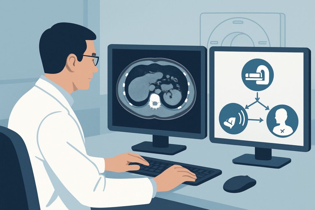
A 28-year-old woman presents to the emergency department with three days of worsening lower abdominal pain, fever to 102.1°F, and nausea. Her white blood cell count is 18,000/μL. On pelvic examination, she has significant cervical motion tenderness and a palpable fullness in the right adnexa. The gynecology team is consulted, and after a contrast-enhanced CT of the abdomen and pelvis confirms a 5 cm, thick-walled, complex fluid collection consistent with a tubo-ovarian abscess (TOA), they initiate broad-spectrum intravenous antibiotics. Now, the critical clinical question arises: Are antibiotics enough, or is procedural intervention required for source control? For this specific scenario—a confirmed tubo-ovarian abscess greater than 3 cm—the American College of Radiology (ACR) rates percutaneous catheter drainage via several routes, including **Transabdominal percutaneous catheter drainage**, as *Usually appropriate*.
Who Fits This Clinical Scenario?
This guidance applies specifically to a woman of childbearing age presenting with a clinical picture highly suggestive of advanced Pelvic Inflammatory Disease (PID), whose diagnostic workup has already progressed to a definitive imaging finding. The key inclusion criteria are:
- A clinical presentation of abdominal or pelvic pain, fever, and leukocytosis.
- Marked tenderness on pelvic examination (e.g., cervical motion tenderness).
- Cross-sectional imaging (typically CT) has already been performed and identifies a well-defined, walled-off fluid collection consistent with a tubo-ovarian abscess.
- The abscess measures greater than 3 cm in its largest dimension.
- The patient has been started on appropriate antibiotic therapy.
This workflow is distinct from other clinical situations that may present similarly. This guidance does not apply to patients with right lower quadrant pain where an appendiceal abscess is the primary concern, nor does it apply to post-surgical collections or abscesses in other locations like the liver or lung. It is also not intended for patients with smaller TOAs (typically less than 3 cm) where a trial of antibiotics alone may be considered, or for patients with ill-defined pelvic inflammation (phlegmon) without a drainable, organized fluid collection.
What Diagnoses Are You Working Up in This Scenario?
At this stage, with a CT scan demonstrating a probable TOA, the primary diagnosis is established with high confidence. The focus of management shifts from diagnosis to treatment, but understanding the underlying pathology and potential mimics is crucial for confirming the intervention plan.
Tubo-Ovarian Abscess (TOA)
This is the leading diagnosis. A TOA is an inflammatory mass involving the fallopian tube, ovary, and occasionally adjacent pelvic organs like the bowel and bladder. It represents a severe complication of Pelvic Inflammatory Disease (PID), where infection ascends from the lower genital tract. The CT finding of a complex, thick-walled, rim-enhancing fluid collection in the adnexa makes this the primary consideration. Source control via drainage is often necessary to facilitate antibiotic efficacy and speed resolution.
Pyosalpinx
This refers to a fallopian tube filled with pus, which can be a precursor to or a component of a larger TOA. On imaging, it may appear as a dilated, fluid-filled, serpentine structure. If it is part of a larger abscess complex, the management strategy is the same. If it is an isolated finding, the need for drainage is still dictated by size and clinical response to antibiotics.
Hemorrhagic Ovarian Cyst
While a hemorrhagic cyst can cause acute pelvic pain and appear as a complex adnexal mass on imaging, it is not typically associated with high fever and marked leukocytosis unless it becomes secondarily infected. The presence of clear infectious signs and symptoms makes a simple hemorrhagic cyst much less likely, though it remains a consideration if the clinical picture is atypical.
Appendiceal or Diverticular Abscess
An abscess originating from the appendix or colon can occasionally be located deep in the pelvis, mimicking a TOA. A careful review of the CT scan for signs like appendiceal inflammation, appendicolith, or colonic diverticula is essential. While the drainage technique is similar, the underlying cause has different surgical and long-term management implications.
Why Is Transabdominal Percutaneous Catheter Drainage the Recommended Study for This Presentation?
For a tubo-ovarian abscess larger than 3 cm, medical management with antibiotics alone has a notable failure rate. The organized, encapsulated nature of the abscess prevents adequate antibiotic penetration, making source control a critical component of treatment. The ACR designates several percutaneous drainage approaches as *Usually appropriate*, highlighting the central role of interventional radiology in managing these complex infections.
The primary goal is to evacuate the purulent material, which reduces the bacterial load, alleviates pressure and pain, and allows antibiotics to work more effectively. Transabdominal percutaneous catheter drainage is a common and effective approach. Under image guidance (typically CT or ultrasound), an interventional radiologist can safely navigate a path through the anterior abdominal wall into the abscess cavity, avoiding bowel, blood vessels, and the bladder. A drainage catheter is then placed and left in situ to allow for continuous drainage over several days.
Other access routes are also rated *Usually appropriate* and are chosen based on the specific location of the abscess and the safest access window:
- Transvaginal Drainage: Ideal for abscesses located deep in the pelvis in the pouch of Douglas, accessible via the vaginal fornix. It avoids an abdominal incision but requires specialized ultrasound equipment.
- Transgluteal Drainage: Used for collections that point posteriorly and are most safely accessed through the gluteal muscles.
- Transrectal Drainage: An alternative for deep pelvic collections abutting the rectum.
In contrast, other management options are considered less optimal for this specific scenario. **Needle aspiration** alone is rated as *May be appropriate* because simply removing fluid once without leaving a catheter is often insufficient for large or viscous abscesses, leading to higher rates of re-accumulation and treatment failure. **Conservative management only** (antibiotics without drainage) is also rated *May be appropriate* but carries a significant risk of failure for abscesses of this size, potentially leading to sepsis, abscess rupture, or the eventual need for more invasive open surgery. Similarly, **Surgical/laparoscopic drainage** is rated *May be appropriate* and is a valid alternative, particularly if percutaneous access is not feasible or if there is a concern for a ruptured abscess, but it is generally more invasive than a percutaneous approach.
What’s Next After Transabdominal Percutaneous Catheter Drainage? Downstream Workflow
Placing a drainage catheter is the beginning, not the end, of the management process. The post-procedure workflow is critical for ensuring a successful outcome.
If the procedure is successful and purulent fluid is drained:
The fluid should immediately be sent for Gram stain, culture, and sensitivity testing to confirm the infectious organism and tailor antibiotic therapy if necessary. The patient’s clinical status—fever curve, white blood cell count, and pain level—should be monitored closely. The drain is connected to a suction bulb or bag, and its output is recorded daily. A significant decrease in drain output and marked clinical improvement are expected within 48-72 hours. Follow-up imaging, often with ultrasound or a limited CT scan with contrast injected through the drain (a “drainogram”), is typically performed in 5-7 days to assess for a decrease in the abscess cavity size. The drain is usually removed at the bedside once output is minimal (e.g., <10-20 mL/day) and the patient has clinically recovered.
If the patient fails to improve:
If fever and leukocytosis persist despite catheter drainage and antibiotics, several possibilities must be considered. The catheter may be malpositioned or occluded, which can be evaluated with a flush or drainogram. There may be other, undrained loculations of the abscess that require a second drainage catheter. Finally, the antibiotic coverage may be inadequate, a determination that can be guided by culture results.
If percutaneous drainage is not technically feasible or fails:
In some cases, a safe percutaneous window to the abscess cannot be found due to overlying bowel or other critical structures. If the patient is not improving on antibiotics alone, the next step is to escalate to surgical consultation. Laparoscopic or open surgical drainage becomes the primary option for source control.
Pitfalls to Avoid (and When to Get Help)
Several common pitfalls can complicate the management of a large TOA. First, delaying the decision for drainage by pursuing an overly long trial of antibiotics alone can increase the risk of sepsis and abscess rupture, a life-threatening emergency. Second, choosing simple needle aspiration over catheter drainage for a large, thick-walled abscess often leads to re-accumulation and prolongs the illness. Third, failing to send the drained fluid for culture misses a key opportunity to optimize antibiotic therapy. Finally, not monitoring drain function and patient response closely can lead to a missed diagnosis of catheter occlusion or an undrained fluid collection. If the patient develops signs of peritonitis or becomes hemodynamically unstable at any point, this constitutes a surgical emergency, and you should escalate immediately for surgical evaluation for exploratory laparotomy.
Related ACR Topics and Tools
This article covers one specific, common scenario in depth. For a comprehensive overview of managing other types of infected collections, or to explore variations on this clinical presentation, the following resources are valuable.
- For breadth across all scenarios in Radiologic Management of Infected Fluid Collections, see our parent guide: Radiologic Management of Infected Fluid Collections: ACR Appropriateness Decoded.
- To explore other clinical presentations and their corresponding ACR recommendations, use the ACR Appropriateness Criteria Lookup.
- For details on the techniques used in image-guided procedures, consult the Imaging Protocol Library.
- To discuss cumulative radiation exposure with patients who have had multiple CT scans, the Radiation Dose Calculator can be a helpful tool.
Frequently Asked Questions
Why is 3 cm the typical size cutoff for considering drainage of a tubo-ovarian abscess?
The 3 cm cutoff is based on clinical studies and experience showing that smaller abscesses have a higher likelihood of resolving with intravenous antibiotics alone. As an abscess grows, its central core becomes more organized and walled-off, which impairs antibiotic penetration. For collections larger than 3-4 cm, the failure rate of medical management alone increases significantly, making procedural drainage for source control a more effective and often necessary strategy to ensure timely recovery and prevent complications.
What’s the difference between transabdominal, transvaginal, and other drainage routes, and how is the best one chosen?
The different routes describe the path the catheter takes to reach the abscess. Transabdominal is through the front of the abdomen, transvaginal is through the top of the vagina, and transgluteal is through the buttock. The best route is determined by the interventional radiologist based on the abscess’s exact location on the CT scan. The goal is to choose the shortest, straightest, and safest path that avoids puncturing bowel, major blood vessels, the bladder, or other vital organs.
Is percutaneous drainage safe in a woman who desires future fertility?
Preserving fertility is a major goal in managing TOA. Percutaneous drainage is generally considered less invasive than open surgery and is aimed at resolving the infection while minimizing damage to the reproductive organs. By effectively treating the abscess, percutaneous drainage can help prevent the severe scarring and adhesion formation that often follows untreated or poorly treated PID and can lead to infertility. However, any severe pelvic infection, regardless of treatment, carries a risk to future fertility.
What if the CT report says ‘complex fluid collection’ but isn’t 100% certain it’s an abscess?
In the context of high fever, pelvic pain, and leukocytosis, a complex adnexal fluid collection is treated as a tubo-ovarian abscess until proven otherwise. The clinical picture is paramount. Percutaneous drainage can be both diagnostic and therapeutic in this situation. Aspiration of purulent material confirms the diagnosis of an abscess, and placement of a drain initiates treatment. If non-purulent fluid is returned (e.g., blood from a hemorrhagic cyst), the drain can be removed, and the diagnosis is clarified.
How long does the drainage catheter typically stay in place?
The duration varies depending on the size of the abscess and the patient’s clinical response, but a typical timeframe is 5 to 10 days. The catheter is left in place until three criteria are met: 1) the patient’s clinical signs of infection (fever, pain, leukocytosis) have resolved, 2) the daily drainage output has decreased to a minimal amount (e.g., less than 10-20 mL per day), and 3) follow-up imaging confirms that the abscess cavity has significantly decreased in size or resolved.
Reviewed by Pouyan Golshani, MD, Interventional Radiologist — May 21, 2026