What Is the Best Management for Bleeding From Small Gastric Varices in Cirrhosis?
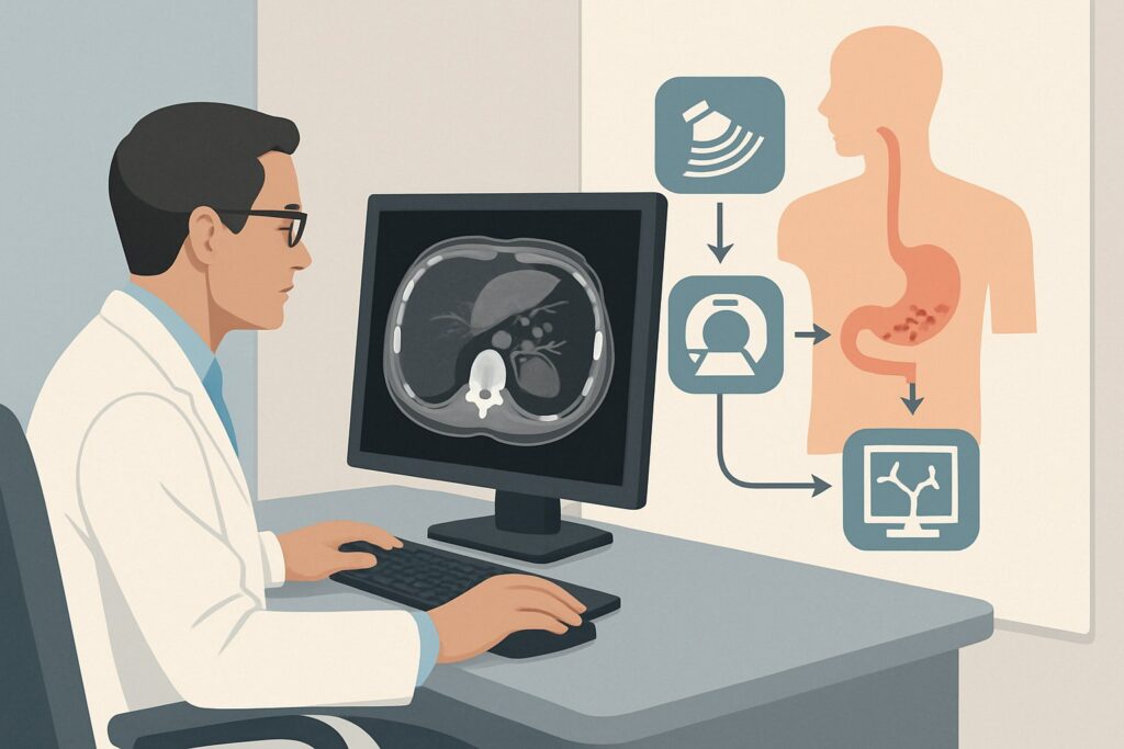
It’s 2 AM, and the GI fellow on call gets a page from the emergency department. A 58-year-old male with a known history of alcohol-related cirrhosis presents with hematemesis. His labs show a hemoglobin of 7.2 g/dL, and his calculated Model for End-stage Liver Disease (MELD) score is 18. An urgent upper endoscopy reveals small, oozing gastric varices in the fundus, and he has moderate ascites on exam. A recent MRI abdomen, done for hepatocellular carcinoma screening, was reviewed and does not show a gastrorenal shunt. The immediate question is not just how to stop the bleeding, but which intervention offers the best balance of efficacy and safety for this specific clinical picture. This article provides a workflow for this exact scenario, guided by the American College of Radiology (ACR) Appropriateness Criteria, which rates Endoscopic management as Usually appropriate.
Who Fits This Clinical Scenario for Gastric Variceal Bleeding?
This guidance is tailored for a precise patient profile. Applying this workflow to a patient who does not meet these criteria can lead to suboptimal or even harmful management decisions.
Inclusion criteria for this workflow:
- Diagnosis: Liver cirrhosis with portal hypertension.
- Presentation: Acute or recent bleeding from gastric varices.
- Variceal Anatomy: The varices are characterized as small and low-flow on endoscopy.
- Patient Status: The patient has moderate ascites and a MELD score of approximately 18, indicating significant but not pre-terminal liver dysfunction.
- Key Anatomic Finding: Prior cross-sectional imaging (like CT or MRI) explicitly shows no evidence of a significant gastrorenal or splenorenal shunt.
Exclusion criteria (patients who require a different workflow):
- Large, High-Flow Varices: Patients with large-caliber, high-flow varices often have complex feeding vessels and a higher risk of catastrophic re-bleeding, frequently making them candidates for more definitive portal decompression like a Transjugular Intrahepatic Portosystemic Shunt (TIPS).
- Presence of a Large Gastrorenal Shunt: If imaging demonstrates a dominant gastrorenal shunt, the patient becomes a candidate for Balloon-occluded Retrograde Transvenous Obliteration (BRTO), a procedure that is specifically contraindicated in this scenario due to the lack of that anatomic pathway.
- Isolated Splenic Vein Occlusion: If the varices are caused by splenic vein thrombosis (left-sided portal hypertension) rather than cirrhosis, the management is entirely different, often involving splenectomy or splenic artery embolization.
What Diagnoses Are You Working Up in This Scenario?
While the immediate source of bleeding is identified as gastric varices, the choice of intervention depends on understanding the underlying pathophysiology and hemodynamic state. The “workup” is less about finding a new diagnosis and more about characterizing the existing one to guide therapy.
Decompensated Cirrhosis with Portal Hypertension
This is the core diagnosis. A MELD score of 18 with moderate ascites confirms decompensation. The gastric varices are a direct consequence of elevated pressures in the portal venous system, which seeks alternative, low-resistance pathways back to the systemic circulation. The management goal is not only to obliterate the bleeding varix but also to do so without dangerously worsening the patient’s fragile liver function.
Sarin Type 1 Gastroesophageal Varices (GOV1) or Type 1 Isolated Gastric Varices (IGV1)
These classifications describe the location and extent of the varices. GOV1 are esophageal varices that extend below the gastroesophageal junction along the lesser curve, while IGV1 are located in the fundus without associated esophageal varices. Small varices of these types are often supplied by the left and posterior gastric veins. Their smaller size and lower flow make them more amenable to targeted obliteration with endoscopic techniques compared to large fundal varices (GOV2) fed by short gastric veins.
Absence of a Spontaneous Portosystemic Shunt
The explicit lack of a gastrorenal shunt is a critical diagnostic finding. Large shunts act as natural, albeit high-risk, decompression pathways. Their absence means the portal pressure is contained, increasing pressure on smaller collaterals like the gastric varices. It also definitively rules out BRTO as a therapeutic option, narrowing the decision tree for the interventional radiologist and gastroenterologist.
Why Is Endoscopic Management a Top Choice for Small, Low-Flow Gastric Varices?
For this specific presentation, the ACR panel rates both Endoscopic management and TIPS as Usually appropriate. However, the clinical nuances of small, low-flow varices often favor endoscopy as the initial approach.
The primary rationale for choosing endoscopic therapy, such as the injection of N-butyl-2-cyanoacrylate (histoacryl glue), is its direct and targeted nature. The endoscopist can visualize the bleeding varix and inject the sclerosant directly into the vessel, causing thrombosis and obliteration at the source. This approach is highly effective for achieving immediate hemostasis in small, low-flow varices without inducing major systemic hemodynamic shifts. It avoids the procedural risks and systemic impact of creating a large portosystemic shunt.
Comparing Alternative Procedures:
- TIPS: While also rated Usually appropriate, TIPS is a more physiologically demanding intervention. It involves creating a shunt between the portal vein and a hepatic vein, effectively decompressing the entire portal system. In a patient with a MELD score of 18 and ascites, this carries a substantial risk of inducing or worsening hepatic encephalopathy. While TIPS is a powerful tool for controlling variceal bleeding and ascites, it is often reserved as a second-line or rescue therapy in this scenario if endoscopic management fails or for patients with larger, higher-risk varices where endoscopy is less likely to succeed.
- BRTO: This procedure is rated Usually not appropriate for one simple reason: it is technically impossible without a suitable gastrorenal or splenorenal shunt. BRTO involves advancing a catheter retrograde from the renal vein into the shunt to block it with a balloon and inject a sclerosant. The scenario’s explicit lack of this anatomy removes BRTO from consideration.
- Partial Splenic Embolization: Rated as May be appropriate, this technique reduces portal inflow by occluding a portion of the splenic artery. It can lower portal pressure and decrease flow to the varices but is generally considered an adjunctive or secondary therapy rather than a primary treatment for active bleeding.
Endoscopic management has the distinct advantage of involving no ionizing radiation, a key consideration in patients who will require repeated imaging over their lifetime.
What’s Next After Endoscopic Management? Downstream Workflow
The patient’s course after the initial intervention determines the subsequent steps. A successful procedure is not the end of management but the beginning of secondary prophylaxis.
- If Endoscopic Management is Successful: If hemostasis is achieved, the patient is admitted to the intensive care unit for close monitoring, resuscitation, and medical management of their liver disease (e.g., octreotide infusion, antibiotics for SBP prophylaxis). The primary focus shifts to secondary prophylaxis to prevent re-bleeding. This typically involves initiation or titration of non-selective beta-blockers. A follow-up endoscopy is often scheduled in the subsequent weeks to confirm varix obliteration and screen for other varices.
- If Endoscopic Management Fails or Re-bleeding Occurs: Failure to control the initial bleed or early re-bleeding is a critical event that necessitates immediate escalation. This patient would then become a prime candidate for the other ‘Usually appropriate’ intervention: TIPS. The initial attempt at a less invasive therapy has failed, and the priority becomes definitive portal decompression to save the patient’s life, accepting the increased risk of hepatic encephalopathy.
- If the Finding is Indeterminate or More Complex: In rare cases, endoscopy might reveal a complex vascular anatomy not appreciated on prior imaging. If the bleeding source is unclear or the varices are larger or higher-flow than anticipated, the next step is typically urgent, high-quality, multiphasic CT angiography. This provides the detailed vascular map needed to plan a definitive radiologic intervention, most often TIPS.
Pitfalls to Avoid (and When to Get Help)
In managing this complex patient, several common errors can compromise outcomes.
- Underestimating the MELD Score’s Significance: A MELD of 18 is not just a number; it signifies a high short-term mortality risk and a low tolerance for procedural stress. All decisions must be weighed against the risk of precipitating further liver decompensation.
- Delaying Intervention: Active variceal bleeding is a medical emergency. Delays in diagnosis, resuscitation, or definitive therapy dramatically increase mortality.
- Using Endoscopic Band Ligation on Gastric Varices: While banding is the standard for esophageal varices, it is generally contraindicated for fundal gastric varices due to a high risk of incomplete obliteration and massive re-bleeding when the band sloughs off. Glue injection is the standard endoscopic technique.
- Not Confirming Lack of Shunt: Relying on an old or poor-quality imaging report can be dangerous. If there is any doubt about the presence of a gastrorenal shunt, a new multiphasic CT should be performed before ruling out BRTO or proceeding with TIPS.
If the patient shows any signs of hemodynamic instability despite initial endoscopic success, or if there is any difficulty in achieving hemostasis, immediate consultation with Interventional Radiology for emergent TIPS is the critical escalation step.
Related ACR Topics and Tools
For a comprehensive overview of all clinical variants and management options, or to explore adjacent clinical scenarios, the following resources are essential.
- For breadth across all scenarios in Radiologic Management of Gastric Varices, see our parent guide: Radiologic Management of Gastric Varices: ACR Appropriateness Decoded.
- Imaging Appropriateness Selector — For exploring different clinical presentations and their recommended imaging pathways.
- Imaging Protocol Library — For detailed technical specifications on imaging studies like multiphasic CT or MRI.
- Radiation Dose Calculator — For discussing cumulative radiation exposure with patients who require frequent imaging.
Frequently Asked Questions
Why is BRTO ‘Usually not appropriate’ for this specific patient?
BRTO (Balloon-occluded Retrograde Transvenous Obliteration) is a procedure that requires a large, naturally occurring shunt—typically a gastrorenal shunt—to access the gastric varices from the venous side. This scenario explicitly states that the patient’s MRI does not demonstrate such a shunt, making the BRTO procedure technically infeasible.
If both endoscopy and TIPS are ‘Usually appropriate’, how do we choose?
The choice is based on a risk-benefit analysis for the individual patient. For small, low-flow varices, endoscopic management (like glue injection) is often preferred first because it is less invasive and has a lower risk of causing systemic complications like hepatic encephalopathy. TIPS is a more definitive but higher-risk procedure, often reserved for cases where endoscopy fails, for re-bleeding, or for patients with larger, high-flow varices from the outset.
Does the presence of moderate ascites change the management plan?
Yes, moderate ascites is a sign of significant portal hypertension and liver decompensation. While it doesn’t change the initial recommendation for endoscopic management of small varices, it makes TIPS a stronger consideration as a secondary option. TIPS is very effective at controlling ascites, which could be an added benefit if the patient ultimately requires it for refractory bleeding.
What if the MELD score was much higher, for example, 25?
A much higher MELD score would significantly increase the procedural risk of any intervention, especially TIPS, which carries a very high risk of acute-on-chronic liver failure and severe encephalopathy in that population. In such cases, the decision-making becomes more complex, often involving a multidisciplinary discussion about the patient’s candidacy for liver transplantation and whether an intervention is a bridge to transplant or a palliative measure.
If the initial endoscopy is successful, is the patient ‘cured’?
No. Successful endoscopic obliteration of a bleeding varix is a treatment for the acute event, not a cure for the underlying portal hypertension. The patient remains at high risk for developing new varices or re-bleeding from other sites. Long-term management with non-selective beta-blockers, surveillance endoscopy, and management of the underlying liver disease is crucial for secondary prevention.
Reviewed by Pouyan Golshani, MD, Interventional Radiologist — May 26, 2026