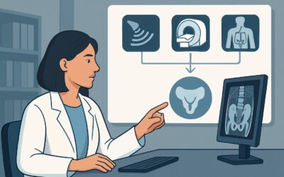
What Is the Initial Imaging for a Patient with Low Risk for Placenta Accreta Spectrum?
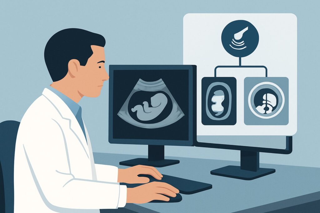
A 32-year-old G2P1 patient presents for her routine 20-week anatomy scan. The sonographer notes an anterior placenta that appears to completely cover the internal cervical os. Reviewing her chart, you confirm she has no history of cesarean delivery, myomectomy, or other uterine surgeries. While placenta previa is the clear initial finding, the possibility of Placenta Accreta Spectrum (PAS) must be considered, even if the pre-test probability is very low. This raises the immediate clinical question: what is the appropriate initial imaging study to confirm placental location and screen for signs of abnormal invasion in this specific low-risk patient? This article details the American College of Radiology (ACR) guided workflow for this scenario, where a `US pregnant uterus transabdominal` is rated Usually Appropriate as the first-line examination.
Who Fits This Clinical Scenario for Placenta Accreta Spectrum Workup?
This guidance applies to pregnant patients, typically undergoing second or third-trimester ultrasound, where the placenta is found to be low-lying or covering the internal cervical os (placenta previa), but who lack the major clinical risk factors for Placenta Accreta Spectrum (PAS). The key to this scenario is the “low-risk” profile. This patient has no history that would significantly raise the index of suspicion for abnormal placental invasion.
Inclusion criteria for this workflow are straightforward:
- A pregnant patient with a sonographically suspected placenta previa or low-lying placenta.
- No known major clinical risk factors for PAS.
The most important exclusion criteria are the risk factors themselves. If your patient has any of the following, they fit a different, higher-risk workflow:
- History of Cesarean Delivery: This is the single greatest risk factor, with risk increasing with the number of prior cesareans. A patient with placenta previa and a history of even one C-section immediately moves into the high-risk category.
- Prior Uterine Surgery: This includes procedures that breach the myometrium, such as myomectomy, hysteroscopic resection of fibroids or septa, or vigorous curettage leading to Asherman syndrome.
- Known Diagnosis of PAS: A patient with a previously established diagnosis of PAS who is undergoing surveillance imaging fits the follow-up scenario, not this initial imaging pathway.
What Diagnoses Are You Working Up in This Low-Risk Scenario?
In a patient without major risk factors, the differential diagnosis is narrow, but the implications of missing a key finding are significant. The primary goal of imaging is to confirm placental location and screen for secondary conditions.
Placenta Previa
This is the most common and most likely diagnosis. The placenta is simply implanted over or very near the internal cervical os. In this low-risk setting, the primary clinical concern is the risk of antepartum or intrapartum hemorrhage. The initial ultrasound serves to definitively confirm the diagnosis and its classification (e.g., complete, marginal, low-lying).
Placenta Accreta Spectrum (PAS)
While the pre-test probability is very low in the absence of risk factors, PAS remains a critical, albeit rare, consideration. Spontaneous or “primary” accreta can occur without prior uterine scarring. The initial ultrasound should include a systematic evaluation for sonographic signs of invasion, such as placental lacunae or loss of the clear space between the myometrium and placenta. In this scenario, the goal is primarily to rule out this consequential diagnosis.
Vasa Previa
This is a distinct but related condition where unprotected fetal vessels run over or in close proximity to the cervix. It is often evaluated concurrently during an ultrasound for placenta previa. Identifying vasa previa is critical, as rupture of these vessels during labor or membrane rupture can lead to rapid fetal exsanguination. Color Doppler imaging is essential for this part of the evaluation.
Normal Variant / “Placental Migration”
Particularly in the second trimester, a diagnosis of placenta previa may be transient. As the lower uterine segment grows and elongates throughout the third trimester, the placental edge often appears to move away from the cervix. The initial ultrasound establishes a baseline for future comparison to assess for this “migration.”
Why Is Transabdominal Ultrasound the Recommended Initial Study for Low-Risk Patients?
The ACR designates `US pregnant uterus transabdominal` as Usually Appropriate for the initial evaluation of a low-risk patient with suspected placenta previa. This recommendation is based on the modality’s high diagnostic utility, safety profile, and accessibility.
The primary role of the transabdominal ultrasound is to provide a global view of the uterus, fetus, and placenta. It is excellent for confirming the presence of placenta previa and assessing the overall placental location (e.g., anterior vs. posterior). It serves as the foundational screening examination. Critically, it involves no ionizing radiation (adult RRL=O 0 mSv) and is safe for both the patient and the fetus at any gestational age.
However, the transabdominal approach alone may not be sufficient. The ACR rates two other ultrasound techniques as May be appropriate for this scenario, and they are often used adjunctively:
- US pregnant uterus transvaginal: This is frequently necessary for a definitive view of the relationship between the leading edge of the placenta and the internal cervical os. A transabdominal view can be obscured by the maternal bladder or fetal position, leading to false-positive diagnoses of previa. Transvaginal imaging provides higher resolution of the lower uterine segment and is considered the gold standard for confirming or refuting the diagnosis of placenta previa.
- US duplex Doppler pregnant uterus: Color and spectral Doppler are essential components of the evaluation. They are used to screen for vasa previa by identifying fetal vessels near the cervix and to look for signs of PAS, such as turbulent flow within placental lacunae or increased vascularity at the uterine-placental interface.
In contrast, MRI is rated as Usually not appropriate for this initial, low-risk presentation. Both `MRI abdomen and pelvis without IV contrast` and `MRI abdomen and pelvis without and with IV contrast` are reserved for cases where ultrasound is indeterminate or when there is a high clinical suspicion for PAS. In a low-risk patient, the diagnostic yield of MRI does not justify its higher cost and lower accessibility compared to a comprehensive ultrasound examination.
What’s Next After Ultrasound? Downstream Workflow
The results of the initial comprehensive ultrasound will guide the subsequent management plan. The decision tree branches based on whether the findings are normal, confirm simple previa, or raise suspicion for a more complex issue.
If the study confirms uncomplicated placenta previa:
The next step is typically serial follow-up ultrasound. A common approach is to repeat the ultrasound around 32 weeks of gestation to assess for placental “migration.” If the previa persists, another scan is often performed around 36 weeks to inform delivery planning. The patient will be counseled on pelvic rest and signs of bleeding. The primary goal shifts from diagnosis to management and planning for a safe delivery, which will likely be a scheduled cesarean section.
If the study is negative (placenta is not previa):
If the comprehensive ultrasound (including transvaginal views) shows the placenta is clearly away from the cervical os, the patient can typically return to routine obstetric care. The initial concern is resolved, and no further specific imaging for placental location is required unless other clinical indications arise.
If the study is suspicious for PAS or vasa previa:
If the ultrasound reveals sonographic signs of accreta (e.g., multiple large lacunae, loss of the retroplacental clear space) or confirms vasa previa, the patient’s risk category changes immediately. This finding triggers an escalation in care. The patient should be referred to a maternal-fetal medicine (MFM) specialist and a center with experience in managing these high-risk conditions. This moves the patient into the High risk for placenta accreta spectrum disorder clinical scenario, where further imaging, often with MRI, may be considered to map the extent of invasion and plan for a multidisciplinary peripartum hysterectomy.
Pitfalls to Avoid (and When to Get Help)
In this low-risk scenario, diagnostic errors often stem from an incomplete or technically limited initial study. Here are several common pitfalls to avoid:
- Relying solely on a transabdominal view: An over-distended bladder or fetal head can compress the lower uterine segment, creating a false appearance of placenta previa. Always proceed to transvaginal imaging if the relationship to the os is unclear.
- Forgetting to use Color Doppler: Failing to activate color Doppler over the cervix is a common oversight that can lead to a missed diagnosis of vasa previa, a true obstetric emergency.
- Assuming low risk means no risk: While the chance of PAS is small, the sonographer and interpreting physician should still perform a systematic search for its signs. A cursory exam focused only on the os is insufficient.
- Misinterpreting a myometrial contraction: A transient focal myometrial contraction can mimic a placenta. If the anatomy is unclear, waiting 20-30 minutes and rescanning can often clarify the finding.
If any sonographic features are suspicious for PAS, or if the imaging is technically limited and inconclusive, the appropriate next step is to escalate care by obtaining a maternal-fetal medicine consultation.
Related ACR Topics and Tools
For a comprehensive overview of all clinical variants related to Placenta Accreta Spectrum Disorder, please consult our parent topic hub article. For further exploration of adjacent scenarios or imaging techniques, the following GigHz resources are available.
- For breadth across all scenarios in Placenta Accreta Spectrum Disorder, see our parent guide: Placenta Accreta Spectrum Disorder: ACR Appropriateness Decoded.
- To explore other clinical presentations, use the ACR Appropriateness Criteria Lookup.
- For details on imaging techniques, visit the Imaging Protocol Library.
- For discussions about cumulative exposure from medical imaging, see the Radiation Dose Calculator.
Frequently Asked Questions
Is a transvaginal ultrasound safe when there is a known placenta previa?
Yes, when performed correctly, transvaginal ultrasound is safe in patients with placenta previa. The probe is inserted carefully into the vaginal canal and does not need to touch the cervix to obtain excellent images of the lower uterine segment and the placental edge’s relationship to the internal os. It provides critical diagnostic information that is often unobtainable with a transabdominal-only approach.
Why isn’t MRI recommended as the first imaging test for a low-risk patient?
In a patient without clinical risk factors for Placenta Accreta Spectrum (PAS), the pre-test probability of the disease is very low. A comprehensive ultrasound is highly effective for diagnosing placenta previa and screening for PAS signs. MRI is more expensive, less accessible, and does not add sufficient diagnostic value in this low-risk setting to justify its use as a first-line tool. It is reserved for cases where ultrasound is inconclusive or when high-risk features are present.
At what gestational age should this initial ultrasound be performed?
The suspicion for placenta previa is often first raised during the routine mid-trimester anatomy scan, typically performed between 18 and 22 weeks. This is the most common time for this initial evaluation to occur. If a patient presents with bleeding later in pregnancy, an ultrasound would be performed then, regardless of gestational age.
What if my patient is low-risk but the ultrasound is indeterminate for accreta?
If a high-quality ultrasound examination (including transvaginal and Doppler imaging) is equivocal or suspicious for PAS, the patient’s risk status should be re-evaluated. The next step is to consult with a maternal-fetal medicine (MFM) specialist. They may recommend a follow-up ultrasound at a tertiary center or proceed to MRI for further characterization, effectively moving the patient into a higher-risk management pathway.
Does an anterior placenta automatically increase the risk for accreta?
No, an anterior placenta by itself does not increase the risk for accreta. The risk elevates dramatically when an anterior placenta previa coexists with a prior uterine scar from a cesarean section, as the placenta is implanted directly over the most vulnerable, scarred part of the uterus. In a patient with no prior uterine surgery, an anterior placenta is a normal variant.
Reviewed by Pouyan Golshani, MD, Interventional Radiologist — May 29, 2026