What Is the Next Imaging Step for a Female Infant with Moderate Antenatal Hydronephrosis?
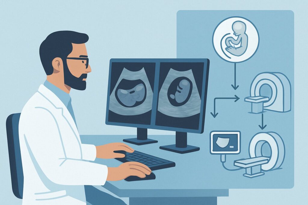
A pediatrician reviews the one-week postnatal ultrasound of a female infant. The prenatal scans had shown hydronephrosis, and this new study confirms it: Society for Fetal Urology (SFU) grade 3 dilation of the left renal pelvis, accompanied by a mildly dilated ureter. The renal parenchyma appears thinned. The immediate question is not just about diagnosis, but about the sequence of the workup. What is the next, most appropriate imaging step to monitor this infant, assess for underlying causes like reflux or obstruction, and guide potential intervention without unnecessary radiation or invasive procedures? This article details the American College of Radiology (ACR) Appropriateness Criteria for this specific scenario, explaining why a follow-up ultrasound is rated Usually Appropriate as the next step in the diagnostic pathway.
Who Fits This Clinical Scenario for Antenatal Hydronephrosis?
This guidance is specifically for a female infant who had hydronephrosis diagnosed on prenatal imaging and whose initial postnatal renal and bladder ultrasound confirms significant findings.
Inclusion criteria for this workflow are:
- Moderate or severe hydronephrosis, defined as SFU grade 3 or 4, or an anterior-posterior renal pelvic diameter (APRPD) greater than 15 mm.
- Associated abnormalities on the initial postnatal ultrasound, such as ureteral dilation (hydroureter), bladder wall thickening, or visible abnormalities of the renal parenchyma (e.g., thinning, increased echogenicity, or cysts).
This workflow is distinct from other related clinical situations. It is crucial to apply this guidance only to patients who fit the criteria above.
Exclusion criteria (patients who follow a different workflow) include:
- Male infants: The differential diagnosis in males is different, with a higher index of suspicion for posterior urethral valves (PUV), which often requires a more urgent and distinct imaging pathway.
- Infants with isolated mild hydronephrosis: Cases with SFU grade 1 or 2 hydronephrosis and an APRPD less than 15 mm, without any other associated abnormalities, typically follow a less intensive monitoring protocol.
- Infants with a normal postnatal ultrasound: If the initial ultrasound after birth shows complete resolution of the antenatally detected hydronephrosis, further imaging is often unnecessary.
What Diagnoses Are You Working Up with Moderate or Severe Hydronephrosis?
In a female infant with significant hydronephrosis and/or hydroureter, the imaging workup aims to differentiate between several potential underlying causes. The goal is to identify conditions that may require surgical intervention or long-term monitoring to preserve renal function.
The most common cause of significant isolated hydronephrosis is a ureteropelvic junction (UPJ) obstruction. This is a functional or anatomic blockage at the point where the kidney’s collecting system (the renal pelvis) joins the ureter. This blockage impedes urine flow, causing it to back up and dilate the kidney. The key clinical question is whether the obstruction is significant enough to impair renal function over time.
Another primary concern, especially when hydroureter is present, is vesicoureteral reflux (VUR). In VUR, urine flows backward from the bladder up the ureters to the kidneys, particularly during voiding. High-grade reflux can cause dilation of the ureters and kidneys and increases the risk of pyelonephritis (kidney infection) and subsequent renal scarring.
A primary megaureter is another possibility. This refers to a dilated ureter that may be due to an obstruction at the ureterovesical junction (where the ureter enters the bladder) or may be non-obstructive and simply due to abnormal development of the ureteral wall. Differentiating between obstructive and non-obstructive megaureter is a key goal of the workup.
Less common but important considerations in a female infant include a ureterocele (a cystic outpouching of the distal ureter into the bladder) or an ectopic ureter, which can be associated with a duplex collecting system. These anomalies can cause both obstruction and reflux.
Why Is a Follow-Up Ultrasound the Recommended Next Step for This Presentation?
For a female infant with moderate to severe hydronephrosis, the ACR designates US kidneys and bladder follow-up in 1-6 months as Usually Appropriate. This non-invasive, radiation-free study serves as the cornerstone of monitoring. It allows for serial assessment of the degree of hydronephrosis, tracking of parenchymal thickness, and evaluation of renal growth. The goal of this initial follow-up is to establish a trend: is the hydronephrosis stable, improving, or worsening? This information is critical for timing further, more specific investigations.
While a follow-up ultrasound is the next step for monitoring, other studies are also rated Usually Appropriate to investigate specific questions raised by the initial findings. The timing of these studies depends on the clinical picture.
- Voiding Cystourethrography (VCUG) or Voiding Urosonography (VUS): These studies are the gold standard for detecting and grading vesicoureteral reflux (VUR). A fluoroscopy voiding cystourethrography (VCUG) uses low-dose X-rays (pediatric radiation relative level ☢☢, 0.03-0.3 mSv) and is widely available. A voiding urosonography (VUS) uses ultrasound with microbubble contrast and involves no ionizing radiation (0 mSv), making it an excellent alternative where available. One of these studies is typically performed early in the workup, especially if hydroureter or recurrent urinary tract infections are present.
- MAG3 Renal Scan: This nuclear medicine study is Usually Appropriate for assessing function. It provides crucial information on differential renal function (how much work each kidney is doing) and quantifies the degree of obstruction by measuring drainage time from the renal pelvis. This study is essential for surgical decision-making in cases of suspected UPJ obstruction. It involves a small amount of radiation (pediatric radiation relative level ☢☢☢, 0.3-3 mSv).
Why are other studies not recommended?
- MRI (Usually Not Appropriate): While MRI provides excellent anatomical detail, it requires sedation in infants, is expensive, and does not offer the functional data on reflux or drainage provided by VCUG and MAG3 scans.
- DTPA Renal Scan (Usually Not Appropriate): The MAG3 tracer is preferred over DTPA in infants because it is more efficiently extracted by the immature kidneys, generally providing higher-quality images and more reliable functional data.
What’s Next After the Follow-Up Ultrasound? Downstream Workflow
The results of the follow-up ultrasound and other initial studies guide the subsequent clinical pathway. The workflow is designed to identify infants who require intervention while avoiding unnecessary procedures in those whose condition is stable or improving.
- If hydronephrosis is worsening or parenchyma is thinning: This finding suggests a significant, ongoing obstruction. The next step is typically a MAG3 renal scan to quantify the differential function and drainage. If the scan confirms a significant obstruction (e.g., differential function <40% in the affected kidney and poor drainage), a referral to a pediatric urologist for potential surgical correction (pyeloplasty) is warranted.
- If hydronephrosis is stable or improving: This is a reassuring sign. The infant can typically be monitored with serial ultrasounds. The frequency of these ultrasounds will decrease over time if stability is maintained. If a VCUG was performed and showed low-grade VUR, the infant may be managed with observation or continuous antibiotic prophylaxis, depending on clinical factors like the occurrence of UTIs.
- If the VCUG is positive for high-grade VUR (Grade IV-V): This finding requires close management by a pediatric urologist. The management strategy may involve continuous antibiotic prophylaxis to prevent UTIs and renal scarring, with regular monitoring. In some cases, surgical correction may be considered.
- If all studies are inconclusive or findings are complex: In rare cases with complex anatomy (e.g., suspected duplex system with an ectopic ureter), an MRI may eventually be considered, but this would be after consultation with pediatric urology and radiology and is not a first-line study.
Pitfalls to Avoid (and When to Get Help)
Navigating the workup for neonatal hydronephrosis requires careful sequencing and interpretation. Here are a few common pitfalls to avoid:
- Prematurely ordering a functional scan: A MAG3 scan performed too early in the first few weeks of life may be inaccurate due to the physiologic immaturity of the neonatal kidneys. It is often best to wait until the infant is 4-6 weeks old.
- Forgetting antibiotic prophylaxis: For infants with high-grade hydronephrosis or known high-grade VUR, prophylactic antibiotics are often recommended to prevent UTIs while the workup is underway. Consult local guidelines or a pediatric specialist.
- Not differentiating from the male scenario: The workup in a female is focused on UPJ obstruction and VUR. In a male, ruling out posterior urethral valves with a VCUG is a higher and more urgent priority.
- Over-reliance on a single ultrasound: The degree of hydronephrosis can fluctuate with the infant’s hydration status. Establishing a trend with serial ultrasounds is more valuable than making a major decision based on one measurement.
If the infant develops a febrile urinary tract infection, shows signs of poor feeding or failure to thrive, or if follow-up imaging demonstrates rapidly worsening hydronephrosis or declining renal function, escalate care promptly to a pediatric urologist.
Related ACR Topics and Tools
This article covers one specific scenario in depth. For a broader view of all clinical variants and the full recommendations, or to explore related imaging tools, please refer to the following resources.
- For breadth across all scenarios in Antenatal Hydronephrosis-Infant, see our parent guide: Antenatal Hydronephrosis-Infant: ACR Appropriateness Decoded.
- ACR Appropriateness Criteria Lookup — for adjacent scenarios
- Imaging Protocol Library — for technique on the recommended study
- Radiation Dose Calculator — for cumulative dose conversations
Frequently Asked Questions
Why is a follow-up ultrasound recommended instead of immediately proceeding to a VCUG or MAG3 scan?
A follow-up ultrasound is recommended to establish a trend. Many cases of moderate hydronephrosis are transient and improve or resolve on their own. Proceeding directly to more invasive or radiation-involving studies may be unnecessary. The follow-up US helps stratify which infants need further investigation (those with worsening hydronephrosis) from those who can be safely monitored.
In this scenario, when should a VCUG be performed?
A VCUG (or VUS) is typically performed early in the workup, often after the initial postnatal ultrasound, especially if there is ureteral dilation (hydroureter) or bladder abnormalities. Its purpose is to rule out vesicoureteral reflux (VUR) as a contributing cause. It is not for monitoring the degree of hydronephrosis itself, but for identifying reflux.
Is there a difference between voiding urosonography (VUS) and a fluoroscopic VCUG?
Yes. Both studies evaluate for vesicoureteral reflux. The main difference is the imaging modality. A fluoroscopic VCUG uses X-rays and has a small amount of radiation. A VUS uses ultrasound with a contrast agent (microbubbles) and involves no ionizing radiation. VUS is an excellent alternative but may be less widely available or require specific pediatric radiology expertise.
What specific finding on the follow-up ultrasound would prompt an urgent MAG3 renal scan?
A significant increase in the degree of hydronephrosis or, more importantly, evidence of progressive renal parenchymal thinning would be a strong indication for a MAG3 scan. These findings suggest that the obstruction is causing ongoing renal damage, and quantifying the function and drainage becomes a priority to guide potential surgical intervention.
Does the workup change if the infant develops a urinary tract infection (UTI)?
Yes. A febrile UTI is a significant event and accelerates the workup. If a VCUG has not yet been performed, it should be done promptly after the infection is treated to assess for VUR, which is a major risk factor for pyelonephritis. The presence of a UTI may also lower the threshold for initiating antibiotic prophylaxis.
Reviewed by Pouyan Golshani, MD, Interventional Radiologist — May 29, 2026