What Is the Next Imaging Step for a Major Fetal Anomaly on Ultrasound?

A 23-week-old G1P0 patient is in your office for a follow-up visit after her routine second-trimester anatomy scan. The initial report from the screening ultrasound is concerning, suggesting a possible complex cardiac anomaly and raising questions about the outflow tracts. The patient is anxious, and you need to determine the most effective next step to clarify the diagnosis, provide an accurate prognosis, and guide management. This clinical decision point—choosing the next imaging study after a suspected major anomaly is found on screening ultrasound—is critical for parental counseling and delivery planning.
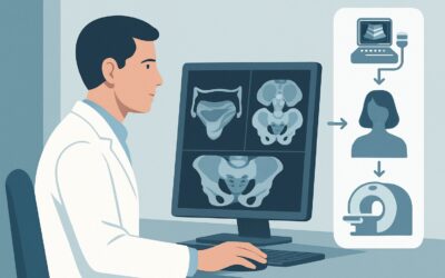
According to the American College of Radiology (ACR) Appropriateness Criteria, several advanced imaging modalities can be considered. For a suspected cardiac defect, a fetal echocardiogram (`US echocardiography fetal`) is rated as Usually Appropriate, providing the detailed functional and anatomic information necessary to manage this specific finding.
Who Fits This Clinical Scenario for Major Fetal Anomalies?
This guidance applies to a specific patient population: a pregnant patient in the second or third trimester who has undergone a screening obstetric ultrasound where a finding suspicious for a major structural anomaly was identified. A major anomaly is defined as one that has significant medical, surgical, or cosmetic consequences for the fetus or newborn. This includes, but is not limited to, suspected congenital heart disease, neural tube defects, abdominal wall defects, or significant renal anomalies. The primary clinical question is which imaging study should be performed next to confirm the finding, delineate the full extent of the anomaly, and search for associated defects.
This workflow is distinct from several related scenarios:
- Initial Screening in Low-Risk or High-Risk Pregnancies: This article is not about the initial anatomy scan itself. It addresses the workup after that initial scan has revealed a potential problem.
- “Soft Markers” for Aneuploidy: This guidance does not apply to the workup of isolated soft markers, such as an echogenic intracardiac focus, mild pyelectasis, or a single umbilical artery. The management of soft markers follows a different diagnostic pathway, often focused on aneuploidy risk assessment rather than immediate advanced structural imaging.
The focus here is on the patient whose screening study has already crossed the threshold of suspicion for a significant, potentially life-altering structural defect.
What Diagnoses Are You Working Up in This Scenario?
When a major anomaly is suspected on a screening ultrasound, the differential diagnosis is broad, but the goal of the next imaging study is to move from suspicion to a specific diagnosis. The findings on the initial scan will guide the differential.
A common and often complex category is congenital heart disease (CHD). The initial scan might show an abnormal four-chamber view, issues with the great vessel outflow tracts, or other signs of cardiac dysfunction. The differential includes specific lesions like Tetralogy of Fallot, transposition of the great arteries, hypoplastic left heart syndrome, and coarctation of the aorta. Confirming the exact type of CHD is paramount, as it directly influences perinatal management, delivery location, and the need for immediate postnatal intervention.
Another critical category includes central nervous system (CNS) anomalies. A screening ultrasound may reveal ventriculomegaly, an abnormal posterior fossa (suggesting Dandy-Walker malformation or Chiari II malformation), or a spinal defect. The differential here includes hydrocephalus, agenesis of thecorpus callosum, and open neural tube defects like myelomeningocele. Advanced imaging is needed to define the precise anatomy, which is crucial for predicting neurologic function and planning for potential fetal or neonatal surgery.
Other major anomalies include abdominal wall defects like omphalocele and gastroschisis, or complex genitourinary anomalies such as posterior urethral valves or multicystic dysplastic kidney. In each case, the follow-up imaging aims to confirm the diagnosis, assess severity, and look for other associated anomalies that might indicate a broader genetic syndrome.
Why Advanced Ultrasound and Fetal MRI Are the Recommended Studies
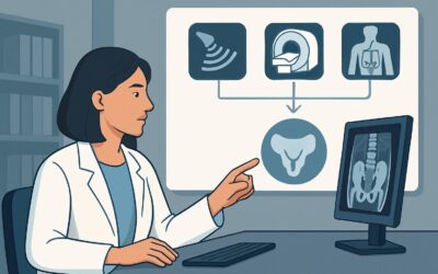
When a major anomaly is suspected, the ACR guidelines designate several imaging modalities as Usually Appropriate, reflecting that the best test depends on the specific organ system in question. All recommended options carry a radiation dose of 0 mSv.
For a suspected cardiac anomaly, US echocardiography fetal is the cornerstone of the workup. This is a highly specialized ultrasound performed by an experienced sonographer and interpreted by a specialist in fetal cardiology or maternal-fetal medicine. It goes far beyond the basic four-chamber and outflow tract views of a screening exam, using techniques like M-mode and Doppler imaging to assess cardiac structure, blood flow patterns, and myocardial function in detail. It is highly sensitive and specific for diagnosing the precise type of congenital heart defect, which is essential for counseling and planning.
For suspected non-cardiac anomalies, particularly those involving the central nervous system, lungs, or complex abdominal/pelvic structures, MRI fetal without IV contrast is also rated Usually Appropriate. Fetal MRI provides superior soft-tissue contrast compared to ultrasound and is not limited by factors like maternal body habitus, fetal position, or oligohydramnios. It is particularly valuable for evaluating the fetal brain, allowing for detailed assessment of gyration, the corpus callosum, and the posterior fossa. It can also quantify lung volumes in cases of congenital diaphragmatic hernia or provide clarity on complex genitourinary anatomy.
A US pregnant uterus transabdominal detailed scan or follow-up scan is also Usually Appropriate. This often serves as the first step, performed at a referral center to confirm the initial finding before proceeding to a more specialized study like fetal echo or MRI.
The one study rated Usually not appropriate is MRI fetal without and with IV contrast. The use of gadolinium-based contrast agents is generally avoided during pregnancy. Gadolinium is known to cross the placenta and enter the fetal circulation, with subsequent excretion into the amniotic fluid. The long-term effects of fetal exposure are not fully understood, and contrast is rarely required to make a diagnosis for a structural fetal anomaly.
What’s Next After Advanced Fetal Imaging? Downstream Workflow
The results of the advanced imaging study are a critical branch point in the patient’s care, initiating a cascade of counseling, planning, and multidisciplinary coordination.
- If the study is positive and confirms a major anomaly: The immediate next step is comprehensive, multidisciplinary counseling. This should involve a maternal-fetal medicine (MFM) specialist and the relevant pediatric subspecialists (e.g., pediatric cardiologist, neurosurgeon, or general surgeon). The team will discuss the diagnosis, prognosis, treatment options (including potential for in-utero intervention), and long-term outcomes. This information allows the family to make informed decisions. Delivery planning becomes paramount, often requiring transfer of care to a tertiary center with an appropriate level of neonatal intensive care (NICU) and immediate access to pediatric surgical services.
- If the study is negative and refutes the initial concern: This provides significant reassurance. However, depending on the nature of the initial finding, a follow-up ultrasound later in gestation may still be recommended to ensure normal interval growth and development. The patient can typically resume routine prenatal care.
- If the study is indeterminate: In some cases, the findings may remain unclear due to technical limitations or the evolving nature of the anomaly. This may necessitate serial imaging (e.g., another ultrasound in 2-4 weeks) to monitor the finding. A second opinion from another fetal imaging expert may also be valuable.
Pitfalls to Avoid (and When to Get Help)
Navigating the workup of a major fetal anomaly requires careful coordination and timely action. Common pitfalls to avoid include:
- Confusing Soft Markers with Major Anomalies: Applying this high-intensity workup to an isolated soft marker can cause unnecessary parental anxiety and resource utilization. Ensure the finding truly represents a major structural concern.
- Delaying Referral: Time is critical. A delay in obtaining the definitive imaging study can limit the time available for comprehensive counseling, genetic testing, and delivery planning, potentially closing the window for certain management options.
- Fragmented Care: Failing to assemble a multidisciplinary team early can lead to conflicting information and suboptimal planning. The MFM specialist should act as the central coordinator for consultations.
If the advanced imaging reveals signs of impending fetal compromise, such as the development of hydrops fetalis (abnormal fluid accumulation), immediate escalation to and consultation with an MFM specialist is required to discuss urgent management strategies.
Related ACR Topics and Tools
For a comprehensive overview of all clinical scenarios related to fetal anomaly screening, including initial imaging for low- and high-risk pregnancies, please see our parent guide. Additional tools can help you apply these guidelines in your practice.
- For breadth across all scenarios in Second and Third Trimester Screening for Fetal Anomaly, see our parent guide: Second and Third Trimester Screening for Fetal Anomaly: ACR Appropriateness Decoded.
- To look up other clinical scenarios, visit the ACR Appropriateness Criteria Lookup tool.
- For details on imaging techniques, explore the Imaging Protocol Library.
- To discuss radiation exposure with patients for other imaging modalities, use the Radiation Dose Calculator.
Frequently Asked Questions
Why is fetal MRI also rated ‘Usually Appropriate’ for major anomalies?
Fetal MRI is rated ‘Usually Appropriate’ because it is the preferred problem-solving tool for complex non-cardiac anomalies. While fetal echocardiography is superior for the heart, MRI provides unparalleled soft-tissue detail of the fetal brain, spine, lungs, and abdomen, making it the test of choice for suspected CNS defects, diaphragmatic hernias, or other complex anatomic abnormalities.
Is there a role for 3D/4D ultrasound in this scenario?
Yes, 3D/4D ultrasound can be a useful adjunct. While the primary diagnosis is typically made with high-resolution 2D ultrasound, fetal echo, or MRI, 3D/4D imaging can be excellent for visualizing surface abnormalities (like cleft lip/palate or spinal defects) and can be a powerful tool for helping parents understand the anomaly. However, it is generally considered supplementary to the core diagnostic studies.
Why is gadolinium contrast ‘Usually Not Appropriate’ in fetal MRI?
Gadolinium-based contrast agents cross the placenta and are excreted by the fetus into the amniotic fluid, where they are re-swallowed, leading to prolonged exposure. The long-term safety and potential for gadolinium deposition in fetal tissues are not well established. Furthermore, for the vast majority of structural fetal anomalies, non-contrast MRI provides sufficient diagnostic information, making the potential risks of contrast outweigh the benefits.
What’s the difference between a ‘detailed’ ultrasound and a ‘follow-up’ ultrasound in this context?
A ‘detailed’ ultrasound (often called a targeted scan) is a comprehensive examination performed at a referral center to specifically evaluate a suspected anomaly. A ‘follow-up’ ultrasound is typically a subsequent scan performed to monitor a known or suspected issue over time, for example, to assess the growth of a structure or check for the development of complications like hydrops. Both are considered ‘Usually Appropriate’ as part of the diagnostic and management pathway.
At what gestational age can a fetal MRI be performed?
Fetal MRI is technically feasible from about 18 weeks gestation onward. However, it is often most informative later in the second and third trimesters, as fetal structures, particularly the brain, become larger and more developed, allowing for more detailed evaluation. The timing of the MRI is a clinical decision based on the specific anomaly suspected and the information needed for counseling and management.
Reviewed by Pouyan Golshani, MD, Interventional Radiologist — May 29, 2026