What Is the Next Step for an Enlarging Solid Pulmonary Nodule in a Smoker?
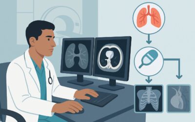
It’s late in your clinic day, and you’re reviewing serial imaging for a 64-year-old patient with a long history of smoking. A chest CT from six months ago showed a 9 mm solid nodule in the right upper lobe. Today’s follow-up scan shows it has grown to 1.2 cm. The nodule is solitary, solid, and clearly enlarging. This progression moves the case beyond simple observation and into the realm of active management. The critical question is how to proceed to a definitive diagnosis. According to the American College of Radiology (ACR) Appropriateness Criteria, the next step for this specific presentation is tissue sampling, with `Percutaneous lung biopsy` rated as `Usually appropriate`.
Who Fits This Clinical Scenario?
This clinical workflow is specifically for an adult patient, typically with a significant smoking history, who presents with a solitary solid pulmonary nodule that has demonstrated interval growth on serial imaging. The current size of the nodule falls between 1 and 3 cm in diameter. The key elements defining this scenario are the combination of nodule morphology (solid), documented growth over time, and a patient risk factor (smoking) that substantially increases the pre-test probability of malignancy.
This guidance does not apply to several similar-but-distinct clinical situations. If the nodule has been stable in size for two or more years, the likelihood of malignancy is very low, and continued observation is often favored. This article also does not cover nodules with different characteristics, such as pure ground-glass or part-solid nodules, which follow separate management algorithms. Finally, patients with a known, active nonpulmonary primary malignancy present a different challenge, as the primary consideration becomes distinguishing a solitary metastasis from a new primary lung cancer.
What Diagnoses Are You Working Up in This Scenario?
When a solid pulmonary nodule enlarges in a person who smokes, the differential diagnosis is heavily weighted toward malignancy, but benign causes must still be considered. The primary goal of obtaining a tissue sample is to differentiate between these possibilities to guide therapy.
Primary Lung Carcinoma: This is the most significant and common concern. Given the risk factors of smoking and documented nodule growth, a new primary lung cancer (such as adenocarcinoma or squamous cell carcinoma) is at the top of the differential. Histologic confirmation is essential for staging, molecular testing, and determining the appropriate treatment plan, which could include surgery, chemotherapy, immunotherapy, or radiation.
Infectious or Inflammatory Granuloma: Benign processes, particularly fungal infections (e.g., histoplasmosis, coccidioidomycosis) or mycobacterial infections (e.g., tuberculosis), can mimic malignancy by presenting as growing solid nodules. A definitive diagnosis via biopsy is crucial to avoid unnecessary oncologic therapy and to initiate appropriate antimicrobial treatment.
Solitary Pulmonary Metastasis: While less common in a patient without a known history of cancer, an enlarging nodule could represent a metastasis from an occult primary tumor elsewhere in the body. The pathology from the biopsy can often suggest the primary source and will trigger a different systemic workup compared to a primary lung cancer.
Carcinoid Tumor: These are well-differentiated neuroendocrine tumors that are less common than other lung cancers but can present as solitary nodules. They typically have a more indolent course but still require tissue diagnosis for confirmation and management, which is often surgical resection.
Why Is Percutaneous Lung Biopsy the Recommended Study for This Presentation?
For an enlarging solid nodule between 1 and 3 cm, the clinical question has shifted from “Is it real?” to “What is it?” The ACR guidelines strongly favor obtaining a tissue diagnosis to answer this question. Both `Percutaneous lung biopsy` and `Endobronchial ultrasound and biopsy` are rated as `Usually appropriate` because they provide a direct path to a histologic diagnosis, which is necessary before initiating treatment.
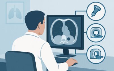
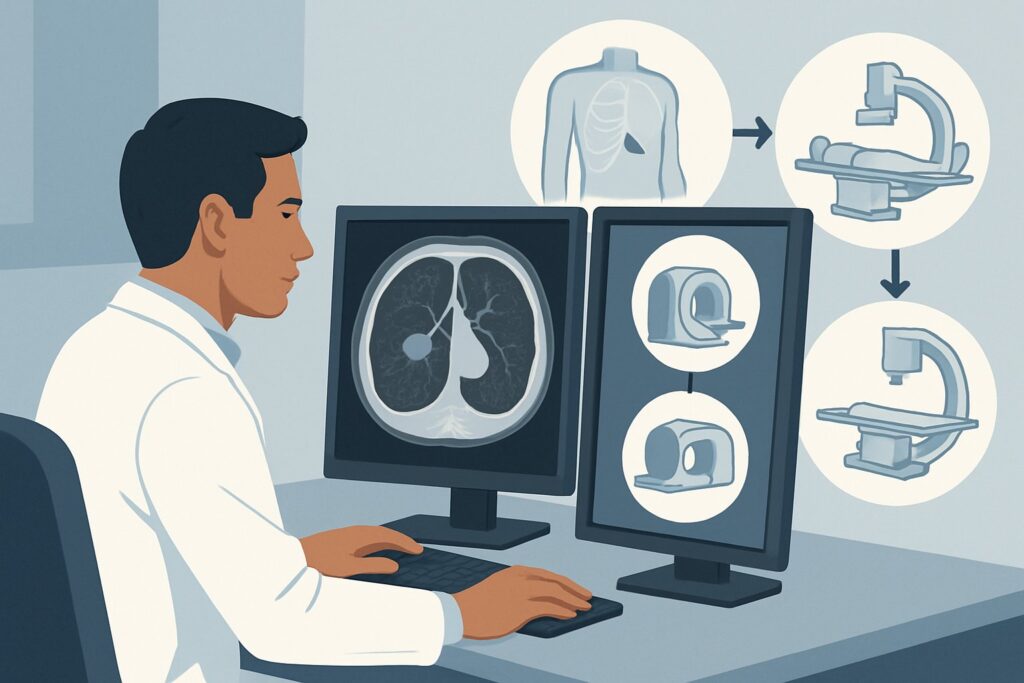
A percutaneous transthoracic needle biopsy, typically performed under Computed Tomography (CT) guidance, is highly effective for nodules located in the periphery of the lungs. It offers a direct, minimally invasive route to the lesion with a high diagnostic yield. Endobronchial ultrasound (EBUS) with transbronchial needle aspiration is an excellent alternative, particularly for nodules that are more central or when there is a need to sample mediastinal or hilar lymph nodes at the same time for staging purposes. The choice between these two equivalent options often depends on the nodule’s exact location, the institution’s technical expertise, and patient-specific factors.
In contrast, other strategies are considered `Usually not appropriate` for this specific scenario:
- Follow-up imaging only: This is `Usually not appropriate` because the nodule has already declared its aggressive potential through documented growth. Further delay in diagnosis could allow a potential malignancy to progress, potentially compromising curative treatment options.
- Surgical management (without prior biopsy): Proceeding directly to surgical resection is `Usually not appropriate`. While surgery may ultimately be the treatment, obtaining a preoperative tissue diagnosis is preferred. This avoids subjecting a patient to the risks of major thoracic surgery for what could be a benign, treatable condition like an infection.
The biopsy procedure itself involves ionizing radiation for guidance, but the dose is localized and managed to be as low as reasonably achievable. For a deeper dive into the principles of low-dose chest CT, the modality used for both initial detection and procedural guidance, our protocol guide covers key techniques: CT Lung Cancer Screening (Low-Dose).
What’s Next After Percutaneous Lung Biopsy? Downstream Workflow
The pathology result from the biopsy is a critical branch point that directs all subsequent management. The downstream workflow depends entirely on whether the diagnosis is malignant, benign, or non-diagnostic.
If the result is positive for malignancy: This finding triggers a full oncologic workup. The next steps typically include a PET/CT scan to assess for metastatic disease and a brain MRI for central nervous system staging. The patient should be referred to a multidisciplinary thoracic oncology team—including thoracic surgery, medical oncology, and radiation oncology—to develop a comprehensive, stage-appropriate treatment plan.
If the result is a specific benign diagnosis: If the biopsy definitively identifies an infectious granuloma or another benign entity, the workflow shifts away from oncology. The patient should be managed according to the specific finding, which may involve consultation with an infectious disease specialist for antimicrobial therapy. A short-interval follow-up CT is often still recommended to ensure the nodule stabilizes or resolves with treatment.
If the result is non-diagnostic: A non-diagnostic or “negative for malignancy” result in this high-risk clinical context cannot be considered conclusive. The pre-test probability of cancer remains high due to the nodule’s growth and the patient’s smoking history. The next step requires careful consideration and multidisciplinary discussion. Options include repeating the biopsy (perhaps with an alternative approach like EBUS if the first was percutaneous) or proceeding to a diagnostic surgical excision (e.g., video-assisted thoracoscopic surgery [VATS] wedge resection).
Pitfalls to Avoid (and When to Get Help)
Navigating the workup of a suspicious pulmonary nodule requires careful attention to detail to avoid common errors. First, do not mistake a non-diagnostic biopsy for a benign result. In a serially enlarging nodule in a smoker, a sample that shows only normal lung tissue or inflammation is insufficient to rule out cancer. Second, always consider the nodule’s location when choosing a biopsy method; a peripheral lesion is best suited for a percutaneous approach, while a central one may be better and more safely accessed via EBUS. Finally, ensure the patient is adequately counseled on procedure-specific risks, such as the risk of pneumothorax with percutaneous biopsy, and that appropriate post-procedure care is arranged. If a biopsy is non-diagnostic or the nodule is in a difficult-to-access location, the case should be escalated for review at a multidisciplinary thoracic tumor board.
Related ACR Topics and Tools
For a comprehensive overview of all clinical variants related to pulmonary nodules, from incidental findings to suspected malignancy, please see our parent guide. It provides a breadth of information that complements this deep-dive article.
- For breadth across all scenarios in Radiologic Management of Pulmonary Nodules and Masses, see our parent guide: Radiologic Management of Pulmonary Nodules and Masses: ACR Appropriateness Decoded.
- To explore other clinical scenarios or refine your imaging orders, use the ACR Appropriateness Criteria Lookup.
- To review technical details for hundreds of imaging studies, visit our Imaging Protocol Library.
- To help discuss radiation exposure with your patients, consult the Radiation Dose Calculator.
Frequently Asked Questions
Why not just go straight to surgery for an enlarging nodule in a smoker?
Proceeding directly to surgery without a tissue diagnosis is rated ‘Usually not appropriate’ by the ACR. This is to avoid the significant risks and recovery time of a major thoracic operation for a condition that might be benign, such as an infection, which could be treated with medication.
What is the difference between a percutaneous biopsy and an EBUS-guided biopsy?
A percutaneous biopsy uses a needle inserted through the skin and chest wall under CT guidance, making it ideal for nodules in the outer (peripheral) parts of the lung. Endobronchial ultrasound (EBUS) uses a specialized bronchoscope passed through the patient’s airway to visualize and biopsy nodules or lymph nodes adjacent to the bronchi, making it better for more central lesions.
If the nodule is 1.5 cm, is follow-up imaging ever an option?
In this specific scenario, where the nodule is serially enlarging, further follow-up imaging is ‘Usually not appropriate.’ The documented growth is the key feature that elevates the risk of malignancy and necessitates a definitive diagnostic step like a biopsy. For a new, stable nodule of that size, follow-up might be an option, but not for one that is actively growing.
What is the risk of a pneumothorax with a percutaneous lung biopsy?
Pneumothorax (a collapsed lung) is the most common complication of percutaneous lung biopsy. The risk varies depending on the nodule’s location and depth, as well as the presence of underlying lung disease like emphysema. While many cases are small and resolve on their own, a significant minority may require a chest tube for re-expansion. Patients should be counseled on this risk beforehand.
Does the nodule’s size within the 1 to 3 cm range change the recommendation?
No, for this specific scenario, the recommendation for tissue sampling holds true for any solid, enlarging nodule within the 1 to 3 cm range. A larger nodule within this range may increase the clinical suspicion for malignancy and the urgency of the workup, but the fundamental next step—obtaining a tissue diagnosis—remains the same.
Reviewed by Pouyan Golshani, MD, Interventional Radiologist — May 30, 2026