What Is the Optimal Imaging for High-Risk Pancreatic Cancer Screening in Adults?
A 58-year-old asymptomatic man presents to your high-risk pancreas clinic for his annual evaluation. His family history is significant for a mother and a brother diagnosed with pancreatic ductal adenocarcinoma (PDAC) before age 60, qualifying him for screening under multiple societal guidelines. He has no abdominal pain, weight loss, or jaundice. You need to select the most effective and safest imaging modality for his surveillance, balancing the need for high-resolution imaging against the risks of cumulative radiation exposure over many years of screening. This article details the American College of Radiology (ACR) workflow for this specific scenario, explaining why certain studies are favored. For this presentation, the ACR rates `MRI abdomen without and with IV contrast` as ‘Usually appropriate’.
Who Qualifies for High-Risk Pancreatic Cancer Screening?
This clinical workflow applies specifically to asymptomatic adults identified as being at high risk for developing pancreatic ductal adenocarcinoma. High-risk status is typically defined by a combination of genetic predisposition and family history. Individuals who fit this scenario often include those with:
- A pathogenic germline variant in a cancer susceptibility gene associated with an increased risk of PDAC (e.g., BRCA1, BRCA2, PALB2, ATM, CDKN2A, or genes associated with Lynch syndrome or Peutz-Jeghers syndrome).
- A strong family history, such as having one or more first-degree relatives with PDAC, particularly if the diagnoses occurred at a young age. This is often categorized as familial pancreatic cancer (FPC).
This guidance is not intended for:
- Symptomatic patients: An adult presenting with new-onset jaundice, unexplained weight loss, or epigastric pain radiating to the back requires a diagnostic workup, not screening. This presentation falls under the ACR variant for clinically suspected PDAC.
- Patients with a known pancreatic mass: Once a mass is identified, the imaging goal shifts to locoregional staging and evaluation for metastatic disease, which are covered in separate ACR guidelines.
- Average-risk individuals: There is no current recommendation for pancreatic cancer screening in the general population.
What Diagnoses Are You Working Up in This Scenario?
In a high-risk screening setting, the primary goal is the early detection of precursor lesions or small, asymptomatic, and potentially resectable cancers. The “differential” in this context is a list of potential findings that require further action.
Pancreatic Ductal Adenocarcinoma (PDAC): The principal target of screening is to identify PDAC at stage I, when it is most amenable to curative-intent surgery. On imaging, this may appear as a subtle, ill-defined, hypoenhancing mass that disrupts the normal pancreatic ductal anatomy.
Intraductal Papillary Mucinous Neoplasm (IPMN): These are common precursor lesions, particularly in high-risk individuals. They appear as cystic lesions that communicate with the pancreatic duct system. Identifying high-risk IPMN features (e.g., main duct involvement, enhancing mural nodule, thickened septa) is a critical component of screening, as these may warrant surgical resection to prevent malignant transformation.
Pancreatic Intraepithelial Neoplasia (PanIN): These are microscopic precursor lesions that are not directly visible on cross-sectional imaging. However, high-grade PanIN can cause secondary changes, such as subtle main pancreatic duct dilation or focal pancreatitis, which may be the only imaging clue to an underlying pre-malignant process.
Other Pancreatic Cysts: Benign or low-risk cysts, such as serous cystadenomas, are also frequently detected. A key role of high-quality imaging is to differentiate these from higher-risk lesions like IPMNs or mucinous cystic neoplasms, thereby avoiding unnecessary invasive procedures.
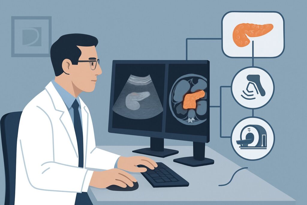
Why Is MRI of the Abdomen the Recommended Screening Study?
For high-risk screening, the ACR designates `MRI abdomen without and with IV contrast` as ‘Usually appropriate’. This recommendation is driven by its superior soft tissue contrast, ability to characterize cystic lesions, and lack of ionizing radiation, a crucial factor in a longitudinal screening program.
The high-resolution, T2-weighted sequences of an MRI, particularly when combined with Magnetic Resonance Cholangiopancreatography (MRCP), provide exquisite detail of the pancreatic ductal system and cystic architecture. This makes MRI highly sensitive for detecting small IPMNs, subtle main duct dilation, and the internal features of cysts that help stratify risk. The addition of IV contrast enhances the detection of small solid masses (early PDAC) and suspicious mural nodules within cysts.
Alternative studies are rated lower for specific reasons in this screening context:
- CT abdomen with IV contrast (multiphase): While also rated ‘Usually appropriate’, its primary drawback is the cumulative radiation dose (☢☢☢☢ 10-30 mSv per scan). For a patient undergoing annual screening for potentially decades, this exposure is a significant concern. While excellent for staging known cancer, its soft tissue contrast for detecting subtle, non-mass-forming precursors can be inferior to MRI.
- US abdomen transabdominal: This is rated ‘Usually not appropriate’ for screening. The pancreas is often obscured by overlying bowel gas, and the sensitivity of transabdominal ultrasound for detecting small pancreatic lesions (<2 cm) is unacceptably low for a high-risk screening program.
The optimal screening study is `MRI abdomen without and with IV contrast with MRCP`, which is also ‘Usually appropriate’ and explicitly includes the non-contrast sequences needed to fully evaluate the biliary and pancreatic ducts. The lack of radiation (O 0 mSv) makes it the preferred modality for repeated surveillance in this asymptomatic population.
What’s Next After MRI? Downstream Workflow
The results of the screening MRI will dictate the subsequent clinical pathway, which often involves a multidisciplinary discussion.
- Negative or Benign-Appearing Findings: If the MRI is entirely normal or shows only simple, benign-appearing cysts without worrisome features, the patient typically continues with routine annual surveillance. The interval may be adjusted based on the specific genetic risk and institutional protocols.
- Positive for a Worrisome Lesion: If the MRI identifies a solid mass suspicious for PDAC, an enhancing mural nodule, or significant main pancreatic duct dilation, the next step is typically Endoscopic Ultrasound (EUS) with Fine-Needle Aspiration (FNA) or Biopsy (FNB). EUS provides high-resolution imaging and allows for tissue sampling to confirm a diagnosis and guide surgical planning.
- Indeterminate Findings: For findings like a small, branch-duct IPMN without high-risk stigmata, the workflow involves continued surveillance. The interval may be shortened (e.g., to 6 months) to assess for stability or growth. If the lesion grows or develops worrisome features on follow-up imaging, the patient would then be referred for EUS. This approach balances the risk of malignancy against the morbidity of invasive procedures.
The goal is to triage patients appropriately, escalating care for high-risk lesions while safely monitoring those with low-risk findings, avoiding unnecessary interventions.
Pitfalls to Avoid (and When to Get Help)
Several pitfalls can compromise the effectiveness of a high-risk pancreatic screening program. A primary error is using an inadequate imaging modality, such as relying on transabdominal ultrasound, which lacks the necessary sensitivity. Another common issue is ordering a non-contrast or single-phase CT, which can easily miss small, hypoenhancing pancreatic adenocarcinomas. It is also crucial to ensure the imaging protocol is optimized for pancreatic evaluation; a generic “MRI abdomen” may not include the high-resolution sequences or MRCP needed to properly assess the ducts and cysts. Finally, failing to adhere to a consistent surveillance schedule can lead to delayed detection of developing lesions. If any high-risk features are identified on imaging, such as a solid mass or a dilated main pancreatic duct with an abrupt cutoff, immediate escalation to a gastroenterologist for EUS or a surgical oncologist is warranted.
Related ACR Topics and Tools
For a comprehensive overview of imaging across all clinical presentations of pancreatic cancer, from initial suspicion to staging and surveillance, please consult our parent topic guide. Additional tools can help you select the right study and understand the technical details.
- For breadth across all scenarios in Screening, Locoregional Assessment, and Surveillance of Pancreatic Ductal Adenocarcinoma, see our parent guide: Screening, Locoregional Assessment, and Surveillance of Pancreatic Ductal Adenocarcinoma: ACR Appropriateness Decoded.
- ACR Appropriateness Criteria Lookup — for adjacent scenarios
- Imaging Protocol Library — for technique on the recommended study
- Radiation Dose Calculator — for cumulative dose conversations
Frequently Asked Questions
Why is MRI preferred over CT for annual pancreatic cancer screening?
MRI is preferred primarily because it does not use ionizing radiation. Since high-risk individuals undergo annual screening for many years, minimizing cumulative radiation exposure is a critical safety consideration. Additionally, MRI with MRCP offers superior soft tissue contrast for characterizing cystic lesions and detecting subtle changes in the pancreatic ducts, which are often the earliest signs of trouble.
Is Endoscopic Ultrasound (EUS) a primary screening tool for high-risk individuals?
While EUS is highly sensitive, it is an invasive procedure with associated risks. Most screening programs use non-invasive imaging like MRI or CT as the primary modality. EUS is typically reserved as a second-line or problem-solving tool to further evaluate abnormalities seen on MRI/CT or to obtain tissue samples via fine-needle aspiration/biopsy.
What if a patient in a screening program has a contraindication to MRI, like a non-compatible pacemaker?
In cases where MRI is contraindicated, a multiphase pancreatic protocol CT abdomen with IV contrast is the best alternative and is also rated ‘Usually appropriate’ by the ACR. The clinical team and patient should discuss the risks and benefits, acknowledging the need for ionizing radiation, and proceed with CT for surveillance.
Does a normal screening MRI mean the patient has no risk of developing pancreatic cancer?
No. A normal (negative) screening exam is reassuring and indicates there is no detectable cancer or high-risk precursor lesion at that time. However, it does not eliminate the patient’s underlying lifetime risk. This is why continued, consistent annual surveillance is essential for this population.
At what age should high-risk screening for pancreatic cancer begin?
The starting age for screening varies based on the specific genetic mutation and family history. Generally, guidelines recommend starting at age 50, or 10 years earlier than the youngest affected first-degree relative. For certain high-risk syndromes like Peutz-Jeghers, screening may begin even earlier. This decision should be made in consultation with a genetic counselor and a high-risk pancreas clinic.
Reviewed by Pouyan Golshani, MD, Interventional Radiologist — May 30, 2026