What Is the Right Imaging for a Critically Ill Patient with Severe Acute Pancreatitis?
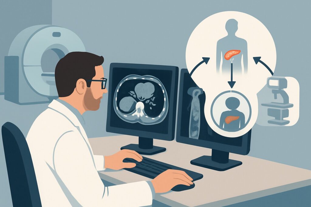
It’s 2 AM in the intensive care unit. Your patient, admitted three days ago with gallstone pancreatitis, is not improving. They meet criteria for Systemic Inflammatory Response Syndrome (SIRS), their BISAP score is climbing, and they are requiring increasing vasopressor support. The primary team has managed the initial resuscitation, but now a critical question emerges: has their inflammation progressed to pancreatic necrosis? Deciding on the next step in management—from antibiotics to potential intervention—hinges on accurately staging the severity of the disease. This is the precise clinical crossroads where choosing the right imaging study is paramount. For this scenario, the American College of Radiology (ACR) rates MRI abdomen without and with IV contrast with MRCP as Usually Appropriate.
Who Fits This Clinical Scenario for Severe Acute Pancreatitis?
This guidance is specifically for a patient with an established diagnosis of acute pancreatitis who, after 48 to 72 hours from symptom onset, is clinically deteriorating or failing to improve. The key inclusion criteria are signs of severe disease, which can be identified through both clinical and objective measures.
Inclusion Criteria:
- Established Diagnosis: The patient has a confirmed diagnosis of acute pancreatitis based on clinical symptoms and elevated lipase/amylase.
- Timeframe: The imaging decision is being made more than 48 to 72 hours after the initial onset of symptoms. Imaging earlier than this can underestimate the extent of potential necrosis.
- Critical Illness: The patient exhibits signs of severe systemic inflammation, such as meeting SIRS criteria, or has high-risk scores on prognostic tools like the Acute Physiology, Age, and Chronic Health Evaluation (APACHE)-II, Bedside Index for Severity in Acute Pancreatitis (BISAP), or the Marshall scoring system.
Exclusion Criteria (Who This Does NOT Apply To):
- Initial Presentation (<48 hours): A patient in the first two days of a first-time presentation of acute pancreatitis falls under a different ACR variant. In that early phase, the primary goal is diagnosis, and the full extent of parenchymal damage may not yet be visible.
- Mild or Improving Pancreatitis: A patient who is clinically stable or improving, with low severity scores and no signs of SIRS, typically does not require advanced cross-sectional imaging to assess for complications.
- Known Necrotizing Pancreatitis with Acute Decompensation: A patient with a previously confirmed diagnosis of necrotizing pancreatitis who experiences a sudden clinical decline (e.g., abrupt drop in hemoglobin) is evaluated under a different workflow focused on specific complications like hemorrhage or infection.
What Diagnoses Are You Working Up in This Critically Ill Patient?
In this scenario, the initial diagnosis of pancreatitis is already known. The purpose of imaging is to identify the life-threatening complications that define severe disease and guide critical care management. The differential is focused on the sequelae of uncontrolled inflammation.
Pancreatic and Peripancreatic Necrosis
This is the most consequential diagnosis to establish. The primary goal of imaging is to differentiate between interstitial edematous pancreatitis (glandular swelling) and necrotizing pancreatitis (non-viable pancreatic tissue). The presence and extent of necrosis dramatically increase morbidity and mortality. Furthermore, determining if necrosis is sterile or has become infected is a pivotal decision point that influences the use of antibiotics and the timing of any potential debridement.
Acute Fluid Collections (APFC vs. ANC)
Severe pancreatitis leads to fluid collections around the pancreas. Imaging helps distinguish between a simple Acute Peripancreatic Fluid Collection (APFC), which contains homogenous fluid, and an Acute Necrotic Collection (ANC), which contains a mixture of fluid and solid necrotic debris. This distinction is critical because ANCs have a much higher risk of infection and may require drainage.
Vascular Complications
The intense inflammatory process can erode into adjacent blood vessels. This can lead to the formation of a pseudoaneurysm (a contained rupture of an artery, most commonly the splenic or gastroduodenal artery) or thrombosis of the splenic or portal veins. A ruptured pseudoaneurysm is a catastrophic, life-threatening hemorrhage that requires immediate intervention.
Biliary and Pancreatic Duct Integrity
For patients with gallstone pancreatitis, a persistent stone obstructing the common bile duct may be driving the ongoing inflammation. Additionally, severe necrosis can lead to a disruption of the main pancreatic duct, creating a “disconnected duct syndrome.” This complication can lead to persistent fluid collections or fistulas and has significant implications for long-term management.
Why Is MRI with MRCP Usually Appropriate for Severe Pancreatitis After 72 Hours?
For the critically ill patient with suspected severe pancreatitis after the initial 48-to-72-hour window, the ACR identifies MRI abdomen without and with IV contrast with MRCP as a Usually Appropriate study. This recommendation is based on its superior diagnostic capabilities for the key clinical questions in this scenario.
The primary advantage of MRI is its exceptional soft-tissue contrast, which allows for a more confident assessment of pancreatic parenchymal viability. By comparing non-contrast images with post-gadolinium contrast images, radiologists can clearly delineate enhancing, viable pancreatic tissue from non-enhancing, necrotic areas. This is often more sensitive than CT, especially for detecting early or patchy necrosis. MRI is also superior in characterizing the contents of peripancreatic fluid collections, reliably distinguishing the simple fluid of an APFC from the complex, debris-filled appearance of an ANC.
The inclusion of Magnetic Resonance Cholangiopancreatography (MRCP) sequences is another key benefit. This non-invasive technique provides detailed, fluid-sensitive images of the biliary tree and pancreatic duct without requiring contrast or radiation. It is highly effective for identifying choledocholithiasis, biliary obstruction, or pancreatic duct disruption—all critical findings that directly influence management.
Finally, an MRI accomplishes this comprehensive evaluation with no ionizing radiation (Adult RRL: O 0 mSv), a significant advantage for patients who may be young or require follow-up imaging.
Why Other Studies Are Rated Lower
- CT abdomen and pelvis with IV contrast: This study is also rated Usually Appropriate and is a very common and acceptable alternative, particularly when MRI is unavailable, contraindicated (e.g., incompatible hardware), or the patient is too unstable for the longer scan time. CT is excellent for detecting necrosis and is faster to acquire. However, its soft-tissue resolution is lower than MRI, making it slightly less sensitive for subtle necrosis or for definitively characterizing fluid collections. It also involves a notable radiation dose (Adult RRL: ☢☢☢ 1-10 mSv).
- US abdomen: This is rated Usually not appropriate in this context. While useful for an initial diagnosis to look for gallstones, its utility in severe pancreatitis is severely limited. The ileus (bowel paralysis) that accompanies severe pancreatitis causes overlying bowel gas to completely obscure the pancreas, rendering the study non-diagnostic for assessing necrosis or deep fluid collections.
What’s Next After the MRI? Downstream Workflow
The results of the MRI will directly guide the next steps in managing the critically ill patient. The report should be used to stratify risk and plan interventions.
- If the MRI shows only interstitial edematous pancreatitis: This is a reassuring finding. The patient, while clinically ill, does not have the added mortality risk of necrosis. Management continues to be supportive care. If they fail to improve, other causes for their SIRS should be investigated.
- If the MRI confirms pancreatic and/or peripancreatic necrosis (ANC): This finding confirms a diagnosis of severe necrotizing pancreatitis.
- Sterile Necrosis: Prophylactic antibiotics are generally not recommended. Management focuses on aggressive ICU support. Nutritional support, often via an enteral tube placed past the duodenum, is critical.
- Suspected Infected Necrosis: If the patient develops new fever, leukocytosis, or gas is seen within the collection on imaging, infected necrosis is suspected. This is an indication for broad-spectrum antibiotics. A CT-guided fine-needle aspiration (FNA) for culture may be considered to confirm infection, though this is debated. If infection is confirmed or highly suspected, drainage (percutaneous or endoscopic) or debridement is often necessary, typically after the collections have had time to mature and “wall-off” (usually around 4 weeks).
- If the MRI shows a vascular complication (e.g., pseudoaneurysm): This is a critical finding that requires immediate consultation with interventional radiology or vascular surgery for potential embolization or surgical repair to prevent catastrophic hemorrhage.
- If MRCP identifies a persistent CBD stone or duct disruption: This requires gastroenterology consultation for potential Endoscopic Retrograde Cholangiopancreatography (ERCP) for stone removal or stenting of the pancreatic duct.
Pitfalls to Avoid (and When to Get Help)
Navigating imaging in severe pancreatitis requires careful timing and technique. Here are common pitfalls to avoid:
- Imaging Too Early: Ordering a CT or MRI within the first 48 hours to “rule out necrosis” is a frequent error. Necrosis is a process that evolves, and an early scan can be falsely reassuring, underestimating the ultimate extent of tissue damage.
- Omitting IV Contrast: Assessing for necrosis is entirely dependent on evaluating tissue enhancement. Ordering a non-contrast study provides no information about parenchymal viability and is not appropriate for this clinical question.
- Misinterpreting Fluid Collections: Not all fluid is the same. Mistaking an Acute Necrotic Collection (ANC) for a simple Acute Peripancreatic Fluid Collection (APFC) can lead to a significant underestimation of disease severity and risk of infection.
- Ignoring Patient Instability: While MRI is diagnostically superior, it requires a stable patient who can tolerate a longer scan time and lie flat. For a patient on maximum vasopressor support or with respiratory compromise, a rapid, high-quality contrast-enhanced CT at the bedside or in a nearby scanner may be the more pragmatic and safer choice.
If a vascular complication like a pseudoaneurysm is suspected or confirmed, escalate immediately to Interventional Radiology. If infected necrosis is confirmed and the patient is septic, escalate to a multidisciplinary team including surgery, gastroenterology, and interventional radiology to plan for drainage or debridement.
Related ACR Topics and Tools
This article covers one specific, complex scenario in depth. For a broader overview of imaging recommendations across all clinical presentations of acute pancreatitis, from initial diagnosis to long-term complications, please see our parent guide.
- For breadth across all scenarios in Acute Pancreatitis, see our parent guide: Acute Pancreatitis: ACR Appropriateness Decoded.
For additional decision support and technical details, the following GigHz tools can help streamline your workflow:
- Imaging Appropriateness Selector — for adjacent scenarios
- Imaging Protocol Library — for technique on the recommended study
- Radiation Dose Calculator — for cumulative dose conversations
Frequently Asked Questions
Why wait 48-72 hours to order an MRI or CT for a critically ill pancreatitis patient?
Pancreatic necrosis is an evolving process. In the first 48 hours, the inflammatory changes can be difficult to distinguish from true non-viability. Imaging performed too early can significantly underestimate the extent of necrosis, providing a false sense of security. Waiting until after 72 hours allows the process to declare itself, leading to a more accurate assessment of disease severity.
If both MRI and CT with contrast are ‘Usually Appropriate’, how do I choose between them?
The choice often depends on patient stability, institutional availability, and the specific clinical question. MRI is generally preferred for its superior soft-tissue contrast for detecting necrosis, characterizing fluid collections, and its lack of radiation. However, if a patient is too unstable for a long MRI scan, or if MRI is not readily available, a contrast-enhanced CT is an excellent and often faster alternative.
Does a finding of sterile necrosis on MRI mean the patient needs surgery?
No. The management of sterile pancreatic necrosis is typically non-operative, focusing on intensive supportive care, including nutritional support and monitoring. Intervention (drainage or surgical debridement) is generally reserved for cases where the necrosis becomes infected, as this is associated with a much higher mortality rate if not addressed.
What is the role of MRCP in this specific scenario?
MRCP (Magnetic Resonance Cholangiopancreatography) is a non-invasive sequence performed during the MRI that creates detailed images of the biliary and pancreatic ducts. In a critically ill patient with pancreatitis, its key roles are to identify a persistent obstructing gallstone in the common bile duct (which may be driving the process) and to assess for pancreatic duct disruption, a serious complication that can lead to persistent fluid collections.
If the patient has renal failure, can I still order a contrast-enhanced MRI?
This is a complex decision requiring a risk-benefit analysis. In patients with acute kidney injury (AKI) or severe chronic kidney disease, there is a risk of nephrogenic systemic fibrosis (NSF) with certain types of gadolinium-based contrast agents. However, modern macrocyclic agents have a much lower risk. The decision should be made in consultation with the radiology department to weigh the diagnostic need against the potential risk and to select the safest available contrast agent.
Reviewed by Pouyan Golshani, MD, Interventional Radiologist — May 26, 2026