What Is the Right Imaging for Follow-Up After Open Thoracoabdominal Aortic Repair?

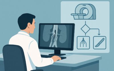
A 67-year-old man sits in your vascular surgery clinic for his one-year follow-up after an extensive open repair of a Crawford Type II thoracoabdominal aortic aneurysm. He feels well, his incisions are healed, and his blood pressure is controlled. The time has come for routine surveillance imaging to ensure the integrity of the complex graft repair. Your task is to order the correct study that can comprehensively evaluate the entire surgical field, from the descending thoracic aorta down to the iliac arteries, without missing subtle but potentially catastrophic complications. This clinical workflow article details the evidence-based approach for this specific scenario. According to the American College of Radiology (ACR) Appropriateness Criteria, the recommended first-line study is CTA chest abdomen pelvis with IV contrast, which is rated Usually Appropriate.
Who Fits This Clinical Scenario for Aortic Surveillance?
This guidance is specifically for patients undergoing routine, asymptomatic surveillance imaging following open surgical repair of a thoracoabdominal aortic aneurysm (TAAA) or dissection. The key inclusion criteria are a history of a conventional, sewn-in graft repair and the absence of new, concerning symptoms like chest pain, back pain, fever, or signs of malperfusion. The goal of imaging is to detect long-term complications before they become clinically apparent.
This workflow is distinct from several closely related clinical situations that require different imaging strategies. This article does not apply to:
- Patients with endovascular repairs (EVAR/TEVAR/FEVAR): Surveillance after stent-graft placement has a different focus, primarily the detection of endoleaks, stent migration, and component separation. That is a separate clinical scenario with its own imaging recommendations.
- Patients with a known, unrepaired aneurysm or dissection: The imaging strategy for monitoring a native, unrepaired aorta focuses on measuring diameter changes and assessing dissection flap morphology, which follows a different ACR variant.
- Pre-operative planning: Imaging performed to plan the initial open or endovascular repair has distinct requirements, including detailed measurements of landing zones, vessel diameters, and branch artery anatomy.
Applying the wrong surveillance protocol to these other scenarios can lead to missed diagnoses or inappropriate radiation exposure.
What Diagnoses Are You Working Up in This Scenario?
Surveillance imaging after open TAAA repair is not a general screen; it is a targeted search for specific, known complications of a major aortic reconstruction. The imaging study must be capable of identifying these potential issues with high confidence.
The most critical finding to rule out is an anastomotic pseudoaneurysm. This is a contained rupture at one of the suture lines where the graft is sewn to the native aorta. If left undetected, it can lead to free rupture and life-threatening hemorrhage. These are often subtle and require high-resolution, contrast-enhanced imaging to detect the characteristic outpouching of contrast at a suture line.
Another key concern is the health of the native aorta proximal or distal to the repair. The underlying disease process that led to the initial aneurysm can cause new aneurysms or dissections to form in the remaining segments of the aorta over time. Surveillance must cover the entire aorta to monitor for this progression.
Graft integrity and infection are also paramount. While less common, graft thrombosis, kinking, or infection can occur. Graft infection is a devastating complication that may present as a subtle, persistent fluid collection or soft tissue thickening around the graft, often best seen on CT.
Finally, the study assesses the patency of reimplanted visceral vessels. During a complex TAAA repair, the celiac, superior mesenteric, and renal arteries are often reimplanted onto the main graft. Imaging confirms that these critical branch vessels remain open and free of stenosis or occlusion.
Why Is CTA of the Chest, Abdomen, and Pelvis the Recommended First Step?
The ACR panel designates CTA chest abdomen pelvis with IV contrast as Usually Appropriate for this scenario because it provides the most reliable and comprehensive evaluation of the potential complications. Its high spatial resolution is ideal for visualizing the fine details of the graft, the anastomotic suture lines, and the origins of reimplanted visceral arteries. The rapid acquisition speed minimizes motion artifacts from breathing and cardiac pulsation, ensuring sharp, clear images of the entire thoracoabdominal aorta.
The rationale for its top rating stems from its ability to directly address the primary clinical questions:
- It is highly sensitive and specific for detecting anastomotic pseudoaneurysms.
- It provides a complete, panoramic view of the entire aorta to assess for new disease in unrepaired segments.
- It clearly delineates the graft material from surrounding tissues, aiding in the detection of perigraft fluid collections that could signify infection.
Alternative Imaging Modalities
While CTA is the primary recommendation, other studies are rated for this scenario, and understanding their limitations is key:
- MRA chest abdomen pelvis without and with IV contrast is also rated Usually Appropriate. It is an excellent alternative that avoids ionizing radiation, a significant benefit for younger patients who will require lifelong surveillance. However, MRA can be limited by metallic artifacts from surgical clips, has longer scan times, and may offer slightly lower spatial resolution for evaluating the anastomoses compared to modern CTA. It is a strong choice for patients with contraindications to iodinated contrast.
- US duplex Doppler aorta abdomen is rated Usually not appropriate. Ultrasound cannot visualize the thoracic portion of the repair, and its view of the abdominal aorta and graft is often severely limited by overlying bowel gas. It is inadequate for comprehensive surveillance after a thoracoabdominal repair.
Radiation and Contrast Considerations
A CTA of the chest, abdomen, and pelvis carries a very high relative radiation level (ACR RRL ☢☢☢☢☢, corresponding to an effective dose of 30-100 mSv). This dose must be considered in the context of the patient’s age and the necessity of lifelong surveillance. However, the risk of missing a life-threatening complication like a pseudoaneurysm generally outweighs the radiation risk. Intravenous contrast is mandatory; a non-contrast study cannot provide the necessary diagnostic information.
Once you’ve decided on CTA, our protocol guide covers the technical parameters for optimal acquisition. For detailed guidance on technique, contrast timing, and interpretation principles, see our complete guide: CT Chest/Abdomen/Pelvis with IV Contrast.
What’s Next After CTA? Downstream Workflow
The results of the surveillance CTA will direct the subsequent clinical pathway. The workflow branches based on whether the findings are negative, positive, or indeterminate.
If the CTA is negative or stable: For a study showing a stable, intact repair with no new aortic pathology, the patient can continue with their established surveillance schedule. The frequency of imaging (e.g., annually, every two years) is determined by the surgeon based on the original pathology, the extent of the repair, and the patient’s overall clinical picture.
If the CTA is positive for a critical finding: The discovery of an anastomotic pseudoaneurysm, a rapidly expanding new aneurysm, or signs of graft infection requires immediate action. This result should prompt an urgent consultation with the vascular surgery team to plan for intervention, which may involve a complex re-operation or an endovascular approach.
If the CTA shows a non-critical positive finding: The development of a small, new aneurysm in a native aortic segment may not require immediate intervention. Instead, the downstream workflow would involve more frequent surveillance imaging (e.g., switching to a 6-month interval) to monitor its growth rate and determine the optimal time for repair.
If the CTA is indeterminate: Findings like a stable, small perigraft fluid collection can be ambiguous. It could represent a benign, chronic seroma or a low-grade, indolent infection. In this case, the next step involves close clinical correlation (e.g., checking inflammatory markers like ESR and CRP) and potentially a short-interval follow-up CTA or a different imaging modality, such as a nuclear medicine tagged white blood cell scan, to specifically evaluate for infection.
Pitfalls to Avoid (and When to Get Help)
Navigating post-operative aortic surveillance requires careful attention to detail to avoid common errors that can compromise patient safety.
A primary pitfall is ordering an incomplete study. The surveillance must cover the entire thoracoabdominal aorta, from the aortic arch through the iliac arteries. Ordering only a “CTA of the abdomen” is insufficient and can miss pathology in the chest.
Another frequent error is ordering the study without IV contrast. A non-contrast CT provides almost no useful information for assessing graft patency, anastomotic integrity, or visceral vessel perfusion and is rated Usually not appropriate.
Ignoring cumulative radiation dose is a concern, especially in younger patients. For stable, long-term follow-up in patients without contraindications, failing to consider MRA as a radiation-free alternative is a missed opportunity to reduce lifetime radiation exposure.
Finally, misinterpreting normal post-operative changes can lead to unnecessary anxiety or interventions. The appearance of the graft and surrounding tissues changes over time, and familiarity with expected findings is crucial. If there is any uncertainty about a finding, especially concerning potential infection or pseudoaneurysm, the correct next step is to escalate with a direct conversation between the radiologist and the referring vascular surgeon.
Related ACR Topics and Tools
This article focuses on a single clinical scenario. For a comprehensive overview of all variants related to TAAA imaging, or to explore the technical details and radiation dose of the recommended studies, the following resources are available.
- For breadth across all scenarios in Thoracoabdominal Aortic Aneurysm or Dissection: Treatment Planning and Follow-Up, see our parent guide: Thoracoabdominal Aortic Aneurysm or Dissection: Treatment Planning and Follow-Up: ACR Appropriateness Decoded.
- To look up appropriateness ratings for thousands of other clinical scenarios, use the ACR Appropriateness Criteria Lookup tool.
- To review detailed imaging techniques for hundreds of studies, visit the Imaging Protocol Library.
- To discuss cumulative radiation exposure with your patients, consult the Radiation Dose Calculator.
Frequently Asked Questions
Why is MRA also ‘Usually Appropriate’ if CTA is the first choice?
MRA is an excellent radiation-free alternative, making it particularly valuable for younger patients requiring lifelong surveillance. While CTA often provides slightly better spatial resolution and is faster, MRA is a strong co-equal option, especially for patients with renal insufficiency or an allergy to iodinated contrast. The choice between them often depends on patient factors, institutional preference, and the specific clinical question.
How often should surveillance imaging be performed after open TAAA repair?
The optimal frequency is not standardized and is typically determined by the operating surgeon. A common approach is a baseline scan within the first 1-3 months post-operatively, followed by an annual scan. The interval may be shortened if there is a known residual dissection, a small untreated aneurysm, or other concerning features.
What if my patient has renal insufficiency? Can I still order a CTA?
For patients with moderate to severe chronic kidney disease, the risk of contrast-induced nephropathy (CIN) is a concern. In these cases, MRA with a macrocyclic gadolinium-based contrast agent is often preferred. If CTA is deemed absolutely necessary, pre-procedural hydration and minimizing contrast volume are critical. The decision should be a shared one between the referring physician, the radiologist, and potentially a nephrologist.
Does the type of graft material (e.g., Dacron) affect the choice of imaging?
No, both CTA and MRA are effective for evaluating standard surgical grafts like Dacron. While MRA can be susceptible to artifact from metallic components, the surgical clips used in open repair are typically made of non-ferromagnetic materials like titanium and cause only minor, localized artifact that does not usually obscure the evaluation of the graft and anastomoses.
If a small, stable fluid collection is seen around the graft on a 2-year follow-up scan, what is the next step?
A chronic, stable, and asymptomatic perigraft fluid collection is often a benign seroma. However, a low-grade infection can have a similar appearance. The next step involves clinical correlation: checking for fever, elevated white blood cell count, and inflammatory markers (CRP, ESR). If there is any clinical suspicion of infection, a tagged WBC scan may be performed. If all clinical and laboratory findings are negative, continued surveillance with short-interval imaging (e.g., in 3-6 months) is a reasonable approach.
Reviewed by Pouyan Golshani, MD, Interventional Radiologist — May 30, 2026