What Is the Right Initial Imaging for a Child with Chest Pain and Suspected PE?
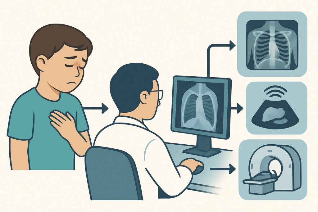
A 15-year-old with a recently casted tibia fracture from a soccer injury presents to the emergency department with acute-onset, sharp right-sided chest pain that worsens with deep breaths. She is tachycardic and her oxygen saturation is 95% on room air. You are concerned about a pulmonary embolism (PE), a rare but serious condition in children. The immediate question is which imaging study to order first to safely and effectively evaluate her symptoms. According to the American College of Radiology (ACR) Appropriateness Criteria, the initial imaging step in this scenario is a study rated as Usually appropriate: `Radiography chest`.
Who Fits the Scenario of a Child with Chest Pain and Suspected Pulmonary Embolism?
This clinical workflow is for any child or adolescent presenting with chest pain where pulmonary embolism is a reasonable consideration in the differential diagnosis. While PE is uncommon in the pediatric population, suspicion should be heightened in the presence of specific risk factors. These include recent major surgery, significant trauma (especially long-bone fractures), prolonged immobilization, the presence of a central venous catheter, a personal or family history of a hypercoagulable state, or, in adolescents, the use of oral contraceptives.
This guidance is distinct from other pediatric chest pain scenarios. It is crucial to differentiate this presentation from:
- Chest wall pain: If the pain is clearly localized, non-pleuritic, and reproducible with palpation of the chest wall, the workup follows the chest wall pain variant, which has a different imaging pathway.
- Known or suspected cardiac disease: If the patient has a known history of congenital heart disease or if clinical signs point towards myocarditis or pericarditis (e.g., friction rub, EKG changes), the workup shifts to a cardiac-focused scenario.
- History of sickle cell disease: In a child with sickle cell disease, acute chest pain and hypoxia raise immediate concern for acute chest syndrome, a distinct clinical entity requiring a specific management approach.
What Diagnoses Are You Working Up When Suspecting Pediatric Pulmonary Embolism?
When a child presents with symptoms concerning for PE, the imaging workup is designed to evaluate for the PE itself while simultaneously assessing for more common alternative diagnoses. The differential diagnosis is broad, and the initial imaging choice reflects this.
Pulmonary Embolism (PE): This is the primary life-threatening diagnosis being considered. While rare, its potential for severe morbidity and mortality makes it a can’t-miss diagnosis. The clinical presentation of pleuritic chest pain, dyspnea, and tachycardia, especially with known risk factors, places PE on the differential.
Pneumonia: An infection of the lung parenchyma is a far more common cause of pleuritic chest pain, fever, and tachypnea in children. A focal consolidation on imaging can confirm this diagnosis and redirect management away from an unnecessary anticoagulation workup.
Pneumothorax or Pneumomediastinum: The sudden onset of sharp chest pain and shortness of breath can be caused by air in the pleural space (pneumothorax) or mediastinum. These conditions can occur spontaneously or after trauma and are critical to identify quickly.
Pericarditis or Myocarditis: Inflammation of the pericardium or myocardium can cause chest pain, though it is often positional or associated with viral prodromes. While not the primary target of a PE workup, severe cases can cause secondary findings like an enlarged cardiac silhouette or pulmonary edema.
Musculoskeletal Pain: Costochondritis and other chest wall syndromes are the most frequent causes of chest pain in children. However, this is a diagnosis of exclusion, and in a patient with risk factors for venous thromboembolism (VTE), more serious causes must be ruled out first.
Why Is a Chest Radiograph the Recommended First Step for Suspected Pediatric PE?
For a child with chest pain and suspected pulmonary embolism, the ACR designates `Radiography chest` as Usually appropriate. This recommendation is based on its dual utility as a screening tool and a necessary prerequisite for more advanced imaging, all while delivering a minimal radiation dose.
The primary role of the initial chest radiograph is not to definitively diagnose the PE, but to assess for common mimics. It is highly effective at identifying alternative causes of chest pain and hypoxia, such as pneumonia, pneumothorax, or significant pleural effusion. Finding one of these may conclude the diagnostic workup for PE and allow for targeted treatment of the actual cause.
Conversely, a normal chest radiograph in a patient with hypoxia and pleuritic pain significantly increases the clinical suspicion for PE. Classic (though insensitive) radiographic signs of PE, such as a Westermark sign (focal oligemia) or a Hampton’s hump (a wedge-shaped opacity), may occasionally be seen. More importantly, a normal radiograph is often required for the proper interpretation of a subsequent ventilation/perfusion (V/Q) scan, should that be the chosen definitive study.
Radiation and Alternative Studies:
- Radiography chest: With a pediatric relative radiation level of ☢ (<0.03 mSv), this study imparts a very low radiation dose, a critical consideration in the pediatric population.
- CTA pulmonary arteries with IV contrast: This study is also rated Usually appropriate and is the definitive test for diagnosing PE. However, it carries a significantly higher radiation dose (☢☢☢☢ 3-10 mSv [ped]) and requires IV contrast, with its associated risks. It is typically reserved as the second step after an unrevealing chest radiograph when clinical suspicion remains high.
- V/Q scan lung: Rated as May be appropriate, this nuclear medicine study has a lower radiation dose than CTPA (☢☢☢ 0.3-3 mSv [ped]) and avoids iodinated contrast. Its major limitation is that it requires a normal baseline chest radiograph to be interpretable and may yield indeterminate results, particularly in patients with underlying lung disease.
- US duplex Doppler lower extremity: This is rated Usually not appropriate as the initial imaging test for chest pain. While it can identify a deep vein thrombosis (DVT) as a potential source of emboli, a negative scan does not rule out PE, as the clot may have already embolized or originated from a different location.
What’s Next After Radiography chest? Downstream Workflow
The results of the initial chest radiograph guide the subsequent diagnostic pathway. The workflow branches based on whether the radiograph reveals an alternative diagnosis or is unrevealing.
If the radiograph is positive for an alternative diagnosis:
- A finding of pneumonia, pneumothorax, or a rib fracture may fully explain the patient’s symptoms. The clinical focus shifts to treating this identified condition, and the PE workup can often be halted, avoiding further radiation and contrast exposure.
If the radiograph is negative or shows non-specific findings (e.g., atelectasis):
- The next step depends on the level of clinical suspicion, which can be stratified using clinical decision rules (e.g., Wells score, adapted for pediatrics) and laboratory testing (e.g., D-dimer).
- High clinical suspicion: Proceed directly to definitive imaging. `CTA pulmonary arteries with IV contrast` is the most common next step, providing a detailed anatomical assessment of the pulmonary vasculature.
- Low to intermediate clinical suspicion: A D-dimer test may be used. If negative, PE is very unlikely, and the workup can often stop. If the D-dimer is positive, or if clinical suspicion remains despite a low pre-test probability, proceed to definitive imaging with either CTPA or a V/Q scan.
If the radiograph is indeterminate:
- In cases where the findings are unclear, the decision to proceed to CTPA is based on the persistent clinical concern for PE. The radiograph has still served its purpose by providing a baseline and ruling out clear-cut alternative diagnoses.
Pitfalls to Avoid (and When to Get Help)
Navigating a suspected pediatric PE workup requires careful attention to avoid common errors.
- Skipping the Initial Radiograph: Jumping directly to CTPA exposes the child to unnecessary radiation and contrast if a simpler diagnosis like pneumothorax could have been found on a plain film.
- Over-reliance on D-dimer: While useful, the D-dimer test has lower specificity in certain populations (e.g., post-operative patients, those with underlying inflammation) and should be interpreted in the context of clinical pre-test probability.
- Ignoring Radiation Dose: Always adhere to the ALARA (As Low As Reasonably Achievable) principle. This involves using pediatric-specific CT protocols and considering lower-dose alternatives like V/Q scans when clinically appropriate.
- Misinterpreting a Normal Radiograph: Remember that a normal chest X-ray is a common finding in PE. It does not rule out the diagnosis; rather, it increases its likelihood in the right clinical setting.
If the patient is hemodynamically unstable or has signs of right heart strain on EKG or bedside ultrasound, escalate care immediately. This may involve consultation with pediatric critical care, cardiology, and interventional radiology for consideration of advanced therapies.
Related ACR Topics and Tools
For a comprehensive overview of all pediatric chest pain scenarios, from musculoskeletal pain to cardiac concerns, please see our parent topic hub article. For tools to assist in ordering the correct study and communicating with patients, the following resources are available.
- For breadth across all scenarios in Chest Pain-Child, see our parent guide: Chest Pain-Child: ACR Appropriateness Decoded.
- ACR Appropriateness Criteria Lookup — for adjacent scenarios
- Imaging Protocol Library — for technique on the recommended study
- Radiation Dose Calculator — for cumulative dose conversations
Frequently Asked Questions
Why not order a CTPA immediately if I’m worried about PE in a child?
While CT Pulmonary Angiography (CTPA) is the definitive test, it involves a significant radiation dose (3-10 mSv) and IV contrast. A chest radiograph is recommended first because it uses a very low dose of radiation (<0.03 mSv) and can often identify a more common cause for the symptoms, like pneumonia or pneumothorax, making the higher-dose CTPA unnecessary.
Is a V/Q scan a good alternative to CTPA for pediatric PE?
A V/Q scan is rated as ‘May be appropriate’ and can be a good alternative, especially in patients with a contraindication to iodinated contrast (like a severe allergy or renal dysfunction). It delivers less radiation than a CTPA. However, it requires a cooperative patient and, crucially, a normal baseline chest radiograph to be reliably interpreted. If the chest radiograph shows other abnormalities, the V/Q scan is more likely to be indeterminate.
What if the chest radiograph is normal but my clinical suspicion for PE remains high?
A normal chest radiograph in the setting of hypoxia and pleuritic chest pain actually increases the likelihood of pulmonary embolism. In this case, the radiograph has successfully ruled out common mimics. The next step is to proceed to definitive imaging with either a CTPA or a V/Q scan, based on institutional preference and patient-specific factors.
Should I order a D-dimer test on every child with suspected PE?
The utility of a D-dimer test depends on your pre-test clinical probability. In patients with low clinical suspicion, a negative D-dimer can be very effective at ruling out PE and preventing further imaging. However, in patients with high clinical suspicion, a D-dimer test is less helpful because it is likely to be positive, and you will need to proceed to definitive imaging regardless of the result. Its use should be guided by validated pediatric clinical decision rules.
Can ultrasound be used to diagnose a pulmonary embolism in a child?
Directly diagnosing PE with ultrasound is not standard practice. While bedside cardiac ultrasound might show signs of right heart strain (a complication of a large PE), and chest ultrasound can sometimes identify peripheral infarcts, these are not primary diagnostic methods. A lower extremity duplex ultrasound to look for DVT is rated ‘Usually not appropriate’ for the initial workup of chest pain, as a negative study does not rule out PE.
Reviewed by Pouyan Golshani, MD, Interventional Radiologist — June 27, 2026