What’s the Best Imaging After an Incomplete Colonoscopy for Colorectal Cancer Screening?
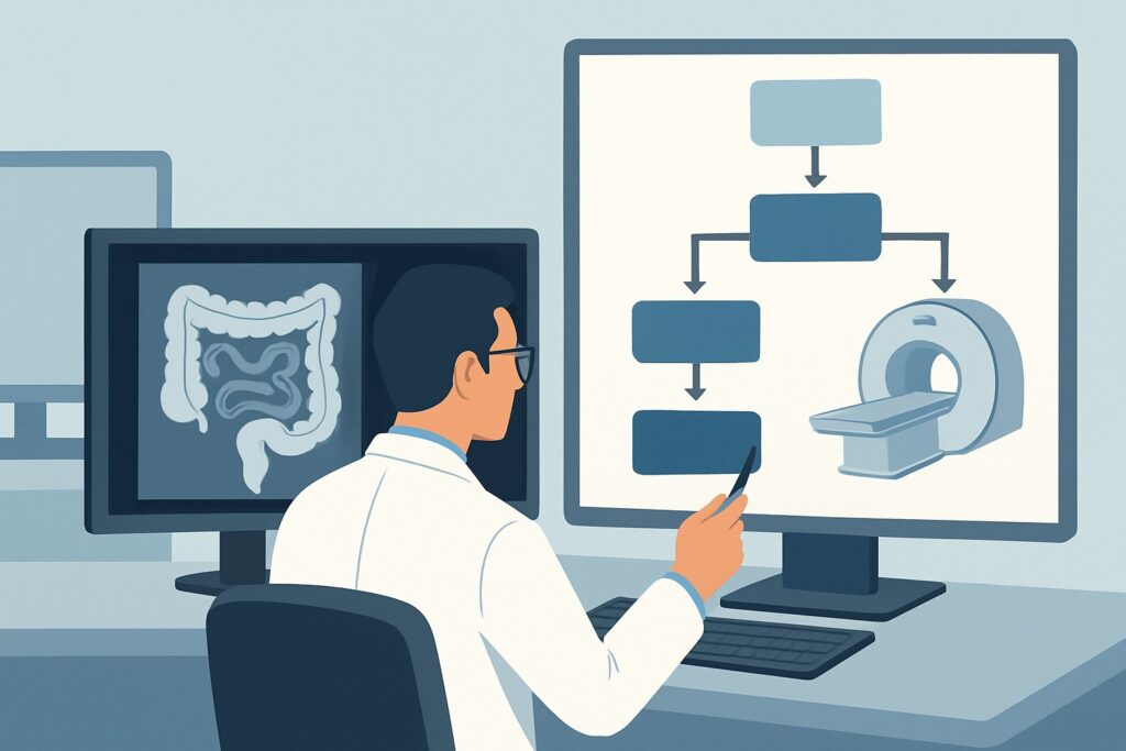
A 62-year-old male patient returns to your clinic for follow-up. His first screening colonoscopy, scheduled last week, was terminated early. The gastroenterologist’s report notes a particularly tortuous sigmoid colon, making it impossible to advance the scope to the cecum despite multiple attempts. The visualized portion of the colon was normal, but the proximal two-thirds remain unevaluated. You now face the critical decision of how to complete his colorectal cancer screening. This article provides a detailed clinical workflow for this exact scenario: selecting the appropriate imaging study for an adult patient at any risk level after an incomplete or intolerable optical colonoscopy. According to the American College of Radiology (ACR) Appropriateness Criteria, the recommended study, rated Usually Appropriate, is CT colonography without IV contrast screening.
Who Fits This Clinical Scenario?
This guidance applies to adult patients of any risk category—average, elevated, or high—who require colorectal cancer (CRC) screening but have had an incomplete optical colonoscopy or are unable to tolerate the procedure. An incomplete colonoscopy is defined as the failure to visualize the entire colon up to the cecum. Common reasons for this include:
- Anatomical challenges, such as a redundant or tortuous colon.
- An obstructing lesion preventing further passage of the endoscope.
- Poor bowel preparation obscuring the view.
- Patient intolerance due to pain or discomfort.
This workflow is also for patients who cannot undergo colonoscopy at all due to significant comorbidities that increase sedation risks or other contraindications. It is crucial to distinguish this scenario from others. This guidance does not apply to:
- Initial screening for patients who can undergo colonoscopy: For average, elevated, or high-risk individuals who are candidates for standard screening methods, the initial approach is different.
- Symptomatic patients: Patients presenting with acute symptoms like rectal bleeding, abdominal pain, or signs of bowel obstruction require a diagnostic workup, not a screening evaluation.
- Surveillance after polypectomy or cancer resection: These patients follow specific surveillance protocols, which may differ from this screening workflow.
What Diagnoses Are You Working Up in This Scenario?
When ordering a completion study after an incomplete colonoscopy, the primary goal remains screening for colorectal neoplasia in the unvisualized segments of the colon. The differential diagnosis, or the list of potential findings you are actively looking for, is focused on detecting precursors to cancer and early-stage disease.
Colorectal Adenomatous Polyps: This is the principal target of CRC screening. The goal is to identify adenomas, particularly those ≥6 mm in size, as they are the precursor lesions for most colorectal adenocarcinomas. Detecting and facilitating the eventual removal of these polyps is the most effective way to prevent cancer.
Colorectal Carcinoma: The most consequential finding is an invasive cancer. If the initial colonoscopy was incomplete due to an obstructing mass, the pre-test probability for carcinoma is significantly higher. In other cases, the goal is to detect early-stage, asymptomatic cancers in the unexamined portion of the colon, when they are most treatable.
Clinically Significant Extracolonic Findings: A unique aspect of cross-sectional imaging like CT colonography is its ability to visualize structures outside the colon. While not the primary indication, the study can reveal important incidental findings such as abdominal aortic aneurysms, renal or adrenal masses, or signs of metastatic disease. These findings can have a major impact on patient management.
Why Is CT Colonography Without IV Contrast Screening the Recommended Study?
For completing colorectal cancer screening after an incomplete or failed optical colonoscopy, the ACR designates CT colonography without IV contrast screening as Usually Appropriate. This recommendation is based on its high diagnostic performance, safety profile, and ability to visualize the entire colon.
CT colonography (CTC), also known as virtual colonoscopy, uses low-dose CT imaging to generate high-resolution 2D and 3D images of the prepared colon. Its sensitivity for detecting adenomas ≥10 mm is comparable to that of optical colonoscopy, and it is highly effective for polyps in the 6–9 mm range. This makes it an excellent non-invasive tool to inspect the segments of the colon that were not reached during the initial procedure.
Alternative studies are rated lower for this specific screening purpose:
- Fluoroscopy barium enema (double- or single-contrast) is rated Usually Not Appropriate. These studies have significantly lower sensitivity for detecting polyps and early-stage cancers compared to CTC. They are now largely considered obsolete for screening.
- CT abdomen and pelvis with IV contrast is also rated Usually Not Appropriate for screening. While it can detect large masses, it is not optimized for evaluating the colonic mucosa. The lack of colonic distention and specific bowel preparation means it will miss most polyps and many flat lesions. The addition of IV contrast and a higher radiation dose is not justified for a screening indication.
The recommended CTC protocol uses a low radiation dose (relative radiation level ☢☢☢☢, 10-30 mSv) and avoids the risks associated with IV contrast. It is crucial to note that CTC requires a full bowel preparation, similar to conventional colonoscopy, to ensure the colon is clean for accurate evaluation.
What’s Next After CT Colonography? Downstream Workflow
The results of the CT colonography will dictate the next steps in patient management, creating a clear decision tree for the referring clinician.
If the study is positive for a significant polyp (typically ≥6 mm): The patient will need to be referred back to gastroenterology for a therapeutic optical colonoscopy to remove the polyp(s). The CTC report provides a “roadmap,” pinpointing the location of the lesion, which can help the endoscopist plan the subsequent procedure. For patients with known difficult anatomy, this may involve using specialized scopes or techniques.
If the study is negative (no polyps ≥6 mm detected): The patient is considered to have had a complete negative screening examination. They can return to the standard recommended screening interval for their risk category (e.g., 5 years for CTC in an average-risk individual, though guidelines vary). No immediate further action is needed.
If the study is indeterminate or shows only diminutive polyps (<6 mm): For very small polyps, management is often to continue routine screening at the standard interval, as the risk of malignancy is extremely low. For indeterminate findings, the decision may involve consultation between the radiologist and the referring physician to decide between short-term follow-up imaging or proceeding to optical colonoscopy.
If a significant extracolonic finding is reported: This requires a separate, dedicated workup. For example, a suspected renal mass would prompt a referral to urology and likely a dedicated diagnostic renal protocol CT or MRI. The management of these incidental findings should follow established guidelines for the specific organ system involved.
Pitfalls to Avoid (and When to Get Help)
Navigating this clinical scenario requires avoiding several common pitfalls to ensure patient safety and diagnostic accuracy.
- Ordering the wrong CT: Do not order a standard “CT Abdomen/Pelvis” for this indication. You must specifically order “CT Colonography” or “Virtual Colonoscopy” to ensure the correct low-dose protocol with colonic insufflation is performed.
- Forgetting the bowel prep: CT colonography is only effective if the patient undergoes a thorough bowel preparation. Ensure the patient receives and understands the prep instructions from the imaging facility.
- Misinterpreting the screening-to-diagnostic transition: If the initial colonoscopy was incomplete because of a truly obstructing lesion, the CTC is less of a screening tool and more of a diagnostic one to assess the lesion’s extent and look for metastatic disease. In such cases, IV contrast may be appropriate, and consultation with the radiologist is advised.
If there is any ambiguity about the initial colonoscopy findings or if the patient develops new symptoms (e.g., bleeding, pain), escalate by consulting directly with the gastroenterologist and radiologist to coordinate the best next step.
Related ACR Topics and Tools
For a comprehensive overview of all clinical variants related to colorectal cancer screening, including initial screening for different risk groups, please refer to our parent topic hub article. Additional GigHz tools can help you apply these principles in your daily practice.
- For breadth across all scenarios in Colorectal Cancer Screening, see our parent guide: Colorectal Cancer Screening: ACR Appropriateness Decoded.
- For adjacent scenarios not covered here, use the ACR Appropriateness Criteria Lookup.
- To understand the technical aspects of various imaging studies, explore the Imaging Protocol Library.
- To discuss radiation exposure with your patients, consult the Radiation Dose Calculator.
Frequently Asked Questions
Does the patient need another bowel prep for the CT colonography?
Yes. CT colonography requires a clean and dry colon for accurate interpretation. The patient will need to undergo a full cathartic bowel preparation, similar to the one used for optical colonoscopy. The imaging center will provide specific instructions.
What if the CT colonography finds a large polyp? Do we need to do another colonoscopy?
Yes. CT colonography is a diagnostic-only procedure; it cannot remove polyps. If a polyp meeting the size criteria for removal (typically 6 mm or larger) is found, the patient must be referred for a therapeutic optical colonoscopy to have it removed.
Is CT colonography a good option for a patient who refuses colonoscopy due to the sedation?
Yes, this is an excellent indication. CT colonography does not require any sedation, making it a suitable alternative for patients who wish to avoid or have contraindications to the sedatives used during optical colonoscopy.
Why is IV contrast not used for a screening CT colonography?
For a screening study, the primary goal is to evaluate the mucosal surface of the colon for polyps. This is best achieved by distending the colon with air or CO2 and using specialized 2D and 3D imaging software. IV contrast does not improve polyp detection and adds unnecessary risk (e.g., allergic reaction, contrast-induced nephropathy) and radiation dose. It is reserved for diagnostic scans where there is a concern for cancer staging or extracolonic pathology.
What is the screening interval after a negative CT colonography?
For an average-risk individual, a negative high-quality CT colonography is typically repeated every 5 years. This interval may be shorter for patients in high-risk categories. Always consult the most current guidelines from major societies like the American Cancer Society (ACS) and the U.S. Multi-Society Task Force on Colorectal Cancer.
Reviewed by Pouyan Golshani, MD, Interventional Radiologist — May 30, 2026