What’s the Best Initial Imaging for Painful Second or Third Trimester Bleeding?
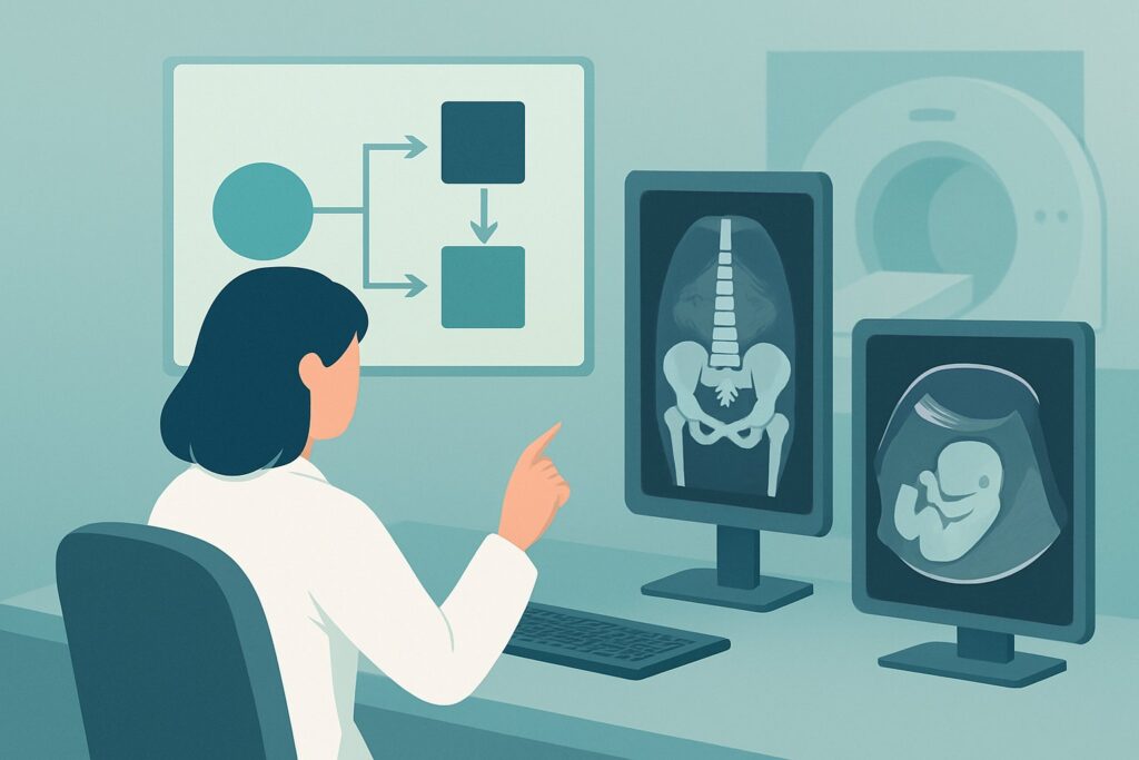
A 32-year-old G2P1 at 34 weeks gestation presents to the emergency department with the sudden onset of severe, constant abdominal pain and dark red vaginal bleeding. Her abdomen is rigid and tender to palpation. You are concerned about the life-threatening causes of painful antepartum hemorrhage, and the immediate next step is to assess both maternal and fetal status while determining the underlying etiology. This clinical scenario requires a rapid, safe, and informative imaging study to guide management. This article details the American College of Radiology (ACR) Appropriateness Criteria workflow for this specific presentation, explaining why certain studies are chosen. For this patient, the ACR rates US duplex Doppler pelvis as ‘Usually Appropriate’ for initial evaluation.
Who Fits This Clinical Scenario?
This guidance applies to a specific and urgent clinical situation: a pregnant patient in the second or third trimester (beyond 20 weeks gestation) who presents with painful vaginal bleeding. The presence of pain is the key differentiator. This includes patients with uterine tenderness, contractions, or a rigid, “board-like” abdomen. The primary goal of initial imaging is to rapidly evaluate for emergent conditions that threaten the mother or fetus, most notably placental abruption.
This workflow is distinct from other presentations of late-term bleeding. It does not apply to patients with:
- Painless vaginal bleeding: This presentation raises a different primary differential, most commonly placenta previa or vasa previa. While ultrasound is also the initial study, the clinical urgency and specific questions being asked of the imaging are different.
- Known placenta previa or vasa previa: Patients with a previously diagnosed placental implantation abnormality who present with bleeding follow a separate management pathway, as the likely cause of bleeding is already suspected.
Correctly identifying your patient’s scenario is critical, as the pre-test probability for certain diagnoses heavily influences the interpretation of imaging results and subsequent clinical decisions.
What Diagnoses Are You Working Up in This Scenario?
When a patient presents with painful vaginal bleeding in the second or third trimester, the differential diagnosis is narrow but includes several high-acuity conditions. The imaging choice is tailored to identify or exclude these possibilities.
Placental Abruption: This is the foremost concern and a true obstetric emergency. It refers to the premature separation of the placenta from the uterine wall. The classic presentation is painful vaginal bleeding, uterine tenderness, and high-frequency contractions. The bleeding occurs behind the placenta, which can dissect between the uterine wall and membranes, leading to external bleeding, or it can be concealed. Abruption can lead to fetal distress, maternal hemorrhage, and disseminated intravascular coagulation (DIC).
Uterine Rupture: While less common, this is a catastrophic event where the uterine wall tears. It is most often associated with a prior uterine scar, such as from a cesarean section. Patients present with sudden, severe abdominal pain, vaginal bleeding, and often a loss of fetal station and signs of fetal distress or demise. Maternal shock can develop rapidly.
Preterm Labor: The onset of labor can present with painful contractions and vaginal bleeding, often from cervical change (the “bloody show”). While this is a diagnosis of exclusion in the setting of significant pain and bleeding, it remains a key part of the differential. Ultrasound helps assess cervical length and fetal presentation.
Non-Obstetric Causes: Rarely, another process can cause abdominal pain that coincides with minor vaginal bleeding. Examples include a degenerating uterine fibroid or, less likely, an intra-abdominal process like appendicitis or nephrolithiasis that triggers uterine irritability.
Why Is a Pelvic Duplex Doppler Ultrasound the Recommended Initial Study?
For a patient with painful second or third trimester bleeding, ultrasound is the cornerstone of initial imaging. The ACR designates US duplex Doppler pelvis, US pregnant uterus transabdominal, and US pregnant uterus transvaginal as ‘Usually Appropriate’. These are not mutually exclusive but are components of a complete obstetric ultrasound evaluation.
The primary advantage of ultrasound is its safety profile: it uses no ionizing radiation (0 mSv), making it entirely safe for the fetus at any gestational age. It is also rapid, portable, and widely available, which is critical in an emergency setting. The examination typically begins with a transabdominal approach to get a global view of the uterus, assess fetal viability and position, locate the placenta, and estimate amniotic fluid volume.
The addition of Duplex Doppler is particularly valuable in this scenario. Doppler imaging assesses blood flow and can help identify a retroplacental hematoma—the hallmark of a placental abruption. While the sensitivity of ultrasound for abruption is variable and often cited as being low (a negative scan does not rule it out), a positive finding is highly specific and confirms the diagnosis, allowing for decisive clinical action.
A transvaginal ultrasound is also ‘Usually Appropriate’ and provides superior resolution of the lower uterine segment and cervix. It is the gold standard for diagnosing placenta previa, which must be ruled out in any patient with third-trimester bleeding before a digital or speculum exam is performed. A transperineal US, rated ‘May be appropriate’, can be a useful alternative for visualizing the cervix if a transvaginal approach is not feasible or is contraindicated.
No other imaging modalities, such as CT or MRI, are recommended for the initial evaluation. They are slower, less available, and in the case of CT, involve ionizing radiation. MRI may have a role in complex, non-emergent cases but is not a first-line tool for an acute presentation.
What’s Next After the Ultrasound? Downstream Workflow
The results of the initial ultrasound, combined with the clinical picture, guide the immediate next steps. The workflow is often parallel, with clinical stabilization and fetal monitoring occurring simultaneously with imaging.
- If the study is positive for placental abruption: A definitive finding of a retroplacental hematoma confirms the diagnosis. The immediate next step is an urgent consultation with obstetrics. Management depends on maternal stability, fetal status (assessed via continuous electronic fetal monitoring), and gestational age. This may range from expectant management in a stable preterm patient with a small abruption to an emergency cesarean delivery for maternal or fetal compromise.
- If the study is negative or inconclusive: This is a common and critical scenario. Ultrasound has limited sensitivity for detecting placental abruption, especially if the abruption is small or the bleeding is isoechoic to the placenta. A negative ultrasound does not rule out abruption. The diagnosis remains a clinical one. If there is high clinical suspicion based on a rigid, tender uterus, non-reassuring fetal heart tracing, or significant bleeding, the patient should be managed as if they have an abruption, regardless of the imaging findings. This involves continuous monitoring, maternal stabilization, and obstetric consultation.
- If the study identifies another cause (e.g., placenta previa): The workflow shifts to managing that specific condition. For instance, if a previously unknown placenta previa is found, the patient will be managed according to protocols for painless bleeding, which typically involves pelvic rest and planning for a cesarean delivery.
Pitfalls to Avoid (and When to Get Help)
In this high-stakes scenario, several clinical and diagnostic pitfalls can lead to adverse outcomes. Awareness is key to avoiding them.
- Over-reliance on a negative ultrasound: The most significant pitfall is ruling out placental abruption based on a normal ultrasound. The decision to intervene must be based on the complete clinical picture, including physical exam, vital signs, and fetal monitoring.
- Delaying clinical intervention for imaging: In an unstable patient with signs of shock or a non-reassuring fetal status, stabilization and preparation for delivery should not be delayed for imaging. A bedside ultrasound can be performed concurrently if it does not impede resuscitation.
- Performing a digital cervical exam prematurely: A digital or speculum examination should never be performed in a patient with third-trimester bleeding until placenta previa has been definitively excluded by ultrasound, as it can provoke catastrophic hemorrhage.
If there are any signs of maternal hemodynamic instability (hypotension, tachycardia) or a non-reassuring fetal heart tracing, escalate immediately to your obstetrics and anesthesia colleagues and consider activating your institution’s massive transfusion protocol.
Related ACR Topics and Tools
The ACR Appropriateness Criteria are a powerful resource for evidence-based imaging decisions. For breadth across all scenarios in Second and Third Trimester Vaginal Bleeding, see our parent guide: Second and Third Trimester Vaginal Bleeding: ACR Appropriateness Decoded.
For additional decision support, the following GigHz tools can help refine your imaging orders and patient conversations:
- ACR Appropriateness Criteria Lookup — for exploring adjacent scenarios or different clinical questions.
- Imaging Protocol Library — for detailed technical guidance on performing the recommended studies.
- Radiation Dose Calculator — for quantifying radiation exposure, particularly useful when considering alternative modalities (though not indicated initially in this scenario).
Frequently Asked Questions
Why is Duplex Doppler specifically mentioned if a standard B-mode ultrasound is also performed?
Duplex Doppler adds functional information by assessing blood flow. While B-mode imaging can identify a retroplacental hematoma (a collection of blood), Doppler can help characterize the flow within it and assess fetal and umbilical circulation, providing more comprehensive data on feto-maternal well-being. It is an integral part of a complete obstetric ultrasound in this high-risk setting.
If ultrasound is negative, should I order an MRI to look for placental abruption?
No, MRI is not recommended for the acute evaluation of suspected placental abruption. While MRI has higher sensitivity for detecting retroplacental hematomas, it is time-consuming, not universally available on an emergency basis, and typically does not change acute management, which is driven by the clinical stability of the mother and fetus. A negative ultrasound with high clinical suspicion should lead to clinical management for abruption, not further imaging.
What if the patient has a known anterior placenta? Does that change the imaging choice?
An anterior placenta can make it more difficult to visualize a retroplacental hematoma with transabdominal ultrasound. However, the initial imaging modality remains ultrasound. The sonographer may use different techniques, such as transducer pressure or placing the patient in a decubitus position, to improve visualization. A transvaginal scan can also be helpful for evaluating the lower edge of an anterior placenta.
Is there any role for CT scan in this scenario?
A CT scan is not appropriate for the initial evaluation of painful third-trimester bleeding. It exposes the fetus to ionizing radiation and offers poor visualization of the placenta and other uterine structures compared to ultrasound. Its only potential role would be in a rare case of a pregnant trauma patient where there is a concern for other intra-abdominal injuries, but it is not a primary tool for evaluating the pregnancy itself.
How does the amount of bleeding correlate with the severity of the abruption?
The amount of visible vaginal bleeding can be a poor indicator of the severity of a placental abruption. A significant amount of blood can be trapped behind the placenta in a ‘concealed abruption,’ leading to severe maternal and fetal compromise with minimal external bleeding. That is why uterine tenderness, rigidity, and fetal status are often more reliable indicators of severity than the volume of vaginal bleeding.
Reviewed by Pouyan Golshani, MD, Interventional Radiologist — May 29, 2026