When to Order Imaging for Acute Elbow and Forearm Pain: ACR Appropriateness Decoded
When to Order Imaging for Acute Elbow and Forearm Pain: ACR Appropriateness Decoded
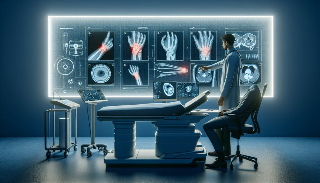
A patient presents to the emergency department after a fall onto an outstretched hand, complaining of severe elbow pain and an inability to fully extend the joint. The physical exam reveals point tenderness over the radial head, but swelling obscures clear landmarks. You suspect a fracture, but could it be a ligamentous injury? The initial decision point—what imaging to order first—is critical for accurate diagnosis and avoiding unnecessary radiation or cost. This is where the American College of Radiology (ACR) Appropriateness Criteria provide an evidence-based framework. This article decodes the ACR guidelines for acute elbow and forearm pain, helping you choose the right initial and follow-up studies based on the clinical context.
What Does ACR Acute Elbow and Forearm Pain Cover?
The ACR Appropriateness Criteria for “Acute Elbow and Forearm Pain” focus on patients presenting with recent-onset pain, typically within days to a few weeks. This guidance applies to both traumatic and non-traumatic etiologies where the primary concern is identifying an acute structural cause, such as a fracture, dislocation, or significant soft-tissue injury (e.g., tendon or ligament rupture). The criteria are designed to guide initial imaging decisions and the immediate next steps when initial radiographs are inconclusive.
It is important to note what this topic does not cover. These recommendations are not intended for clinical scenarios involving chronic elbow pain (e.g., longstanding epicondylitis), suspected septic arthritis, osteomyelitis, inflammatory arthropathies like rheumatoid arthritis, or primary bone tumors. Each of these conditions has its own dedicated ACR Appropriateness Criteria document that should be consulted for specific imaging recommendations.
What Imaging Should I Order for Acute Elbow and Forearm Pain? Recommendations by Clinical Scenario
The ACR panel provides clear, scenario-based recommendations to guide imaging selection. The optimal study depends on the initial clinical presentation and the findings of any preliminary imaging.
For an adult with acute elbow or forearm pain requiring initial imaging, the first and most appropriate step is radiography. The ACR rates Radiography area of interest as “Usually appropriate.” This is the foundational study for evaluating bony alignment, joint effusions, and obvious fractures. At this initial stage, more advanced modalities like ultrasound (US), Magnetic Resonance Imaging (MRI), Computed Tomography (CT), or bone scans are all rated “Usually not appropriate” as they are not cost-effective and often unnecessary for the initial assessment.
For an adult with a suspected fracture where initial radiographs are normal or indeterminate, the next step depends on the clinical suspicion and desired timeframe for diagnosis. The ACR provides two “Usually appropriate” options. A Radiography area of interest repeat in 10-14 days is a valid strategy, as it allows time for an occult fracture line to become more visible due to bone resorption or callus formation. Alternatively, if a more immediate diagnosis is required to guide management, a CT area of interest without IV contrast is also “Usually appropriate.” CT provides excellent detail of cortical and trabecular bone, making it highly sensitive for detecting occult fractures that are not visible on initial radiographs. MRI and US are generally not indicated in this specific scenario.
For an adult with suspected tendon, ligament, or muscle injury where radiographs are normal or indeterminate, the imaging focus shifts from bone to soft tissues. In this case, both US area of interest and MRI area of interest without IV contrast are rated “Usually appropriate.” Ultrasound is a powerful, dynamic tool for evaluating specific tendons (like the distal biceps) and ligaments, allowing for real-time assessment. MRI offers a more comprehensive, global view of all soft tissue structures, including muscles, ligaments, tendons, and cartilage, as well as bone marrow edema. The choice between them often depends on institutional expertise and the specific structure in question. CT is not suitable for primary soft-tissue evaluation and is rated “Usually not appropriate.”
ACR Imaging Recommendations Table
| Clinical Scenario | Top Procedure(s) | ACR Rating | Adult RRL | Pediatric RRL |
|---|---|---|---|---|
| Adult. Acute elbow or forearm pain. Initial imaging. | Radiography area of interest | Usually appropriate | Varies | Varies |
| Adult. Acute elbow or forearm pain. Suspect fracture. Radiographs normal or indeterminate. Next imaging study. | Radiography area of interest repeat in 10-14 days CT area of interest without IV contrast | Usually appropriate | Varies | Varies |
| Adult. Acute elbow or forearm pain. Suspect tendon or ligament or muscle injury. Radiographs normal or indeterminate. Next imaging study. | US area of interest MRI area of interest without IV contrast | Usually appropriate | O 0 mSv | O 0 mSv [ped] |
Adult vs. Pediatric Acute Elbow and Forearm Pain Imaging: Radiation Dose Tradeoffs
While the primary recommendations for imaging modalities are often similar between adults and children, the approach to radiation safety is paramount in pediatric patients. The principle of ALARA (As Low As Reasonably Achievable) guides every decision. For elbow and forearm imaging, many of the relative radiation levels (RRLs) are marked as “Varies,” indicating that the dose from radiography or CT depends heavily on the specific protocol, number of views, and patient size. However, it is critical that pediatric-specific protocols are used to minimize exposure.
For modalities that do not use ionizing radiation, such as MRI and ultrasound, the RRL is zero. The notation “[ped]” next to the pediatric RRL for these studies signifies that while there is no radiation dose, pediatric-specific considerations—such as the need for sedation in MRI or using high-frequency transducers in ultrasound for better superficial structure visualization—are still important. Given the higher lifetime attributable risk of radiation-induced malignancy, clinicians should have a lower threshold for choosing non-ionizing modalities like US or MRI as a follow-up to inconclusive radiographs in children, especially when soft-tissue injury is the primary concern.
Imaging Protocol Details for Acute Elbow and Forearm Pain
Once you have decided on the right study based on the ACR criteria, ensuring it is performed correctly is the next critical step. A well-designed imaging protocol is essential for diagnostic accuracy, whether it’s obtaining the correct radiographic views to detect a subtle radial head fracture or using the optimal MRI sequences to visualize the ulnar collateral ligament. While specific protocol guides for this topic are not yet available, the GigHz library is a comprehensive resource for general imaging techniques. For detailed information on study parameters, patient positioning, and contrast administration, consult our main library.
Tools to Help You Order the Right Study
Navigating imaging guidelines can be complex, especially when dealing with nuanced clinical presentations. GigHz offers several tools designed to support evidence-based decision-making at the point of care.
Our ACR Appropriateness Criteria Lookup provides a searchable interface to the complete, up-to-date ACR guidelines, covering hundreds of clinical variants beyond acute elbow pain. It helps you quickly find the official recommendations for virtually any clinical scenario you encounter.
The Imaging Protocol Library is a detailed repository of technical specifications for a wide range of CT, MRI, and other imaging studies. It serves as a valuable reference for radiologists, technologists, and ordering clinicians to understand the technical aspects of each exam.
For discussions with patients about radiation exposure, the Radiation Dose Calculator is a useful tool. It helps estimate cumulative effective dose from various imaging studies, facilitating informed conversations about the risks and benefits of medical imaging.
Why is radiography the first-line imaging for acute elbow pain?
Radiography (X-ray) is the recommended initial imaging modality because it is fast, widely available, inexpensive, and highly effective at identifying the most common and urgent causes of acute elbow pain, such as fractures and dislocations. It also provides a good overview of bony alignment and can reveal secondary signs of injury, like a joint effusion (the “fat pad sign”), which can indicate an occult fracture even if the fracture line itself is not visible.
When should I order a CT scan for acute elbow pain?
A CT scan is most appropriate when there is a high clinical suspicion for a fracture, but initial radiographs are negative or inconclusive. CT excels at visualizing complex or subtle fractures, particularly of the radial head, coronoid process, or olecranon. It is considered “Usually appropriate” as the next step in this specific scenario. It is not recommended for the initial evaluation or for primary assessment of soft tissues like ligaments or tendons.
Is MRI or Ultrasound better for a suspected tendon or ligament injury?
Both MRI and ultrasound are rated “Usually appropriate” for suspected soft-tissue injuries after normal radiographs, and the best choice can depend on the clinical question and local expertise. Ultrasound is excellent for dynamic evaluation of a specific structure, such as assessing the integrity of the distal biceps tendon during arm rotation. MRI provides a more comprehensive, static overview of all the soft tissues, cartilage, and bone marrow, making it ideal for detecting multiple injuries or when the exact location of the pathology is unclear.
What is the “fat pad sign” and why is it important?
The anterior and posterior fat pads are small collections of fat located within the elbow joint capsule. Normally, the posterior fat pad is hidden in the olecranon fossa and is not visible on a lateral radiograph of a flexed elbow. A joint effusion (swelling and fluid within the joint), often caused by an intra-articular fracture, will push these fat pads away from the bone, making them visible. A visible posterior fat pad on a lateral X-ray is highly suggestive of an intra-articular fracture, even if one is not directly seen, and often warrants further investigation or treatment as an occult fracture.
Is a contrast-enhanced MRI or CT ever needed for acute elbow pain?
For the scenarios covered in this ACR guideline—acute traumatic or non-traumatic pain—intravenous (IV) contrast is generally not necessary. The ACR rates both contrast-enhanced MRI and CT as “Usually not appropriate” for the initial workup of fracture or soft-tissue injury. Contrast is typically reserved for specific indications not covered by this topic, such as evaluating for infection (abscess), inflammatory arthritis (synovitis), or tumors.
Reviewed by Pouyan Golshani, MD, Interventional Radiologist — May 12, 2026