When to Order Imaging for Blunt Chest Trauma-Suspected Cardiac Injury: ACR Appropriateness Decoded
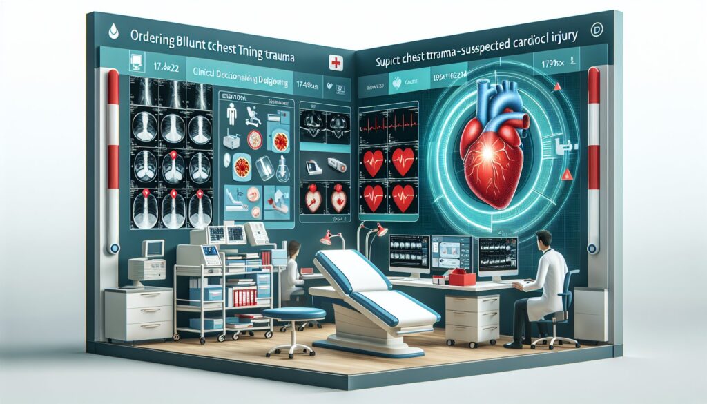
When to Order Imaging for Blunt Chest Trauma-Suspected Cardiac Injury: ACR Appropriateness Decoded
It’s a busy shift in the emergency department when a patient arrives following a high-speed motor vehicle collision. They have significant chest wall bruising and are tender over the sternum. Their electrocardiogram (ECG) shows non-specific ST changes, and you’re concerned about a potential blunt cardiac injury. Do you start with a transthoracic echocardiogram (TTE), or go straight to a computed tomography (CT) scan of the chest? Making the right call quickly is critical for identifying life-threatening conditions like pericardial tamponade, myocardial rupture, or great vessel injury. This guide decodes the American College of Radiology (ACR) Appropriateness Criteria for this exact scenario, helping you choose the most effective imaging pathway based on the patient’s hemodynamic stability.
What Does ACR Blunt Chest Trauma-Suspected Cardiac Injury Cover?
This ACR guideline focuses on the initial imaging evaluation of adult and pediatric patients who have sustained significant blunt trauma to the chest, raising suspicion for an injury to the heart or great vessels. The recommendations are stratified based on a key clinical factor: the patient’s hemodynamic stability. This distinction is crucial, as the urgency, choice, and logistics of imaging differ substantially between a stable and an unstable patient.
The criteria apply to scenarios where a blunt cardiac injury (BCI) is on the differential diagnosis. This includes myocardial contusion, chamber or septal rupture, valvular injury, pericardial effusion or tamponade, and coronary artery or great vessel injury. These guidelines do not apply to patients with penetrating chest trauma, as the injury patterns and diagnostic algorithms for those cases are distinct. They also do not cover the follow-up imaging or management of a diagnosed cardiac injury, focusing instead on the initial diagnostic workup in the acute setting.
What Imaging Should I Order for Blunt Chest Trauma-Suspected Cardiac Injury? Recommendations by Clinical Scenario
The ACR provides clear, evidence-based recommendations tailored to the patient’s clinical status. The primary branch point is whether the patient is hemodynamically stable or unstable.
For a hemodynamically stable patient with suspected cardiac injury following blunt trauma, several imaging modalities are rated as Usually Appropriate. A resting transthoracic echocardiogram (US echocardiography transthoracic resting) is a cornerstone of the evaluation. It is non-invasive, uses no ionizing radiation, can be performed at the bedside, and provides a direct assessment of cardiac function, wall motion, valvular integrity, and the presence of a pericardial effusion. A standard chest radiograph is also Usually Appropriate to evaluate for other thoracic injuries like pneumothorax, hemothorax, rib fractures, or a widened mediastinum. If there is a high suspicion for aortic or other significant intrathoracic injury, CT chest with IV contrast, CTA chest with IV contrast, or CT chest without and with IV contrast are all considered Usually Appropriate to provide detailed anatomic information.
For a hemodynamically unstable patient with suspected cardiac injury following blunt trauma, the list of Usually Appropriate studies is similar, but the urgency is higher. A resting transthoracic echocardiogram remains a key initial study, often performed as part of the Focused Assessment with Sonography for Trauma (FAST) exam, to rapidly identify conditions like cardiac tamponade. A portable chest radiograph is also essential. If the patient can be safely transported, CT chest with IV contrast or CTA chest with IV contrast are Usually Appropriate for a comprehensive evaluation of cardiac, vascular, and other thoracic structures. In this unstable scenario, CT heart function and morphology with IV contrast also becomes Usually Appropriate, offering gated imaging for more detailed functional assessment if the institution and patient’s condition permit. In both stable and unstable patients, cardiac MRI and nuclear imaging studies (SPECT, PET) are Usually Not Appropriate in the acute setting due to longer acquisition times and logistical challenges.
ACR Imaging Recommendations Table
| Clinical Scenario | Top Procedure | ACR Rating | Adult RRL | Pediatric RRL |
|---|---|---|---|---|
| Suspected cardiac injury following blunt trauma, hemodynamically stable patient. | US echocardiography transthoracic resting | Usually appropriate | O 0 mSv | O 0 mSv [ped] |
| Suspected cardiac injury following blunt trauma, hemodynamically unstable patient. | US echocardiography transthoracic resting | Usually appropriate | O 0 mSv | O 0 mSv [ped] |
Adult vs. Pediatric Blunt Chest Trauma-Suspected Cardiac Injury Imaging: Radiation Dose Tradeoffs
When evaluating children with suspected blunt cardiac injury, minimizing radiation exposure is a primary concern. The principle of As Low As Reasonably Achievable (ALARA) is paramount due to the increased lifetime risk of malignancy from ionizing radiation in younger patients. For this reason, non-ionizing modalities like ultrasound are heavily favored as the initial imaging test whenever clinically appropriate. A transthoracic echocardiogram carries a relative radiation level (RRL) of zero and is a first-line, Usually Appropriate study for both adults and children.
When CT is necessary, pediatric protocols are tailored to reduce the dose. The ACR designates the pediatric RRL for a CT chest as ☢ ☢ ☢ ☢ (3-10 mSv), a higher tier than the adult ☢ ☢ ☢ (1-10 mSv), even with an overlapping mSv range. This higher icon reflects the greater biological sensitivity of developing tissues to radiation. Clinicians must weigh the diagnostic necessity of a CT scan against the long-term risks, ensuring the study is clearly indicated to evaluate for injuries that cannot be adequately assessed with ultrasound or radiography.
Imaging Protocol Details for Blunt Chest Trauma-Suspected Cardiac Injury
Once you’ve decided on the right study, the specific imaging protocol is critical for diagnostic accuracy. A CTA of the chest requires precise contrast bolus timing to optimally opacify the aorta and great vessels, which differs from a standard contrast-enhanced CT designed for parenchymal or mediastinal evaluation. Similarly, a comprehensive echocardiogram includes specific views and Doppler assessments that go beyond a basic FAST exam. Standardizing these protocols ensures that the images acquired provide the necessary information to confirm or exclude critical injuries. For detailed technical guidance on these and other imaging procedures, clinicians can consult a comprehensive protocol resource.
Tools to Help You Order the Right Study
Navigating imaging guidelines during a critical case can be challenging. GigHz provides a suite of reference tools designed to support clinical decision-making at the point of care.
For clinical questions beyond blunt cardiac trauma, the ACR Appropriateness Criteria Lookup allows you to search the full ACR guidelines for thousands of clinical scenarios. To ensure your imaging order is executed correctly, the Imaging Protocol Library provides detailed, step-by-step protocols for a wide range of CT, MRI, and ultrasound examinations. For discussions with patients and families about radiation, or for tracking a patient’s cumulative exposure, the Radiation Dose Calculator is a valuable resource for quantifying and contextualizing medical radiation.
Why is a transthoracic echocardiogram (TTE) the first-line study for suspected blunt cardiac injury?
A TTE is considered a primary imaging tool because it is non-invasive, uses no ionizing radiation, is widely available, and can be performed quickly at the patient’s bedside (point-of-care ultrasound, or POCUS). It directly visualizes cardiac structures, allowing for rapid assessment of ventricular function, wall motion abnormalities (suggesting contusion), valvular disruption, and, most critically, the presence of pericardial effusion and signs of tamponade, which is a life-threatening emergency.
When should I order a CT or CTA instead of or in addition to an echocardiogram?
A CT or CTA is indicated when there is a high suspicion for injury to the aorta or other great vessels, which an echocardiogram may not fully visualize. A widened mediastinum on a chest radiograph, a severe mechanism of injury (e.g., deceleration), or specific physical exam findings can prompt a CTA. CT also provides a comprehensive evaluation of the lungs, pleura, chest wall, and spine, making it the modality of choice when multiple significant intrathoracic injuries are suspected.
Why is cardiac MRI usually not appropriate in the acute trauma setting?
Cardiac MRI (CMR) is a powerful tool for assessing myocardial tissue characteristics, including edema from a contusion. However, it is rated as ‘Usually Not Appropriate’ in the acute trauma setting for several practical reasons. MRI scans are lengthy, require the patient to lie still in a confined space, and necessitate transport to a specialized scanner. These factors make it unsuitable for hemodynamically unstable or uncooperative trauma patients who require continuous monitoring and potential intervention.
What is the clinical significance of a myocardial contusion?
A myocardial contusion is essentially a bruise of the heart muscle. While often clinically silent, it can lead to complications such as arrhythmias, cardiogenic shock, or late-term ventricular aneurysm formation. The diagnosis is often presumptive, based on the mechanism of injury, ECG changes, and elevated cardiac biomarkers (e.g., troponin). Imaging, particularly echocardiography, can support the diagnosis by showing regional wall motion abnormalities.
Is an abnormal ECG or troponin level required before ordering imaging for suspected cardiac injury?
No, an abnormal ECG or elevated troponin is not an absolute prerequisite for ordering imaging. While these findings significantly increase the suspicion for blunt cardiac injury and often trigger an imaging workup, a normal ECG and troponin do not definitively rule it out, especially immediately after the traumatic event. The decision to image should be based on the overall clinical picture, including the mechanism of injury, physical exam findings, and hemodynamic stability.
Reviewed by Pouyan Golshani, MD, Interventional Radiologist — May 12, 2026