When to Order Imaging for Occupational Lung Diseases: ACR Appropriateness Decoded

When to Order Imaging for Occupational Lung Diseases: ACR Appropriateness Decoded
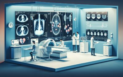
A 68-year-old retired stonemason presents to your clinic with progressive dyspnea and a dry cough that has worsened over the last year. His occupational history is significant for decades of exposure to silica dust. You suspect an occupational lung disease, but the next step in the diagnostic pathway is critical. Should you start with a simple chest radiograph, or is the higher resolution of a computed tomography (CT) scan warranted from the outset? Choosing the right initial imaging study is key to efficient diagnosis, avoiding unnecessary radiation, and guiding patient management. This guide decodes the American College of Radiology (ACR) Appropriateness Criteria for occupational lung diseases to help you make evidence-based decisions for these complex presentations.
What Does ACR Occupational Lung Diseases Cover?
The ACR Appropriateness Criteria for Occupational Lung Diseases provide guidance for imaging patients with a known or suspected history of workplace exposure to dusts, chemicals, or other agents that can cause respiratory illness. The scope of this guideline is focused on the common clinical scenarios encountered in practice, including:
- Screening and surveillance of asymptomatic workers with known hazardous exposures.
- Initial diagnostic imaging for patients presenting with symptoms suggestive of interstitial lung disease (ILD), such as silicosis, asbestosis, or coal workers’ pneumoconiosis.
- Evaluation of patients with suspected occupational airway diseases, like occupational asthma or byssinosis.
- Workup of a suspected thoracic neoplasm (e.g., lung cancer, mesothelioma) in a patient with a confirmed underlying occupational lung disease.
These criteria do not apply to acute toxic inhalation injuries, community-acquired pneumonia, or the evaluation of interstitial lung diseases without a clear occupational exposure history. The recommendations are designed to guide the selection of the most appropriate initial and subsequent imaging studies based on the specific clinical context.
What Imaging Should I Order for Occupational Lung Diseases? Recommendations by Clinical Scenario
The ACR panel provides specific recommendations tailored to the patient’s presentation and the clinical question at hand. The choice of imaging modality depends heavily on whether the goal is screening, initial diagnosis, or further characterization of known disease.
For occupational exposure, screening, and surveillance of lung disease in an initial imaging setting, a standard Radiography chest is rated Usually appropriate. This low-dose examination is effective for detecting many of the parenchymal and pleural changes associated with common pneumoconioses. A CT chest without IV contrast is rated May be appropriate and is typically reserved for cases where radiography is inconclusive or for specific high-risk surveillance programs.
When there is a clinical suspicion of interstitial lung disease (ILD) due to occupational exposure, both Radiography chest and CT chest without IV contrast are rated Usually appropriate for initial imaging. While a chest radiograph is a reasonable first step, a non-contrast CT provides far greater detail of the lung parenchyma, which is essential for identifying and characterizing patterns like reticulation, ground-glass opacities, and honeycombing. If the initial radiograph is suspicious for ILD, the clear next step is a CT chest without IV contrast, which is rated Usually appropriate for this scenario. The detailed protocol for this study can be found in our CT Chest Without Contrast guide.
Similarly, for a patient with an occupational exposure and suspected airway disease, both Radiography chest and CT chest without IV contrast are again rated Usually appropriate. A chest radiograph can exclude alternative diagnoses, while a CT, particularly with expiratory images, can reveal air trapping, a key sign of small airways disease.
In the distinct scenario of a patient with confirmed occupational lung disease and a suspected thoracic neoplasm, the imaging strategy shifts. A CT chest with IV contrast is rated Usually appropriate to evaluate the primary lesion, assess for mediastinal and hilar lymphadenopathy, and detect metastatic disease. For definitive diagnosis, an Image-guided transthoracic needle biopsy is also rated Usually appropriate. In this context, FDG-PET/CT and MRI may also be appropriate to aid in staging or assessing local invasion, while a non-contrast CT is less ideal as it cannot fully evaluate vascular or mediastinal structures. For more details on contrast-enhanced protocols, see our guide on CT Chest/Abdomen/Pelvis with IV Contrast.
ACR Imaging Recommendations Table
| Clinical Scenario | Top Procedure | ACR Rating | Adult RRL | Pediatric RRL |
|---|---|---|---|---|
| Occupational exposure, screening, and surveillance of lung disease. Initial imaging. | Radiography chest | Usually appropriate | ☢ <0.1 mSv | ☢ <0.03 mSv [ped] |
| Occupational exposure, suspected interstitial lung disease. Initial imaging. | Radiography chest | Usually appropriate | ☢ <0.1 mSv | ☢ <0.03 mSv [ped] |
| Occupational exposure, suspected interstitial lung disease based on radiography. Next imaging study. | CT chest without IV contrast | Usually appropriate | ☢ ☢ ☢ 1-10 mSv | ☢ ☢ ☢ ☢ 3-10 mSv [ped] |
| Occupational exposure, suspected airway disease. Initial imaging. | Radiography chest | Usually appropriate | ☢ <0.1 mSv | ☢ <0.03 mSv [ped] |
| Confirmed occupational lung disease, suspected thoracic neoplasm. | CT chest with IV contrast | Usually appropriate | ☢ ☢ ☢ 1-10 mSv | ☢ ☢ ☢ ☢ 3-10 mSv [ped] |
Adult vs. Pediatric Occupational Lung Diseases Imaging: Radiation Dose Tradeoffs
While significant occupational lung disease is rare in the pediatric population, environmental exposures (e.g., from a parent’s work clothes or proximity to industrial sites) can occur. The ACR guidelines provide distinct relative radiation level (RRL) categories for pediatric patients, reflecting a commitment to the ALARA (As Low As Reasonably Achievable) principle. For example, a chest CT that falls in the 1-10 mSv range for adults is placed in a higher radiation category (3-10 mSv) for children. This highlights the increased lifetime risk of radiation-induced malignancy in younger patients and underscores the importance of justifying every study involving ionizing radiation. For children, non-ionizing alternatives should always be considered, though for detailed lung evaluation, CT remains the primary advanced modality. Careful protocol optimization, including dose reduction techniques, is mandatory when performing CT scans in pediatric patients.
Imaging Protocol Details for Occupational Lung Diseases
Once you’ve decided on the right study, the specific imaging protocol is crucial for diagnostic accuracy. High-resolution techniques for CT are essential for evaluating interstitial lung disease, while contrast timing is key for assessing potential neoplasms. Our protocol guides cover technique, contrast administration, and interpretation principles for the studies recommended above:
Tools to Help You Order the Right Study
Navigating imaging guidelines can be complex, especially when managing multiple clinical variables. To streamline this process, GigHz offers several integrated tools. The ACR Appropriateness Criteria Lookup provides direct access to the full ACR guidelines for hundreds of clinical scenarios beyond occupational lung diseases. Once a study is chosen, the Imaging Protocol Library offers detailed, step-by-step protocols to ensure the examination is performed correctly. For communicating radiation exposure to patients and tracking cumulative dose, the Radiation Dose Calculator is a valuable resource for translating mSv into more understandable terms.
Frequently Asked Questions
Why is a non-contrast CT preferred for suspected interstitial lung disease?
For evaluating interstitial lung disease (ILD), the primary goal is to visualize fine details of the lung parenchyma, such as reticulation, ground-glass opacities, traction bronchiectasis, and honeycombing. Intravenous contrast is not necessary to see these findings and can sometimes create artifacts or obscure subtle ground-glass changes. A high-resolution, non-contrast CT protocol is the gold standard for characterizing ILD patterns.
When is an MRI useful for occupational lung disease?
MRI is rated Usually not appropriate for the primary diagnosis of most occupational lung diseases because it provides poor visualization of the lung parenchyma compared to CT. However, it is rated May be appropriate in the workup of a suspected thoracic neoplasm. In this specific context, MRI can be superior to CT for assessing chest wall invasion, involvement of the brachial plexus in superior sulcus tumors, or diaphragm invasion.
What are the key imaging findings of asbestosis?
Asbestosis is an interstitial fibrosis resulting from asbestos inhalation. The hallmark findings on CT include pleural plaques (focal areas of pleural thickening, which may be calcified), diffuse pleural thickening, and signs of pulmonary fibrosis. The fibrosis typically starts in the lower lobes and subpleural regions, manifesting as subpleural curvilinear lines, parenchymal bands, and, in advanced cases, honeycombing.
How does silicosis appear on a chest CT?
Silicosis is caused by inhaling crystalline silica dust. The classic CT appearance is of multiple, small, well-defined nodules (typically 2-5 mm) with a predilection for the upper lobes and posterior lung zones. These nodules can coalesce over time to form large masses, a condition known as progressive massive fibrosis (PMF). Hilar and mediastinal lymph node enlargement is common, and “eggshell” calcification of these nodes is a highly specific, though not always present, sign.
Is PET/CT useful for evaluating occupational lung disease?
FDG-PET/CT is rated Usually not appropriate for the initial diagnosis or surveillance of benign occupational lung diseases. While inflammatory processes can be FDG-avid, the findings are non-specific. Its primary role, rated as May be appropriate, is in the evaluation of a suspected thoracic neoplasm in a patient with known occupational lung disease. It is used for staging known malignancies, differentiating malignant from benign nodules or masses, and assessing treatment response.
What is the difference between screening and surveillance imaging?
Screening refers to the testing of an asymptomatic population at high risk for a disease to detect it at an early stage (e.g., screening all former shipyard workers with a history of asbestos exposure). Surveillance refers to the periodic monitoring of individuals with a known exposure or established disease to track its progression or detect complications, such as the development of a malignancy. For occupational lung disease, the initial imaging modality for both—a chest radiograph—is often the same.
Reviewed by Pouyan Golshani, MD, Interventional Radiologist — May 12, 2026