When to Order Imaging for Palpable Breast Masses: ACR Appropriateness Decoded
When to Order Imaging for Palpable Breast Masses: ACR Appropriateness Decoded
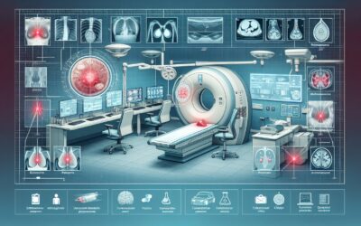
A patient presents with a new, palpable breast mass. It’s a common and often anxiety-provoking clinical scenario that requires a clear, evidence-based imaging workup. The initial choice of imaging modality is critical and depends heavily on the patient’s age and specific clinical context. Ordering the wrong initial study can lead to diagnostic delays, unnecessary radiation exposure, and increased patient anxiety. This guide decodes the American College of Radiology (ACR) Appropriateness Criteria for palpable breast masses, providing a scannable, authoritative reference to help you choose the right study for your patient.
What Does the ACR Guideline for Palpable Breast Masses Cover?
The ACR Appropriateness Criteria for Palpable Breast Masses addresses the initial imaging evaluation and subsequent steps for adult female patients who present with a focal, palpable lump. The guidelines are stratified by patient age and the findings of initial imaging studies, providing a clear diagnostic pathway.
This topic specifically covers:
- Initial imaging for women under 30, from 30 to 39, and 40 years of age or older.
- Next steps after initial imaging yields negative, benign, probably benign, or suspicious findings (corresponding to Breast Imaging Reporting and Data System, or BI-RADS, categories 1 through 5).
These criteria do not apply to generalized breast lumpiness, cyclical nodularity, or diffuse breast pain without a discrete, palpable mass. They also do not cover the evaluation of male breast masses, nipple discharge, or asymptomatic patients undergoing screening, which are addressed in separate ACR guidelines.
What Imaging Should I Order for Palpable Breast Masses? Recommendations by Clinical Scenario
The appropriate imaging pathway for a palpable breast mass is primarily determined by the patient’s age, with different recommendations for women under 30, between 30 and 39, and 40 or older.
For women younger than 30 years of age, the initial imaging evaluation begins with ultrasound. For this scenario, the ACR rates US breast as Usually appropriate. This approach avoids ionizing radiation in a younger population whose breast tissue is typically denser and more sensitive to radiation. Mammography is generally reserved for cases where ultrasound findings are suspicious. If an ultrasound is suspicious or highly suggestive of malignancy (BI-RADS 4 or 5), then Digital breast tomosynthesis (DBT) diagnostic and Mammography diagnostic become Usually appropriate to fully characterize the lesion and evaluate the rest of the breast tissue. An Image-guided core biopsy is also rated Usually appropriate in this context to obtain a definitive diagnosis.
For women 30 to 39 years of age, both ultrasound and mammography are considered primary options. The ACR rates US breast, Digital breast tomosynthesis diagnostic, and Mammography diagnostic as Usually appropriate for the initial workup of a palpable mass in this age group.
For women 40 years of age or older, the initial workup starts with mammography. For this initial evaluation, Digital breast tomosynthesis diagnostic and Mammography diagnostic are rated Usually appropriate. If the mammogram is negative (BI-RADS 1) or shows a benign finding (BI-RADS 2) at the site of the palpable concern, a targeted US breast is the recommended next step. For a negative mammogram, US breast is Usually appropriate; for a benign mammogram, it May be appropriate to ensure the palpable finding corresponds to the benign feature on the mammogram. If initial mammography findings are suspicious or highly suggestive of malignancy (BI-RADS 4 or 5), a targeted US breast is Usually appropriate to assess for a sonographic correlate, which can be used to guide a biopsy.
Across all age groups, if imaging findings are probably benign (BI-RADS 3), short-interval follow-up is typically recommended over immediate biopsy. Advanced imaging modalities like breast MRI and nuclear medicine studies (FDG-PET, Sestamibi) are Usually not appropriate for the initial evaluation of a palpable mass.
ACR Imaging Recommendations Table for Palpable Breast Masses
| Clinical Scenario | Top Procedure(s) | ACR Rating | Adult RRL | Pediatric RRL |
|---|---|---|---|---|
| Adult female, younger than 30 years of age. Palpable breast mass. Initial imaging. | US breast | Usually appropriate | O 0 mSv | O 0 mSv [ped] |
| Adult female, younger than 30. Palpable mass. US findings are suspicious or highly suggestive of malignancy (BI-RADS 4 or 5). Next imaging study. | Digital breast tomosynthesis diagnostic; Mammography diagnostic; Image-guided core biopsy breast | Usually appropriate | Varies | Varies |
| Adult female, younger than 30. Palpable mass. US findings probably benign (BI-RADS 3). Next imaging study. | (Short-term follow-up is typical; no further imaging is rated appropriate) | Usually not appropriate | Varies | Varies |
| Adult female, younger than 30. Palpable mass. US findings benign (BI-RADS 2). Next imaging study. | (No further imaging is rated appropriate) | Usually not appropriate | Varies | Varies |
| Adult female, younger than 30. Palpable mass. US findings negative (BI-RADS 1). Next imaging study. | (No further imaging is rated appropriate) | Usually not appropriate | Varies | Varies |
| Adult female, 30 to 39 years of age. Palpable breast mass. Initial imaging. | US breast; Digital breast tomosynthesis diagnostic; Mammography diagnostic | Usually appropriate | Varies | Varies |
| Adult female, 40 years of age or older. Palpable breast mass. Initial imaging. | Digital breast tomosynthesis diagnostic; Mammography diagnostic | Usually appropriate | ☢ ☢ 0.1-1mSv | |
| Adult female, 40 or older. Palpable mass. Mammography findings are suspicious or highly suggestive of malignancy (BI-RADS 4 or 5). Next imaging study. | US breast | Usually appropriate | O 0 mSv | O 0 mSv [ped] |
| Adult female, 40 or older. Palpable mass. Diagnostic mammography, DBT, and US findings are probably benign (BI-RADS 3). Next imaging study. | (Short-term follow-up is typical; no further imaging is rated appropriate) | Usually not appropriate | Varies | Varies |
| Adult female, 40 or older. Palpable mass. Mammography findings are benign (BI-RADS 2) at the site of palpable mass. Next imaging study. | US breast | May be appropriate | O 0 mSv | O 0 mSv [ped] |
| Adult female, 40 or older. Palpable mass. Mammography findings are negative (BI-RADS 1). Next imaging study. | US breast | Usually appropriate | O 0 mSv | O 0 mSv [ped] |
Adult vs. Pediatric Considerations for Palpable Breast Masses Imaging: Radiation Dose Tradeoffs
The evaluation of palpable breast masses differs significantly based on age, primarily due to variations in breast tissue density and the principle of As Low As Reasonably Achievable (ALARA) for radiation exposure. In adolescents and women under 30, breast tissue is generally denser, which can reduce the sensitivity of mammography. More importantly, the developing breast tissue in younger patients is more susceptible to the carcinogenic effects of ionizing radiation. For these reasons, ultrasound, which uses no ionizing radiation, is the strongly preferred initial imaging modality.
For women over 40, breast tissue typically becomes less dense due to fatty replacement, increasing the diagnostic utility of mammography. At this age, the benefits of using mammography to detect cancer generally outweigh the risks associated with the low radiation dose. The ACR guidelines reflect this tradeoff by recommending mammography as the initial study for this age group, while still emphasizing ultrasound as a critical tool for problem-solving and evaluating palpable findings that are occult or equivocal on mammograms.
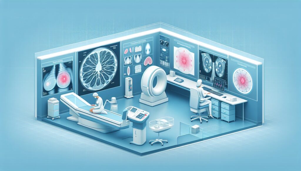
Imaging Protocol Details for Palpable Breast Masses
Once you’ve decided on the right study, the specific imaging protocol is essential for diagnostic accuracy. Key considerations include the field of view for mammography, transducer frequency for ultrasound, and contrast timing for MRI when indicated for other purposes. Our detailed protocol guides cover technique, patient positioning, and interpretation principles for the studies recommended in these guidelines.
Tools to Help You Order the Right Study
Navigating imaging guidelines can be complex. GigHz provides a suite of tools designed to support clinical decision-making and streamline the imaging workflow for physicians and trainees.
The ACR Appropriateness Criteria Lookup tool provides quick access to the full spectrum of ACR guidelines, covering thousands of clinical variants beyond palpable breast masses. It helps you confirm the right study for any indication in seconds.
For detailed procedural steps, our Imaging Protocol Library offers standardized, scannable guides for hundreds of imaging exams. This resource is invaluable for ensuring that the ordered study is performed correctly for optimal diagnostic quality.
To help with patient communication about radiation exposure, the Radiation Dose Calculator allows you to estimate effective dose for various studies and track cumulative exposure for your patients, facilitating informed consent and shared decision-making.
Why is ultrasound the first-line imaging choice for women under 30 with a palpable breast mass?
Ultrasound is recommended as the initial imaging modality for women under 30 for two primary reasons. First, younger women tend to have denser breast tissue, which can obscure findings on a mammogram, making ultrasound a more sensitive tool in this population. Second, and more importantly, breast tissue in younger patients is more sensitive to ionizing radiation. Using ultrasound first adheres to the ALARA (As Low As Reasonably Achievable) principle by avoiding radiation exposure unless a suspicious finding on ultrasound warrants further evaluation with mammography.
If a mammogram is negative (BI-RADS 1) but a mass remains palpable, what is the next step?
A palpable mass with a negative mammogram is a common clinical scenario. The breast tissue may be dense, obscuring the mass. According to the ACR criteria for women 40 and older, the next step is a targeted breast ultrasound. This is rated as Usually appropriate. The ultrasound is performed to specifically evaluate the area of the palpable concern to determine if a sonographic correlate, such as a cyst or solid nodule, is present.
What is the role of breast MRI in the initial workup of a palpable mass?
For the initial diagnostic workup of a palpable breast mass in an average-risk woman, breast MRI is Usually not appropriate. While highly sensitive, MRI has lower specificity, which can lead to false positives and unnecessary biopsies. Its primary roles in breast imaging are for high-risk screening (e.g., in patients with known genetic mutations), determining the extent of disease in newly diagnosed breast cancer, and evaluating for implant integrity, not for the initial characterization of a palpable finding.
What is the difference between a diagnostic and a screening mammogram in this context?
A screening mammogram is performed on asymptomatic women to detect early, non-palpable breast cancers. It typically consists of a standard set of images. A diagnostic mammogram is used to evaluate a specific problem, such as a palpable lump or an abnormal finding on a screening study. It is tailored to the patient’s specific issue and may include additional views, such as spot compression or magnification, and is directly supervised by a radiologist. For a palpable mass, a diagnostic mammogram (or DBT) is always the appropriate choice, while a screening study is Usually not appropriate.
When is a biopsy indicated after initial imaging of a palpable mass?
A biopsy is indicated when imaging findings are suspicious or highly suggestive of malignancy, corresponding to BI-RADS categories 4 or 5. For a patient under 30 with a BI-RADS 4 or 5 finding on ultrasound, an image-guided core biopsy is rated Usually appropriate. For patients 40 and older, a suspicious mammographic finding would first be evaluated with ultrasound to identify a target for biopsy. The goal of the biopsy is to obtain a tissue sample for pathologic analysis to make a definitive diagnosis.
Reviewed by Pouyan Golshani, MD, Interventional Radiologist — May 12, 2026