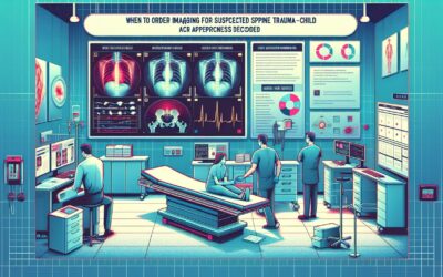
When to Order Imaging for Scoliosis-Child: ACR Appropriateness Decoded
When to Order Imaging for Scoliosis-Child: ACR Appropriateness Decoded
A 12-year-old presents for a well-child check, and a routine Adams forward bend test reveals thoracic asymmetry. You suspect adolescent idiopathic scoliosis, the most common type, but you recall that certain presentations warrant a deeper look for underlying pathology. The immediate question is what imaging, if any, is the right first step. Do you start with plain films? Is an MRI necessary to evaluate the spinal cord? Ordering the correct initial study is critical for accurate diagnosis and management while minimizing unnecessary radiation exposure in a young patient. This guide decodes the American College of Radiology (ACR) Appropriateness Criteria for Scoliosis-Child to help you make the right call.
What Does ACR Scoliosis-Child Cover?
The ACR Appropriateness Criteria for Scoliosis-Child provide evidence-based guidelines for the initial imaging evaluation of children and adolescents with suspected or newly diagnosed scoliosis. The recommendations are tailored to specific clinical scenarios based on the patient’s age and the suspected etiology of the spinal curvature.
This topic specifically addresses four common clinical variants:
- Initial imaging for suspected congenital scoliosis in a child.
- Initial imaging for early-onset idiopathic scoliosis in a child from birth to 9 years of age.
- Initial imaging for adolescent idiopathic scoliosis (10 to 17 years of age) without neurologic risk factors.
- Initial imaging for adolescent idiopathic scoliosis (10 to 17 years of age) with neurologic risk factors (e.g., pain, abnormal reflexes, rapid progression).
These guidelines do not cover imaging for post-operative follow-up, scoliosis secondary to neuromuscular conditions (like cerebral palsy or muscular dystrophy), or scoliosis in adults. The focus is strictly on the initial diagnostic workup in the pediatric population.
What Imaging Should I Order for Scoliosis-Child? Recommendations by Clinical Scenario
The ACR panel’s recommendations hinge on the patient’s age and clinical presentation, balancing the need for detailed anatomical information with the principle of As Low As Reasonably Achievable (ALARA) radiation dose.
For a child with suspected congenital scoliosis, the initial workup involves evaluating both the bony anatomy and the underlying neural structures. Accordingly, both Radiography complete spine and MRI complete spine without IV contrast are rated Usually appropriate. Radiographs define the vertebral anomalies, while MRI is crucial for identifying associated spinal cord abnormalities, which are common in this condition. CT of the spine without contrast May be appropriate, particularly for pre-operative planning, but carries a higher radiation dose.
Similarly, for a child (0 to 9 years) with early-onset idiopathic scoliosis, the ACR rates both Radiography complete spine and MRI complete spine without IV contrast as Usually appropriate. The higher incidence of underlying intraspinal pathology in this younger age group justifies the routine use of MRI for initial evaluation, even in the absence of specific neurologic red flags.
The approach changes for adolescents. For an adolescent (10 to 17 years) with suspected adolescent idiopathic scoliosis and no risk factors, Radiography complete spine is the sole imaging modality rated Usually appropriate. In this common scenario, the pretest probability of an underlying spinal cord abnormality is low, so MRI is deemed Usually not appropriate as a first-line study.
However, if that same adolescent has risk factors—such as significant pain, abnormal neurologic exam findings, an atypical curve pattern (e.g., left-sided thoracic curve), or rapid progression—the recommendations align with those for younger children. For adolescent idiopathic scoliosis with risk factors, both Radiography complete spine and MRI complete spine without IV contrast are Usually appropriate to rule out an underlying cause for the scoliosis.
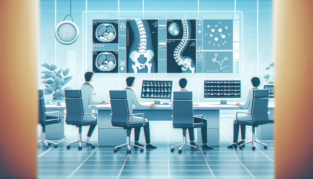
ACR Imaging Recommendations Table
| Clinical Scenario | Top Procedure | ACR Rating | Adult RRL | Pediatric RRL |
|---|---|---|---|---|
| Child. Congenital scoliosis. Initial imaging. | Radiography complete spine | Usually appropriate | ☢ ☢ ☢ 0.3-3 mSv [ped] | |
| Child (0 to 9 years of age). Early onset idiopathic scoliosis. Initial imaging. | Radiography complete spine | Usually appropriate | ☢ ☢ ☢ 0.3-3 mSv [ped] | |
| Adolescent (10 to 17 years of age). Adolescent idiopathic scoliosis. No risk factors. Initial imaging. | Radiography complete spine | Usually appropriate | ☢ ☢ ☢ 0.3-3 mSv [ped] | |
| Adolescent (10 to 17 years of age). Adolescent idiopathic scoliosis. Risk factors. Initial imaging. | Radiography complete spine | Usually appropriate | ☢ ☢ ☢ 0.3-3 mSv [ped] |
Adult vs. Pediatric Scoliosis-Child Imaging: Radiation Dose Tradeoffs
This ACR topic is exclusively pediatric, so direct adult comparisons are not applicable. However, the core principle guiding the recommendations is radiation safety in a young, developing population. Children are more radiosensitive than adults, and they have a longer lifespan over which the potential stochastic effects of radiation can manifest. Therefore, the ALARA principle is paramount.
The guidelines reflect this by consistently recommending radiography as the primary modality for assessing bony alignment and curve magnitude (Cobb angle). Modern techniques, including low-dose protocols and biplanar slot-scanning systems, can significantly reduce the radiation dose compared to older methods. When cross-sectional imaging is required to evaluate for complex bony anatomy or intraspinal pathology, the ACR consistently favors MRI over CT. MRI uses no ionizing radiation, making it the ideal choice for evaluating the spinal cord and soft tissues in children. CT is generally reserved for specific indications where detailed bone definition is essential for surgical planning and is rated as May be appropriate or Usually not appropriate in most initial diagnostic scenarios.
Imaging Protocol Details for Scoliosis-Child
Once you’ve decided on the right study, the specific imaging protocol is essential for diagnostic quality. Our protocol guides cover key considerations like patient positioning, sequence selection, and contrast administration for studies recommended in the ACR criteria. For related procedural guidance, see our detailed protocol articles:
Tools to Help You Order the Right Study
Navigating imaging guidelines can be complex, but several tools can help streamline the process. For scenarios beyond Scoliosis-Child, the ACR Appropriateness Criteria Lookup provides direct access to the full library of ACR guidelines, helping you find evidence-based recommendations for hundreds of clinical presentations.
To ensure the studies you order are performed correctly, the Imaging Protocol Library offers detailed, step-by-step protocols for a wide range of MRI, CT, and ultrasound examinations. This resource is designed to help standardize imaging techniques and improve diagnostic accuracy.
Finally, for discussing radiation exposure with patients and their families, the Radiation Dose Calculator is a valuable tool. It helps estimate cumulative radiation dose from various imaging studies, facilitating informed conversations about the risks and benefits of medical imaging.
Why is MRI recommended for early-onset scoliosis but not for uncomplicated adolescent scoliosis?
The rate of underlying spinal cord abnormalities (like a syrinx, tethered cord, or Chiari malformation) is significantly higher in children with early-onset scoliosis (diagnosed before age 10) and congenital scoliosis. The ACR recommends MRI as a standard part of the initial workup in these younger patients to proactively screen for these conditions. In contrast, the incidence of such abnormalities in adolescents with a typical presentation of idiopathic scoliosis is very low, making routine MRI unnecessary and not cost-effective.
What are the “risk factors” that warrant an MRI in adolescent idiopathic scoliosis?
Risk factors, or “red flags,” are clinical findings that suggest the scoliosis may not be truly idiopathic and could be secondary to an underlying neurologic or other pathology. These include: significant, non-mechanical back pain; a new or progressive neurologic deficit (e.g., weakness, numbness, changes in gait, bowel/bladder dysfunction); abnormal reflexes on physical exam; or an atypical curve pattern, such as a painful curve, a left-sided thoracic curve, or an unusually rapid progression of the curve.
Is there ever a role for CT in the initial evaluation of pediatric scoliosis?
For the initial diagnosis, CT is rarely the first choice due to its use of ionizing radiation. The ACR rates it as May be appropriate (Disagreement) for congenital scoliosis, where it can provide superior bony detail of complex vertebral anomalies that may not be fully characterized by radiographs. It is most often used in a pre-operative setting to help surgeons plan complex spinal reconstructions, rather than for the initial diagnosis.
Why is a bone scan rated “Usually Not Appropriate” for all scenarios?
A nuclear medicine bone scan (scintigraphy) is a functional study that detects areas of increased bone turnover. While it is sensitive for detecting infection, inflammation, or tumors, these are not common causes of scoliosis. A bone scan is not an appropriate tool for evaluating the spinal anatomy or curvature itself and imparts a relatively high radiation dose. It is generally reserved for cases where there is a specific clinical suspicion for conditions like osteoid osteoma, spondylolysis, or infection, which are distinct from the standard scoliosis workup.
Reviewed by Pouyan Golshani, MD, Interventional Radiologist — May 12, 2026