Which Fertility-Sparing Therapy is Best for Symptomatic Uterine Fibroids?
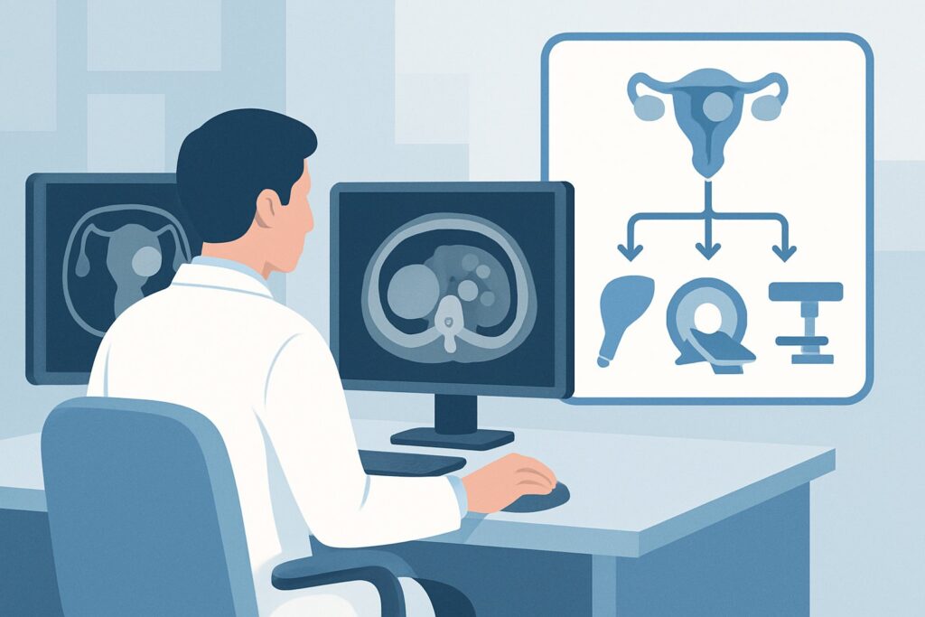
A 32-year-old G0P0 presents to your clinic with progressively heavier menstrual cycles and a constant feeling of pelvic pressure. She is otherwise healthy but notes the symptoms are now interfering with her work and quality of life. An initial pelvic ultrasound confirms multiple uterine fibroids. She is in a long-term relationship and is firm in her desire to have children in the next few years. You are now faced with selecting the most appropriate initial therapy that addresses her symptoms while preserving her fertility. This article details the clinical workflow for this specific scenario, guided by the American College of Radiology (ACR) Appropriateness Criteria. For this patient, several fertility-sparing interventions, including hysteroscopic myomectomy, are rated Usually Appropriate.
Who Fits This Clinical Scenario?
This guidance applies specifically to reproductive-age patients with symptomatic uterine fibroids who wish to preserve their fertility. The key inclusion criteria are:
- Age: Premenopausal/reproductive age.
- Symptoms: The patient must be symptomatic from the fibroids, with either heavy uterine bleeding (menorrhagia) or bulk-related symptoms. Bulk symptoms include pelvic pressure, pain, a feeling of fullness, or impingement on adjacent organs causing urinary frequency or constipation.
- Goal: An explicit desire to preserve the uterus for potential future pregnancy.
- Timing: This is for the selection of initial therapy, not for treatment failure or recurrence.
It is critical to distinguish this presentation from closely related scenarios that follow different management pathways. This advice does not apply if the patient is postmenopausal, as the risks and benefits of intervention change significantly. It also does not apply if the primary symptom is reproductive dysfunction (e.g., infertility or recurrent pregnancy loss) without significant bleeding or bulk symptoms, which is a distinct ACR variant. Finally, if significant concurrent adenomyosis is known or highly suspected, the therapeutic options and their expected efficacy may differ, warranting a separate evaluation.
What Diagnoses Are You Confirming and Characterizing?
While the diagnosis of “uterine fibroids” has likely been made by initial imaging (typically ultrasound), the crucial next step is to precisely characterize the fibroids to guide therapeutic selection. The goal is to determine the size, number, and exact location of the leiomyomas, as this dictates which fertility-sparing options are feasible and most likely to succeed. The key considerations are:
Submucosal Fibroids (FIGO Types 0, 1, 2): These are the most common culprits for heavy menstrual bleeding as they protrude into and distort the endometrial cavity. Identifying their presence, size, and degree of intramural extension is paramount, as they are the primary targets for hysteroscopic myomectomy. A saline-infusion sonohysterogram or pelvic MRI provides the best detail for this assessment.
Intramural and Subserosal Fibroids (FIGO Types 3-7): These fibroids are located within the uterine wall or on its outer surface. They are the typical cause of bulk-related symptoms. While they can contribute to heavy bleeding by increasing the overall surface area of the endometrium, they are not accessible via a hysteroscopic approach. Their presence would steer the therapeutic choice toward options like laparoscopic/open myomectomy or uterine artery embolization.
Adenomyosis: This condition, where endometrial tissue exists within the myometrium, is a significant diagnostic confounder. It often co-exists with fibroids and can cause similar symptoms of heavy, painful bleeding and an enlarged, bulky uterus. Pelvic MRI is highly sensitive for diagnosing adenomyosis and is crucial for counseling the patient, as interventions targeted only at fibroids may not fully resolve symptoms if significant adenomyosis is present.
Endometrial Polyps or Hyperplasia: While fibroids may be the obvious finding, it’s important to rule out other intracavitary pathology that can cause abnormal uterine bleeding. These can often be evaluated during the same diagnostic workup, for instance, with sonohysterography or hysteroscopy.
Why Are Multiple Fertility-Sparing Options Recommended?
For a reproductive-age patient with symptomatic fibroids who desires fertility, the ACR panel designates five therapeutic approaches as Usually Appropriate. This reflects the fact that the optimal choice is not one-size-fits-all but depends heavily on fibroid characteristics (size, location, number) and patient-specific factors. There is no radiation dose associated with these procedures.
Hysteroscopic Myomectomy is a minimally invasive surgical procedure ideal for treating submucosal fibroids (FIGO types 0 and 1) that are the primary cause of heavy bleeding. Because it is performed transcervically without abdominal incisions, recovery is rapid. It directly removes the offending lesion, often leading to immediate symptom improvement. Its appropriateness is contingent on the fibroid being accessible from within the uterine cavity.
Laparoscopic or Open Myomectomy is the standard surgical approach for removing intramural and subserosal fibroids. This is the best option for patients whose primary symptoms are bulk-related or for those with large or numerous fibroids not amenable to other techniques. It is a more invasive procedure but is highly effective and considered the gold standard for fertility preservation when significant fibroid removal is needed.
Uterine Artery Embolization (UAE) is a minimally invasive, image-guided procedure where the blood supply to the fibroids is blocked, causing them to shrink. It is highly effective for both bleeding and bulk symptoms and treats all fibroids in the uterus simultaneously. While pregnancy is possible after UAE, its long-term effects on fertility and placental function are still debated, making shared decision-making essential for patients desiring future pregnancy.
MR-guided High-Frequency Focused Ultrasound Ablation (MRgFUS) is a non-invasive thermal ablation technique that uses focused ultrasound waves to destroy fibroid tissue. It is an excellent option for patients with a limited number of accessible fibroids who wish to avoid surgery. However, not all patients are candidates based on fibroid location and other anatomical factors.
In contrast, procedures like Endometrial Ablation and Hysterectomy are rated Usually Not Appropriate for this specific patient. Endometrial ablation destroys the uterine lining to control bleeding but results in sterility and is contraindicated in patients desiring fertility. Hysterectomy is definitive but removes the uterus entirely, permanently ending any possibility of future pregnancy.
What’s Next? Downstream Clinical Workflow
The results of the detailed characterization of the fibroids (typically with pelvic MRI or sonohysterography) will guide the next steps and the final therapeutic choice.
- If Predominantly Submucosal Fibroids (FIGO 0-1): The patient is an excellent candidate for hysteroscopic myomectomy. The next step is referral to a gynecologic surgeon with expertise in operative hysteroscopy. This is often the most direct and least invasive path to symptom resolution for this fibroid subtype.
- If Predominantly Intramural/Subserosal Fibroids: The patient should be counseled on the options of laparoscopic/open myomectomy, UAE, or potentially MRgFUS. The choice will depend on fibroid size and number, surgical risk, and a detailed discussion of the patient’s reproductive goals and timeline. Referral to both a gynecologic surgeon and an interventional radiologist for consultation is often the best approach to facilitate shared decision-making.
- If a Mix of Fibroid Types: A multi-modal approach may be necessary. For example, a patient might undergo hysteroscopic myomectomy to control bleeding from a submucosal fibroid, followed by UAE at a later time to manage bulk symptoms from intramural fibroids.
- If Significant Adenomyosis is Confirmed: The conversation shifts. While a myomectomy may still be performed, the patient must be counseled that her bleeding and pain may not fully resolve due to the underlying adenomyosis. Medical management may play a larger role, and the expected success of any single procedure must be managed carefully.
Pitfalls to Avoid (and When to Get Help)
A primary pitfall is proceeding to therapy based only on a standard transabdominal/transvaginal ultrasound. While excellent for initial detection, it often lacks the detail to definitively map submucosal fibroids or diagnose concurrent adenomyosis. Relying on it alone can lead to selecting a suboptimal therapy.
Another common error is failing to offer a consultation with both gynecologic surgery and interventional radiology. Patients often receive information about only one type of procedure based on the specialty of their initial provider. A comprehensive discussion requires input from all relevant experts.
Finally, do not underestimate the impact of adenomyosis. Attributing all symptoms to fibroids when significant adenomyosis is present on MRI can lead to patient dissatisfaction when symptoms persist post-procedure. If MRI confirms extensive adenomyosis co-existing with fibroids, a frank discussion about expected outcomes and the potential need for medical management is critical before any intervention.
Related ACR Topics and Tools
For a comprehensive overview of all clinical variants related to this topic, please see our parent guide. The tools below can assist in navigating adjacent scenarios, understanding procedural techniques, and discussing radiation safety with patients.
- Management of Uterine Fibroids: ACR Appropriateness Decoded
- ACR Appropriateness Criteria Lookup
- Imaging Protocol Library
- Radiation Dose Calculator
Frequently Asked Questions
Why is MRI often needed before choosing a therapy for fibroids?
While ultrasound is excellent for detecting fibroids, a pelvic MRI provides superior detail on their exact size, number, and location (submucosal, intramural, subserosal). It is also the best imaging modality for diagnosing concurrent adenomyosis. This detailed mapping is essential for determining if a patient is a candidate for less invasive options like hysteroscopic myomectomy or MR-guided focused ultrasound and for surgical planning for a laparoscopic or open myomectomy.
Can a patient get pregnant after Uterine Artery Embolization (UAE)?
Yes, pregnancies have been reported after UAE. However, the procedure is known to carry some risk of affecting ovarian function and may be associated with higher rates of certain obstetric complications. For this reason, while it is rated ‘Usually Appropriate’ by the ACR, it requires a detailed shared decision-making conversation with any patient who strongly desires future fertility, often weighing it against surgical myomectomy.
If a patient has both heavy bleeding and bulk symptoms, which therapy is best?
The best therapy depends on the location of the fibroids causing the symptoms. If a submucosal fibroid is causing bleeding and separate intramural fibroids are causing bulk, a combined or staged approach might be needed. Alternatively, a single therapy like UAE or a comprehensive myomectomy could address both symptom types simultaneously. A detailed MRI is critical to making this determination.
What is the difference between a myomectomy and a hysterectomy?
A myomectomy is a surgical procedure that removes only the uterine fibroids, leaving the uterus intact. It is a fertility-sparing procedure. A hysterectomy is the surgical removal of the entire uterus, which is a definitive treatment for fibroid symptoms but results in the inability to carry a pregnancy. For the patient in this scenario, a hysterectomy is ‘Usually Not Appropriate’ because of the stated desire to preserve fertility.
Are there effective medical management options for this patient?
Yes, medical management is also rated as ‘Usually Appropriate’ by the ACR for this scenario. Options can include hormonal therapies (like oral contraceptives or progestin-releasing IUDs) and non-hormonal agents (like tranexamic acid) to control heavy bleeding. While they can be effective for symptom control, they do not shrink or remove the fibroids and symptoms typically return after cessation of therapy. They are often used as a bridge to a definitive procedure or for patients wishing to delay intervention.
Reviewed by Pouyan Golshani, MD, Interventional Radiologist — May 30, 2026