Which Imaging Best Stages Nonmuscle Invasive Bladder Cancer Before Treatment?
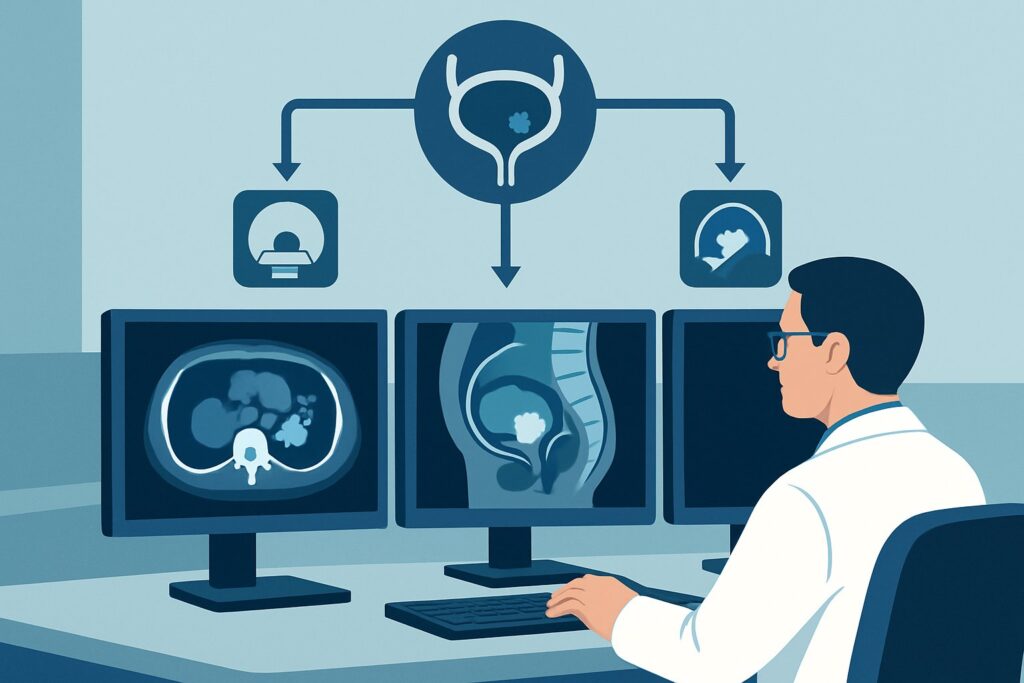
A 68-year-old patient is in your urology clinic for follow-up. Last week’s cystoscopy for hematuria revealed a 2 cm papillary tumor, and the biopsy confirmed high-grade Ta urothelial carcinoma. The pathology report notes that no muscularis propria was present in the specimen. You plan to proceed with a repeat resection and intravesical Bacillus Calmette-Guérin (BCG) therapy, but a critical question remains: is there occult muscle invasion that the initial biopsy missed? Understaging the cancer would lead to inadequate treatment. This article details the clinical workflow for imaging-based pretreatment staging in this exact scenario: an adult with presumed nonmuscle invasive bladder cancer (NMIBC). For this presentation, the American College of Radiology (ACR) finds that MRI pelvis without and with IV contrast is Usually appropriate.
Who Fits This Clinical Scenario for Bladder Cancer Staging?
This guidance applies to adult patients with a new diagnosis of urothelial carcinoma of the bladder that, based on initial cystoscopy and biopsy, appears to be nonmuscle invasive. This includes tumors staged as Ta (noninvasive papillary carcinoma), T1 (tumor invades subepithelial connective tissue), or carcinoma in situ (CIS). A key feature of this scenario is the clinical uncertainty about the true depth of invasion, which is common when the initial biopsy specimen is small or lacks detrusor muscle for a definitive pathologic assessment.
This workflow is specifically for staging the primary bladder tumor and adjacent structures. It is distinct from other related, but different, clinical situations:
- Known Muscle Invasive Bladder Cancer (MIBC): If the biopsy definitively shows tumor invading the muscularis propria (stage T2 or higher), the patient fits the ACR variant for MIBC. The imaging goals shift from local staging to assessing for extravesical extension and distant metastases, often requiring more extensive body imaging.
- Upper Tract Urothelial Cancer (UTUC): If the tumor is located in the renal pelvis or ureter, not the bladder, a different staging approach is required. This falls under the ACR variant for UTUC, which prioritizes imaging of the entire collecting system.
- Post-treatment Surveillance: This guidance is for initial, pretreatment staging. Imaging for surveillance after treatment for bladder cancer follows separate protocols.
What Diagnoses Are You Working Up in This Scenario?
In pretreatment staging of presumed NMIBC, imaging is not used to discover the cancer—that is already known from biopsy. Instead, imaging aims to differentiate between stages that have profoundly different treatments and prognoses. The key differential is the depth of tumor invasion.
True Nonmuscle Invasive Bladder Cancer (Ta, T1, CIS): This is the expected finding and the basis for conservative, bladder-sparing treatments like transurethral resection of bladder tumor (TURBT) and intravesical immunotherapy or chemotherapy. Imaging serves to confirm the confinement of the tumor to the mucosa and submucosa and to rule out deeper, more aggressive disease.
Occult Muscle Invasion (Stage ≥T2): This is the most critical distinction to make. Up to 25% of tumors initially staged as T1 on biopsy are upstaged to muscle-invasive disease on subsequent resection or cystectomy. Missing this diagnosis and treating the patient for NMIBC can lead to disease progression and a lost opportunity for curative therapy (radical cystectomy). Imaging aims to detect subtle tumor extension into the deep muscularis propria layer of the bladder wall.
Perivesical Fat Invasion (Stage ≥T3): While less common in tumors that appear non-invasive on cystoscopy, macroscopic extension through the bladder wall into the surrounding fat is a possibility. Identifying this preoperatively is crucial as it signifies locally advanced disease that requires neoadjuvant chemotherapy followed by radical surgery.
Adjacent Organ Involvement (Stage T4): In rare cases, a tumor that appears small on the surface may have an aggressive, infiltrative component that extends into the prostate, seminal vesicles, uterus, or pelvic sidewall. Cross-sectional imaging is the only non-surgical way to detect this advanced stage of disease prior to definitive treatment planning.
Why Is MRI of the Pelvis Recommended for Staging Nonmuscle Invasive Bladder Cancer?
The ACR designates MRI pelvis without and with IV contrast as Usually appropriate for this scenario because of its superior soft-tissue resolution, which is essential for evaluating the layers of the bladder wall. The primary goal is to distinguish T1 (submucosal invasion) from T2 (muscularis propria invasion). Multiplanar T2-weighted images can often delineate the slightly hyperintense submucosa from the distinctly hypointense (dark) muscularis propria. An intact, dark muscularis propria layer beneath the tumor is a strong indicator of non-invasive disease.
Dynamic contrast-enhanced (DCE) sequences are also critical. Urothelial carcinoma typically enhances earlier and more avidly than the normal bladder wall. Disruption of the muscularis propria by enhancing tumor is the key finding that suggests muscle invasion. The standardized Vesical Imaging Reporting and Data System (VI-RADS) leverages these MRI features to provide a risk score for muscle invasion, improving communication between radiologists and urologists.
Why are other studies rated lower for this specific question?
- CT Urography (CTU): While also rated Usually appropriate, CTU’s primary strength is evaluating the entire urothelial tract for synchronous tumors, not the detailed local staging of the bladder wall. Its soft-tissue resolution is inferior to MRI, making the distinction between T1 and T2 disease more difficult. It also involves significant ionizing radiation (☢☢☢☢ 10-30 mSv), whereas MRI has none (O 0 mSv).
- Pelvic Ultrasound: Rated as May be appropriate, ultrasound is non-invasive and uses no radiation. However, its ability to resolve the bladder wall layers is limited and highly operator-dependent. It is generally not considered reliable enough for definitive T-staging, though it can identify large, obviously invasive masses.
When ordering, specifying a multiparametric bladder MRI protocol ensures the necessary T2-weighted, diffusion-weighted, and dynamic contrast-enhanced sequences are performed for an accurate assessment.
What’s Next After Pelvic MRI? Downstream Workflow for Bladder Cancer Staging
The results of the staging MRI directly influence the subsequent management plan, guiding the decision between bladder preservation and more aggressive surgery. The workflow typically follows the VI-RADS score.
- If MRI confirms likely NMIBC (VI-RADS 1-2): A finding of an intact muscularis propria layer on MRI provides confidence to proceed with bladder-sparing therapy. The standard next step is a repeat, more thorough TURBT to ensure all visible tumor is removed, followed by a course of intravesical therapy (e.g., BCG) for high-risk NMIBC.
- If MRI suggests likely muscle invasion (VI-RADS 4-5): This result is a major red flag, indicating that the initial biopsy likely understaged the cancer. This patient’s care path immediately shifts to that of muscle-invasive disease. The patient should be counseled about radical cystectomy, and neoadjuvant chemotherapy is typically recommended before surgery. A repeat TURBT may still be performed to confirm invasion pathologically before proceeding with major surgery.
- If MRI is indeterminate (VI-RADS 3): An equivocal result requires a multidisciplinary discussion. The most common next step is a comprehensive repeat TURBT, with the specific goal of obtaining deep samples of the bladder wall at the tumor base to provide a definitive pathologic stage. Further management depends on that pathology result.
If the MRI (or a CTU performed for upper tract evaluation) identifies a suspicious lesion in the ureters or renal pelvis, the workflow must also incorporate a workup for synchronous upper tract urothelial cancer, typically starting with ureteroscopy and biopsy.
Common Pitfalls in Staging Nonmuscle Invasive Bladder Cancer
Navigating the initial staging of NMIBC requires careful integration of pathology and imaging. Several pitfalls can lead to incorrect staging and suboptimal treatment.
- Imaging too soon after biopsy: Performing an MRI immediately after a TURBT can be misleading. Post-procedural inflammation and edema can mimic tumor, potentially leading to overstaging. Most guidelines recommend waiting 2 to 4 weeks after resection before performing a staging MRI.
- Relying on an inadequate initial biopsy: If the first biopsy does not contain detrusor muscle, its ability to rule out muscle invasion is limited. Forgoing staging imaging in this context and assuming the disease is non-invasive is a common and dangerous error.
- Ignoring upper tract evaluation: While pelvic MRI is best for local staging, patients with bladder cancer are at risk for synchronous tumors in the upper urinary tract. A complete workup often requires dedicated upper tract imaging, such as with CTU or MRU, particularly for high-grade disease or tumors near the ureteral orifices.
If imaging findings are discordant with pathology or the clinical picture is complex, escalation to a multidisciplinary genitourinary tumor board is the appropriate next step.
Related ACR Topics and Tools
For a comprehensive overview of imaging across all presentations of urothelial cancer, this depth piece is best used alongside its parent topic hub article. The following GigHz tools can also support clinical decision-making for this and adjacent scenarios.
- For breadth across all scenarios in Pretreatment Staging of Urothelial Cancer, see our parent guide: Pretreatment Staging of Urothelial Cancer: ACR Appropriateness Decoded.
- To review other clinical presentations, consult the ACR Appropriateness Criteria Lookup.
- For technical details on the recommended study, see the Imaging Protocol Library.
- To discuss radiation exposure from alternative studies like CT, use the Radiation Dose Calculator.
Frequently Asked Questions
Is a CT scan an acceptable alternative to MRI for staging nonmuscle invasive bladder cancer?
A CT Urogram (CTU) is rated ‘Usually appropriate’ by the ACR and is an excellent study for evaluating the entire urinary system for multifocal disease. However, for the specific question of local bladder wall invasion (distinguishing T1 from T2), MRI is generally superior due to its better soft-tissue contrast. CT may be chosen if the patient has a contraindication to MRI or if evaluating the upper tracts is the primary concern.
Does every patient with a new diagnosis of NMIBC need an MRI?
Not necessarily. For very small, low-grade, solitary Ta tumors, a urologist may reasonably proceed with a complete TURBT and intravesical therapy without preoperative cross-sectional imaging. However, for high-grade tumors (like high-grade Ta or any T1 tumor) or when the initial biopsy is inadequate (no muscle in the specimen), staging MRI is highly recommended to rule out occult muscle invasion before committing to bladder-sparing therapy.
What is VI-RADS and how does it affect my patient’s care?
VI-RADS stands for Vesical Imaging Reporting and Data System. It is a standardized 1-to-5 scoring system for bladder MRI that assesses the likelihood of muscle invasion. A low score (1-2) suggests NMIBC, while a high score (4-5) strongly suggests muscle-invasive disease. It helps standardize communication and guides the next steps, such as proceeding with intravesical therapy versus moving toward radical cystectomy.
Should the MRI be performed before or after the transurethral resection (TURBT)?
The optimal timing is debated. An MRI before any resection provides a pristine view of the tumor and bladder wall but may be logistically difficult. An MRI after TURBT is more common but should be delayed for at least 2-4 weeks to allow post-surgical inflammation to resolve, as this inflammation can mimic tumor and lead to overstaging.
If the MRI is negative for muscle invasion, is a repeat TURBT still necessary?
Yes, for high-risk nonmuscle invasive bladder cancer (i.e., any T1 tumor, high-grade Ta, or CIS), a repeat TURBT is the standard of care, even with a negative MRI. The goal of the second resection is both therapeutic (to ensure complete removal of the tumor) and diagnostic (to confirm the pathologic stage). The MRI provides crucial staging information, but it does not replace the need for a proper pathologic assessment.
Reviewed by Pouyan Golshani, MD, Interventional Radiologist — May 30, 2026