Which Imaging Study Best Assesses a JIA Flare in a Child’s Appendicular Joint?

A 9-year-old with a known diagnosis of oligoarticular juvenile idiopathic arthritis (JIA) returns to your rheumatology clinic for follow-up. For the past six months, her right knee swelling had been well-controlled on methotrexate, but over the last two weeks, she has developed recurrent pain, morning stiffness, and a visible effusion. You need to determine if this represents active synovitis requiring a change in therapy or if there are other processes at play, such as developing structural damage. This article details the American College of Radiology (ACR) workflow for choosing the right follow-up imaging study in this specific scenario. For this presentation, the ACR rates US area of interest as Usually appropriate, offering a radiation-free method to directly visualize inflammation.
Who Fits This Clinical Scenario?
This guidance applies specifically to a pediatric patient with an established diagnosis of idiopathic arthritis who requires follow-up imaging for an appendicular joint—such as a knee, ankle, wrist, or elbow. The key distinction is that this is not an initial workup; the diagnosis of JIA is already confirmed, and the clinical question relates to disease activity, response to treatment, or evaluation of new or worsening symptoms in a previously identified location.
This workflow is intended for assessing disease status, not for screening or for a new, undifferentiated presentation. Be aware of clinical mimics that require a different approach:
- Initial Diagnosis: If you are evaluating a child with joint pain for the first time and JIA is suspected but not confirmed, this is a different scenario. See the ACR criteria for initial imaging in suspected idiopathic arthritis.
- Axial Skeleton Involvement: This guidance does not apply to follow-up of the spine or sacroiliac joints. Those locations have their own dedicated ACR appropriateness criteria variants due to different anatomy and imaging considerations.
- Suspected Septic Arthritis: If there is high fever, severe focal pain, inability to bear weight, and elevated inflammatory markers suggesting infection, the workup is urgent and follows a different pathway, often involving joint aspiration.
What Diagnoses Are You Working Up in This Scenario?
In a follow-up setting for JIA, the diagnostic possibilities are narrower than in an initial workup. The primary goal of imaging is to characterize the current state of the known disease and guide therapeutic adjustments.
Active Synovitis or Tenosynovitis
This is the most common and critical finding to identify. Imaging aims to detect and quantify signs of active inflammation, such as synovial hypertrophy, joint effusion, and increased blood flow (hyperemia) on Doppler ultrasound. Identifying active disease confirms a flare and typically prompts an escalation in medical therapy, such as adjusting a DMARD or biologic agent.
Progressive Structural Damage
A consequential concern in long-term JIA management is the development of irreversible joint damage. Imaging can assess for early signs of cartilage loss, bone erosions, or joint space narrowing. While radiographs are often used for long-term monitoring of bone changes, cross-sectional imaging like MRI or ultrasound can detect earlier, more subtle signs of damage.
Subclinical Disease Activity
Sometimes, a joint may be inflamed at a microscopic level without obvious clinical signs. Imaging, particularly with sensitive techniques like power Doppler ultrasound or contrast-enhanced MRI, can uncover this subclinical inflammation. Detecting it can be crucial for preventing future joint damage, as it may influence the decision to treat more aggressively even with minimal clinical symptoms.
Complications of Disease or Treatment
Less commonly, imaging may be needed to evaluate for complications. For example, in patients who have required corticosteroid therapy, avascular necrosis is a potential concern, though rare. Imaging helps differentiate this from a standard disease flare.
Why Is Ultrasound of the Area of Interest a Recommended Study?
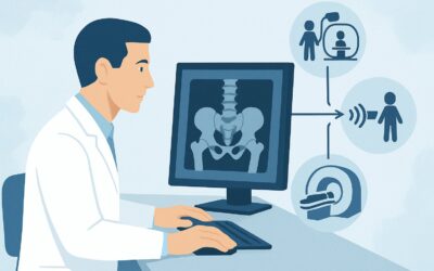
For follow-up of a specific appendicular joint in a child with known JIA, both US area of interest and MRI area of interest without and with IV contrast are rated as Usually appropriate by the ACR. However, ultrasound often serves as the more practical and efficient first-line modality for several reasons.
Ultrasound provides excellent high-resolution visualization of soft tissues and fluid. It is highly sensitive for detecting the key features of a disease flare: synovial hypertrophy (thickening of the joint lining) and joint effusions. The addition of power Doppler is critical, as it can detect the increased blood flow of active synovitis, helping to distinguish active inflammation from inactive, fibrotic synovial thickening. A major advantage of ultrasound is its lack of ionizing radiation (0 mSv), a crucial consideration in pediatric patients who may require multiple imaging studies over their lifetime. Furthermore, it is dynamic, allowing for real-time assessment of joint movement and tendon integrity, and does not require sedation, which is often necessary for young children undergoing a lengthy MRI.
While MRI with and without contrast is equally appropriate, it serves a slightly different role. MRI is superior for evaluating bone marrow edema, cartilage integrity, and early erosions that may not be visible on other modalities. It provides a more comprehensive anatomical overview. The choice between US and MRI often depends on the specific clinical question:
- If the primary question is active synovitis, US is an excellent, fast, and accessible choice.
- If the primary question is cartilage loss, bone erosion, or complex anatomy, MRI is the superior study.
Other modalities are rated lower for this scenario. Radiography area of interest is rated May be appropriate (Disagreement) because while it is useful for monitoring long-term bony changes and growth disturbances, it is insensitive for detecting active soft-tissue inflammation. A plain film can be normal during a significant synovitis flare. Modalities like whole-body bone scans or PET/CT are Usually not appropriate due to high radiation dose (pediatric RRL ☢☢☢☢) and low specificity for synovitis.
What’s Next After Ultrasound? Downstream Workflow
The results of the follow-up ultrasound directly inform the next steps in clinical management and potential further imaging. The workflow branches based on the key findings.
If the US confirms active synovitis (e.g., synovial hypertrophy with positive power Doppler signal):
This finding, correlated with the clinical examination, confirms a disease flare. The next step is typically a consultation with pediatric rheumatology to adjust the treatment plan. This may involve increasing the dose of a current medication, adding a new disease-modifying antirheumatic drug (DMARD), or switching to a different biologic agent. Repeat imaging is generally not needed in the short term unless the patient fails to respond to the new therapy.
If the US is negative for active synovitis:
When the ultrasound shows no effusion, synovial hypertrophy, or Doppler signal, it suggests active inflammation is unlikely to be the cause of the child’s symptoms. The clinical focus should shift to non-inflammatory causes, such as mechanical pain, overuse injury, or centralized pain syndromes. If suspicion for underlying cartilage or bone pathology remains high despite a negative ultrasound, an MRI area of interest without and with IV contrast may be considered as the next step to evaluate for bone marrow edema or subtle cartilage defects.
If the US is indeterminate or shows unexpected findings:
Occasionally, ultrasound may reveal complex fluid collections, significant tendon sheath thickening without clear synovitis, or findings suggestive of early erosive changes. In these cases, MRI is the ideal problem-solving tool to provide more definitive characterization and guide further management.
Pitfalls to Avoid (and When to Get Help)
Navigating follow-up imaging in pediatric JIA requires careful consideration to avoid common errors that can lead to misinterpretation or unnecessary procedures.
- Ignoring Clinical Context: Imaging findings must be interpreted alongside the clinical picture. Subclinical inflammation on ultrasound may not require treatment escalation in an otherwise asymptomatic and fully functional child. Conversely, persistent pain with a normal ultrasound warrants a search for other causes.
- Over-reliance on Radiographs for Flares: Do not use plain X-rays as the primary tool to assess for an acute flare of synovitis. They are insensitive to soft tissue changes and a normal result can provide false reassurance.
- Forgetting the Learning Curve: Pediatric musculoskeletal ultrasound is a highly operator-dependent skill. Ensure the study is performed by a sonographer and interpreted by a radiologist experienced in pediatric imaging to avoid misinterpreting normal pediatric anatomy, like physiologic blood flow near growth plates, as pathology.
- Not Comparing with Priors: The value of follow-up imaging is maximized when compared to previous studies. Always ensure prior images are available to the interpreting radiologist to assess for interval change.
If imaging results are discordant with a strong clinical suspicion of a flare, or if complex structural changes are suspected, direct consultation between the ordering clinician and a pediatric radiologist is invaluable.
Related ACR Topics and Tools
For a comprehensive overview of all clinical scenarios related to imaging in pediatric idiopathic arthritis, from initial workup to follow-up of different body parts, please see the parent topic article. Additional tools can help you apply these guidelines in your practice.
- For breadth across all scenarios in Joint Pain: Idiopathic Arthritis-Child, see our parent guide: Joint Pain: Idiopathic Arthritis-Child: ACR Appropriateness Decoded.
- To look up other clinical presentations, use the ACR Appropriateness Criteria Lookup.
- For detailed procedural techniques, consult the Imaging Protocol Library.
- To discuss radiation exposure with families, the Radiation Dose Calculator can help frame the conversation.
Frequently Asked Questions
Why is ultrasound often preferred over MRI if both are rated ‘Usually Appropriate’ for JIA follow-up?
Ultrasound is often preferred for its practical advantages: it does not use ionizing radiation, is typically faster, does not require sedation in young children, is less expensive, and allows for dynamic assessment. It is excellent for answering the primary question in a flare: ‘Is there active synovitis?’ MRI is reserved for cases where there is a specific concern for bone marrow edema, cartilage damage, or when the ultrasound is inconclusive.
When should I order a radiograph (X-ray) for JIA follow-up?
Radiographs are best used for long-term monitoring of chronic structural changes, such as bone erosions, joint space narrowing, and growth disturbances. They are not sensitive for detecting active synovitis and should not be the first-line test to evaluate an acute flare of pain and swelling.
Is IV contrast necessary for a follow-up MRI in JIA?
Yes, for assessing disease activity, contrast is crucial. The ACR rates ‘MRI area of interest without and with IV contrast’ as ‘Usually appropriate,’ while ‘MRI without IV contrast’ is only ‘May be appropriate.’ Gadolinium-based contrast highlights areas of synovial inflammation (synovitis), which is a key indicator of a disease flare.
What if multiple joints are flaring? Is whole-body MRI a good option?
No, for this purpose, whole-body MRI is rated ‘Usually not appropriate’ by the ACR. The recommendation is to perform focused imaging of the clinically symptomatic joints. While whole-body MRI has a role in other pediatric conditions (e.g., screening for osteomyelitis), it is not the standard for assessing polyarticular JIA flares, where targeted ultrasound or MRI of the most affected joints is more effective.
Does a normal ultrasound rule out a JIA flare?
Not definitively. While a high-quality ultrasound that shows no signs of synovitis makes a significant flare less likely, clinical assessment remains paramount. Pain in JIA can also be non-inflammatory (e.g., mechanical or centralized). If symptoms persist despite a normal ultrasound, the clinical picture should guide further investigation or management.
Reviewed by Pouyan Golshani, MD, Interventional Radiologist — May 30, 2026