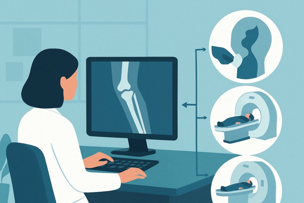
Which Imaging Study Best Evaluates a Limping Child with Suspected Lower Extremity Infection?
A 4-year-old presents to the emergency department with a fever and refusal to bear weight on her right leg for the past two days. On examination, her right shin is warm, swollen, and exquisitely tender to palpation. Her C-reactive protein (CRP) is elevated. You suspect an infectious process like osteomyelitis, but you need to differentiate it from a soft tissue infection or abscess. This clinical scenario requires a definitive imaging study to guide urgent management, including potential surgical intervention. For a young child with an acute limp, localized lower extremity symptoms, and concern for infection, the American College of Radiology (ACR) Appropriateness Criteria rate MRI lower extremity area of interest (not pelvis or hip) without IV contrast as Usually Appropriate.
Who Fits This Clinical Scenario for a Limping Child?
This specific imaging workflow is designed for a well-defined patient presentation. The guidance applies to children up to age 5 who present with an acute limp where the clinical signs and symptoms are clearly localized to a specific part of the lower extremity, such as the tibia, fibula, ankle, or foot. Crucially, there must be a clinical concern for infection, supported by findings like fever, localized erythema, warmth, swelling, or elevated inflammatory markers (e.g., CRP, Erythrocyte Sedimentation Rate).
It is essential to distinguish this presentation from similar but distinct clinical situations that require different imaging pathways:
- Symptoms Localized to the Hip: If the child’s pain, tenderness, or limited range of motion is localized to the hip, this is a separate and more urgent workup. Suspected septic arthritis of the hip is a surgical emergency, and the imaging algorithm differs. This article does not apply.
- Nonlocalized Symptoms: If the child cannot or will not localize the pain, or if the symptoms are diffuse throughout the limb or body, the differential diagnosis is much broader. This nonlocalized presentation follows a different ACR variant.
- No Concern for Infection: If there is a clear history of trauma and no systemic signs of infection (no fever, normal inflammatory markers), the primary concern shifts to occult fracture (like a toddler’s fracture), and the imaging approach changes accordingly.
This article focuses exclusively on the patient with a localized, non-hip lower extremity complaint where infection is the leading concern.
What Diagnoses Are You Working Up in This Scenario?
When a young child presents with a focal, tender, and warm lower limb causing a limp, the differential diagnosis centers on musculoskeletal infections that require prompt and accurate identification. The choice of imaging is driven by the need to distinguish between these possibilities, as their management differs significantly.
The most pressing concern is osteomyelitis, an infection of the bone. In young children, it most commonly affects the metaphyses of long bones like the tibia and femur. Early diagnosis is critical to prevent bone destruction, growth plate damage, and chronic infection. Clinical signs can be clear, but imaging is needed to confirm the diagnosis and define the extent of involvement.
A subperiosteal abscess is a critical complication of osteomyelitis, where pus collects beneath the periosteum (the membrane covering the bone). This is a key finding to identify on imaging because it often requires surgical drainage in addition to antibiotic therapy. MRI is exceptionally sensitive for detecting these fluid collections.
If the symptoms are centered on a joint, septic arthritis becomes a primary consideration. This is an infection within the joint space and is a true orthopedic emergency, as it can rapidly destroy cartilage. While more common in the hip and knee, it can affect any joint, including the ankle.
Finally, soft tissue infections like cellulitis (infection of the skin and subcutaneous tissue) or pyomyositis (infection and abscess formation within muscle) can mimic bone infection. Differentiating a deep soft tissue abscess from osteomyelitis is a central goal of imaging, as one may be managed with antibiotics alone while the other may require surgical debridement.
Why Is MRI of the Lower Extremity the Recommended Initial Study?
For a young child with a suspected lower extremity infection, Magnetic Resonance Imaging (MRI) provides the most comprehensive diagnostic information without using ionizing radiation. The ACR rates both MRI lower extremity area of interest (not pelvis or hip) without IV contrast and the variant without and with IV contrast as Usually Appropriate.
MRI offers superior soft tissue contrast, making it highly sensitive for the earliest signs of osteomyelitis, such as bone marrow edema, which can appear days before any changes are visible on radiographs. It can precisely delineate the extent of infection within the bone and identify complications like a subperiosteal abscess or extension into an adjacent joint (septic arthritis). Furthermore, MRI excels at distinguishing bone infection from overlying cellulitis or pyomyositis, directly guiding whether medical or surgical management is most appropriate.
The use of intravenous contrast can be beneficial for identifying abscesses, defining non-viable tissue (sequestrum), and assessing the vascularity of infected areas, but a non-contrast study is often sufficient for the initial diagnosis of osteomyelitis. The decision to use contrast is typically made in consultation with the radiologist.
Why are other studies rated lower for this specific scenario?
- Radiography (X-rays): Rated May be appropriate. While often obtained as a first-line test in the emergency setting, radiographs have very low sensitivity for early osteomyelitis. Bony changes like periosteal reaction or lytic lesions may not be visible for 7 to 14 days after the onset of infection. X-rays are primarily useful for ruling out other causes of pain, such as a fracture or a bone tumor, but a normal radiograph does not exclude osteomyelitis.
- Ultrasound: Rated May be appropriate. Ultrasound is a valuable, radiation-free tool for assessing soft tissues and joints. It is excellent for detecting joint effusions (suggesting septic arthritis) and identifying drainable fluid collections like soft tissue or subperiosteal abscesses. However, it cannot visualize bone marrow and therefore cannot be used to rule out osteomyelitis.
- Computed Tomography (CT): Rated Usually not appropriate. CT provides poor soft tissue contrast compared to MRI and is less sensitive for detecting early bone marrow edema. Its primary disadvantage in this pediatric population is the use of ionizing radiation, which should be avoided whenever a superior, non-radiating alternative like MRI is available.
What’s Next After MRI? Downstream Workflow
The results of the MRI will dictate the subsequent clinical pathway. The findings create clear decision points for management, consultation, and potential further imaging.
- If the MRI is positive for osteomyelitis or a subperiosteal abscess: This confirms the diagnosis and requires immediate action. The patient should be admitted for intravenous antibiotics. An urgent consultation with pediatric orthopedic surgery is necessary, especially if a drainable subperiosteal or intraosseous abscess is identified, as this often requires surgical incision and drainage.
- If the MRI is negative for bone or joint infection but shows cellulitis or pyomyositis: This finding still requires treatment, typically with intravenous antibiotics. The need for surgical consultation depends on whether a drainable muscular abscess (pyomyositis) is present. Simple cellulitis can often be managed medically without surgical intervention.
- If the MRI is entirely negative: This is a reassuring finding that makes a significant musculoskeletal infection unlikely. The clinical team should then reconsider the differential diagnosis. This may involve evaluating for non-infectious inflammatory conditions, occult trauma not visible on initial radiographs, or referred pain. If the limp and systemic symptoms persist without a diagnosis, the workup may shift to that of a child with nonlocalized symptoms.
- If the MRI is indeterminate: In rare cases, the findings may be equivocal. At this point, a discussion between the clinical team and the radiologist is crucial. Depending on the specific findings and clinical suspicion, next steps could include a short-interval follow-up MRI, a different imaging modality like a three-phase bone scan (though rated Usually not appropriate for initial imaging due to radiation), or proceeding with empiric treatment and close clinical monitoring.
Pitfalls to Avoid (and When to Get Help)
Navigating this clinical scenario requires careful attention to avoid common diagnostic and management errors.
- Pitfall 1: Over-reliance on normal radiographs. A normal X-ray in the first week of symptoms does not rule out osteomyelitis. Delaying advanced imaging like MRI based on a negative X-ray can lead to significant delays in diagnosis and treatment.
- Pitfall 2: Not specifying the area of interest. When ordering the MRI, be as specific as possible about the location of maximal tenderness. Ordering a generic “MRI of the lower extremity” can be too broad; specifying “MRI of the right tibia and ankle” focuses the study and ensures the correct sequences are performed.
- Pitfall 3: Delaying imaging while waiting for labs. While inflammatory markers are helpful, they are not perfectly sensitive or specific. If clinical suspicion for osteomyelitis or septic arthritis is high, imaging should proceed expeditiously, even with equivocal lab results.
- Pitfall 4: Sedation challenges. Young children often require sedation or general anesthesia for an MRI. Coordinate with radiology and anesthesiology early to avoid logistical delays, especially for an urgent study.
If the child appears septic, has rapidly progressing symptoms, or if there is any concern for hip involvement, escalate immediately to a pediatric orthopedic surgeon and consider initiating empiric antibiotics after blood cultures are drawn.
Related ACR Topics and Tools
For a comprehensive overview of all clinical variants and imaging modalities related to the acutely limping child, or to explore the technical details of specific imaging studies, the following resources are available:
- For breadth across all scenarios in Acutely Limping Child Up To Age 5, see our parent guide: Acutely Limping Child Up To Age 5: ACR Appropriateness Decoded.
- To explore adjacent clinical scenarios and their corresponding ACR ratings, use the ACR Appropriateness Criteria Lookup tool.
- For detailed procedural techniques on MRI and other modalities, consult the Imaging Protocol Library.
- To discuss radiation exposure from alternative studies like CT with families, the Radiation Dose Calculator can help frame the conversation.
Frequently Asked Questions
Why is MRI recommended over a three-phase bone scan for suspected osteomyelitis in a young child?
While a bone scan is sensitive for osteomyelitis, the ACR rates it as ‘Usually not appropriate’ for initial imaging in this scenario. MRI is preferred because it provides superior anatomic detail, allowing it to differentiate bone infection from soft tissue infection, identify abscesses, and assess for joint involvement. Critically, MRI does not use ionizing radiation, whereas a pediatric bone scan involves a significant radiation dose (3-10 mSv).
Should I always order the MRI with and without IV contrast?
Not necessarily. The ACR rates both ‘MRI without IV contrast’ and ‘MRI without and with IV contrast’ as ‘Usually Appropriate.’ A non-contrast MRI is often sufficient to diagnose osteomyelitis by showing bone marrow edema. Contrast is most helpful for delineating the margins of an abscess for surgical planning or assessing tissue viability. The decision can be made in consultation with the radiologist, but a non-contrast study is a valid and often sufficient first choice.
What if my hospital has limited or no immediate access to pediatric MRI?
If MRI is not readily available, a combination of other modalities may be used. Radiographs should be obtained to rule out fracture. Ultrasound is excellent for evaluating for a joint effusion or a drainable subperiosteal fluid collection. If these are negative but clinical suspicion for osteomyelitis remains high, the patient should be transferred to a center with pediatric MRI capabilities. Starting empiric antibiotics after obtaining blood cultures is reasonable while arranging for definitive imaging.
Can I use whole-body MRI if I’m not sure where the infection is located?
No, for this specific scenario where symptoms are localized to the lower extremity, whole-body MRI is rated ‘Usually not appropriate.’ It is reserved for different clinical scenarios, such as a child with fever and multifocal bone pain or nonlocalized symptoms where conditions like chronic recurrent multifocal osteomyelitis (CRMO) are being considered. For a focal presentation, a targeted MRI is more efficient and provides higher-resolution images of the area of concern.
Do elevated inflammatory markers like CRP guarantee an infection?
No. While elevated inflammatory markers strongly support an infectious or inflammatory process, they are not specific. Other conditions, such as trauma, inflammatory arthritis, or even malignancy, can cause an elevated CRP or ESR. However, in the clinical context of fever, focal bone tenderness, and a limp in a young child, a high CRP significantly increases the pre-test probability of a musculoskeletal infection and strengthens the indication for an MRI.
Reviewed by Pouyan Golshani, MD, Interventional Radiologist — May 29, 2026