Which Imaging Study Best Evaluates Suspected Mandibular Injury After Trauma?
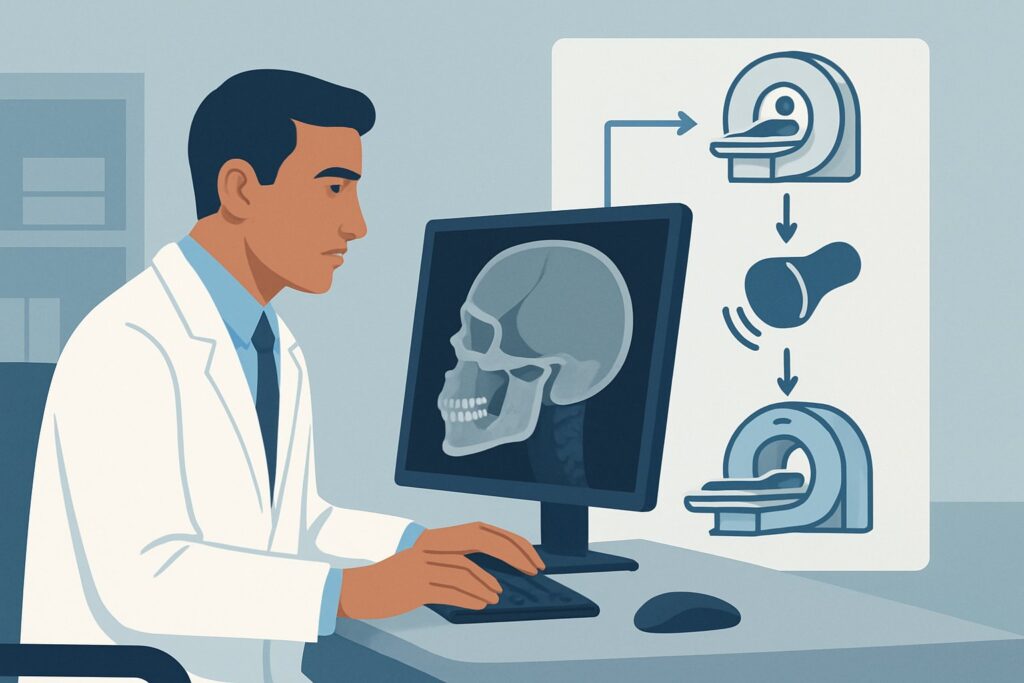
A 24-year-old male presents to the emergency department after a fistfight, complaining of severe jaw pain and an inability to fully open his mouth. On examination, you note tenderness over the angle of his right mandible, and when he tries to bite down, his teeth do not align as they normally would. There is a small amount of blood along the gingival margin of his lower molars. You suspect a mandibular fracture, but the exact location and complexity are unclear. This clinical scenario requires a definitive imaging choice to guide immediate consultation and management. This article details the American College of Radiology (ACR) recommended workflow for this specific presentation, where the primary concern is injury to the mandible. For this patient, a CT maxillofacial without IV contrast is rated Usually appropriate.
Who Fits This Clinical Scenario?
This guidance applies to patients who have undergone facial trauma and present with signs or symptoms specifically pointing to a mandibular injury after the primary survey is complete. The key clinical indicators include:
- Trismus: Difficulty or pain when opening the mouth, often due to muscle spasm (splinting) from an underlying fracture.
- Malocclusion: The patient’s subjective feeling or objective finding that their teeth no longer fit together correctly.
- Gingival or Mucosal Hemorrhage: Bleeding from the gums or inside of the mouth, which can signal an open fracture extending through a tooth socket.
- Dental Injury: Loose, fractured, or displaced teeth, which are frequently associated with fractures of the underlying alveolar bone of the mandible.
This workflow is distinct from other facial trauma scenarios. If the patient’s primary signs point elsewhere, a different imaging pathway may be indicated. For example, a patient with tenderness isolated to the nasal bridge and epistaxis would fit the suspected nasal injury scenario. Similarly, a patient with facial elongation, pain with upper jaw manipulation, and midface instability would be evaluated for a suspected midface or Le Fort-type fracture, which has its own specific considerations.
What Diagnoses Are You Working Up in This Scenario?
When a patient presents with signs of mandibular injury, your imaging choice is meant to confirm or exclude several key diagnoses. The differential is focused on bony and dental-alveolar structures, as these findings will directly influence the need for surgical consultation.
Mandibular Fracture is the most common and significant diagnosis in this setting. The mandible can fracture in multiple locations, and CT is excellent at identifying the precise pattern. Common sites include the condyle/subcondylar region (often from an indirect blow to the chin), the angle, the body, the parasymphysis, and the symphysis. Fractures can be simple, comminuted (multiple fragments), or open (communicating with the oral cavity or skin), and identifying these features is critical for management by an Oral and Maxillofacial Surgery (OMFS) or Otolaryngology (ENT) specialist.
Dentoalveolar Fracture involves the tooth-bearing portion of the mandible. This injury may involve the alveolar bone, the periodontal ligament, and the teeth themselves. While some dental injuries are evident clinically, imaging helps define the extent of bony involvement, which can affect tooth viability and the stability of the mandibular arch.
A less common but important consideration is Temporomandibular Joint (TMJ) Dislocation or Injury. A direct blow can cause the mandibular condyle to dislocate from the glenoid fossa. While often a clinical diagnosis, imaging is crucial when a dislocation is associated with a condylar fracture, as this complicates reduction and subsequent management.
Why Is CT Maxillofacial without IV Contrast the Recommended Study?
For a patient with suspected mandibular injury characterized by trismus, malocclusion, or intraoral bleeding, the ACR designates CT maxillofacial without IV contrast as Usually appropriate. This recommendation is based on the modality’s high diagnostic accuracy for the primary clinical questions in this scenario.
CT provides superb, multiplanar visualization of the complex anatomy of the mandible, far surpassing other modalities in detecting and characterizing fractures. It is highly sensitive for identifying fracture lines, assessing the degree of displacement or angulation, and detecting comminution. This is particularly crucial for fractures of the mandibular condyle and subcondylar region, which are notoriously difficult to visualize on plain films due to superimposed bony structures.
In contrast, Radiography mandible (including panoramic views like a Panorex) is rated as May be appropriate. While it can be a useful screening tool in low-resource settings or for very low-suspicion injuries, it has significant limitations. Plain radiographs are less sensitive for non-displaced fractures and can easily miss condylar head fractures. The superimposition of the cervical spine and contralateral mandible often obscures key areas, potentially leading to a missed diagnosis.
Modalities with intravenous contrast, such as CT maxillofacial with IV contrast, are rated Usually not appropriate for the initial evaluation of a suspected isolated mandible fracture. The addition of IV contrast provides no significant benefit for assessing bony detail and exposes the patient to the risks of an allergic-like reaction and contrast-induced nephropathy. Contrast is reserved for cases where there is a specific concern for an associated vascular injury, such as a rapidly expanding hematoma or suspected carotid or vertebral artery dissection from a high-energy mechanism.
The radiation dose for a non-contrast maxillofacial CT is relatively low (adult relative radiation level ☢☢, 0.1-1 mSv), balancing the high diagnostic yield with radiation safety principles. Once you’ve decided on the study, understanding the technical aspects is key. While the specifics vary, the principles of non-contrast head and facial CT are covered in our protocol guide: CT Brain Without Contrast.
What’s Next After CT Maxillofacial without IV contrast? Downstream Workflow
The results of the maxillofacial CT will guide your next steps, primarily determining the need for specialist consultation and the urgency of intervention.
If the study is positive for a mandibular fracture: The essential next step is to consult the appropriate surgical service, typically OMFS or ENT, depending on institutional practice. The imaging report, along with your clinical findings, will help them decide on management. Non-displaced or minimally displaced fractures may be managed conservatively with a soft diet and close follow-up. Displaced, open, or comminuted fractures, especially those causing significant malocclusion, often require surgical intervention, such as open reduction and internal fixation (ORIF).
If the study is negative for fracture: Re-evaluate the patient. Persistent malocclusion or trismus despite a negative CT may suggest a non-bony injury, such as TMJ internal derangement, severe muscular contusion and spasm (myofascial pain), or an occult dental injury not well-visualized on CT. These patients often benefit from analgesia, a soft diet, and outpatient follow-up with their primary care physician, dentist, or an OMFS specialist for further evaluation of persistent symptoms.
If the study is indeterminate or shows subtle findings: A direct conversation with the interpreting radiologist is invaluable. They can perform 3D reconstructions from the CT data, which can be extremely helpful in clarifying subtle or complex fracture patterns for surgical planning. If a dentoalveolar fracture is suspected but not clearly defined, specialized dental imaging like cone-beam CT may be considered in the outpatient setting.
Pitfalls to Avoid (and When to Get Help)
In the workup of a suspected mandibular injury, several common pitfalls can lead to delayed or missed diagnoses. First, always consider the mechanism of injury; high-energy trauma sufficient to fracture the mandible can also cause a concomitant cervical spine injury, which must be clinically or radiographically cleared. Second, do not overlook the dental examination. A CT scan is for bones, but tooth avulsion, subluxation, or fracture requires dental consultation. Third, in patients with severe fractures, particularly bilateral parasymphyseal fractures, be vigilant for posterior displacement of the tongue and potential airway compromise. If you note airway compromise, expanding hematoma, or neurologic deficits, escalate immediately to a senior physician and consider a CTA to rule out vascular injury.
Related ACR Topics and Tools
This article focuses on a single clinical scenario. For a comprehensive overview of all facial trauma presentations, or to explore adjacent clinical questions, the following resources are available:
- For breadth across all scenarios in Imaging of Facial Trauma Following Primary Survey, see our parent guide: Imaging of Facial Trauma Following Primary Survey: ACR Appropriateness Decoded.
- To search other clinical scenarios and their ACR-recommended imaging pathways, use the ACR Appropriateness Criteria Lookup.
- To review technical details for various imaging studies, explore the Imaging Protocol Library.
- For discussions with patients about radiation exposure, the Radiation Dose Calculator can help frame the conversation.
Frequently Asked Questions
Why not just start with a simple mandible X-ray or a Panorex view?
While mandible radiography is rated as ‘May be appropriate’ by the ACR, it has known limitations. It is less sensitive than CT for non-displaced fractures and provides poor visualization of the mandibular condyles due to superimposed anatomy. CT is the recommended first-line study when there is a high clinical suspicion of fracture, such as the presence of malocclusion or trismus, because of its superior diagnostic accuracy.
Is IV contrast ever needed for a suspected mandible fracture?
For an isolated, uncomplicated mandible fracture, IV contrast is ‘Usually not appropriate’ as it does not improve the visualization of bone. However, contrast is indicated if there is a concurrent concern for vascular injury. This might be suspected in cases of high-energy trauma, penetrating injury to the neck or face, an expanding hematoma, or a bruit on examination. In those specific cases, a CT Angiography (CTA) would be the appropriate study.
What if the patient has isolated dental trauma without malocclusion or trismus?
If the injury is confined to the teeth (e.g., a chipped or loose tooth) without signs pointing to a larger mandibular fracture (like malocclusion, trismus, or significant jawline deformity), a maxillofacial CT may not be necessary. These patients are often best evaluated with dedicated dental imaging and a consultation with a dentist or oral surgeon.
How does the imaging workup change if the patient is a child?
The principles of diagnosis are the same in children, but there is a heightened emphasis on minimizing radiation exposure (ALARA principle). While CT remains the most accurate modality, the decision to proceed should be carefully weighed. The radiation dose for a pediatric maxillofacial CT (relative radiation level ☢☢☢, 0.3-3 mSv) is higher than for adults. In cases of low suspicion, a clinician might start with plain radiographs. However, if a fracture is strongly suspected, CT is often still necessary for definitive diagnosis and surgical planning, using pediatric-specific low-dose protocols.
Does a negative CT scan completely rule out a significant mandibular injury?
A modern, high-quality maxillofacial CT scan has a very high negative predictive value for bony fractures. It is extremely unlikely to miss a clinically significant fracture. However, a negative CT does not rule out other injuries, such as temporomandibular joint (TMJ) internal derangement (e.g., disc displacement), ligamentous injury, significant muscular contusion, or purely dental injuries. If symptoms like pain, trismus, or a sensation of malocclusion persist despite a negative CT, clinical follow-up is essential to evaluate for these other causes.
Reviewed by Pouyan Golshani, MD, Interventional Radiologist — May 29, 2026