Which Imaging Study Is Best for Horner Syndrome with Cranial Nerve Signs?
A 58-year-old patient is in your neurology clinic with a three-week history of a noticeably droopy left eyelid and a smaller pupil on that side. On further questioning, he also reports intermittent double vision and a new, subtle numbness on the left side of his face. The onset was gradual, not sudden. This constellation of a nonacute Horner syndrome plus additional cranial nerve signs points toward a central nervous system lesion. You need to select the initial imaging study that can best visualize the brainstem, skull base, and cranial nerves to uncover the cause.
This article provides a detailed clinical workflow for this specific scenario, based on the American College of Radiology (ACR) Appropriateness Criteria. For this presentation, the initial imaging study of MRI head without and with IV contrast is rated as May be appropriate, representing the most logical first step in the diagnostic pathway.
## Who Fits This Clinical Scenario?
This guidance applies to a specific patient population: an adult presenting with a nonacute (i.e., subacute or insidious) onset of Horner syndrome who also has concurrent neurological signs or symptoms that localize to the brain or cranial nerves.
Inclusion criteria for this workflow:
- Patient: Adult.
- Onset: Nonacute, meaning the symptoms developed over days, weeks, or longer, rather than suddenly.
- Core Finding: Horner syndrome (a classic triad of ptosis, miosis, and anhidrosis, though the triad may be incomplete).
- Localizing Signs: Additional, distinct neurological findings such as diplopia (cranial nerves III, IV, VI), facial numbness or weakness (CN V, VII), hearing loss or vertigo (CN VIII), or signs of brainstem dysfunction like ataxia, dysarthria, or dysphagia.
Exclusion criteria (patients who fit a different workflow):
- Acute Onset with Pain or Trauma: A sudden onset of Horner syndrome, particularly if associated with head or neck pain, raises immediate concern for a vascular emergency like a carotid or vertebral artery dissection. This is a separate, more urgent clinical scenario.
- Localizing Spinal Cord Signs: If the patient presents with Horner syndrome accompanied by arm weakness, a sensory level on the trunk, or other signs of myelopathy, the investigation should focus on the spinal cord.
- Isolated, Nonlocalizable Horner Syndrome: An adult with a nonacute Horner syndrome but no other neurological signs or symptoms falls into a different diagnostic category, where the search for a lesion must cover the entire three-neuron sympathetic pathway from the brainstem to the eye.
## What Diagnoses Are You Working Up in This Scenario?
The presence of localizing cranial nerve or brain signs narrows the differential diagnosis significantly, focusing the investigation on intracranial pathology. The imaging strategy is designed to identify structural lesions affecting both the descending sympathetic pathway and adjacent neurological structures.
Brainstem Stroke
A classic cause is a lateral medullary syndrome (Wallenberg syndrome), typically from an infarct in the territory of the posterior inferior cerebellar artery (PICA). While often acute, a stuttering or progressive presentation can mimic a nonacute onset. The infarct damages the descending sympathetic fibers, vestibular nuclei, spinal trigeminal nucleus, and other nearby structures, explaining the combination of Horner syndrome, vertigo, and ipsilateral facial numbness.
Brainstem or Skull Base Tumor
A primary or metastatic neoplasm can compress or invade the brainstem and skull base. A meningioma, schwannoma of a lower cranial nerve, or nasopharyngeal carcinoma with intracranial extension could produce this clinical picture. The gradual onset is highly characteristic of a slow-growing mass lesion.
Demyelinating Disease
Multiple sclerosis (MS) can present with a demyelinating plaque strategically located in the brainstem. Such a lesion can disrupt the sympathetic pathway and involve adjacent cranial nerve nuclei or white matter tracts, causing a wide variety of associated neurological deficits. The nonacute onset fits well with the typical presentation of a new MS lesion.
Cavernous Sinus Pathology
Less commonly, a lesion within the cavernous sinus—such as a tumor, aneurysm, or inflammatory process (e.g., Tolosa-Hunt syndrome)—can cause Horner syndrome by affecting the sympathetic plexus traveling with the internal carotid artery. It can simultaneously involve cranial nerves III, IV, V1, V2, and VI, which all traverse this space, leading to ophthalmoplegia and facial numbness.
## Why MRI of the Head Is the Primary Study for Horner Syndrome with Cranial Nerve Signs
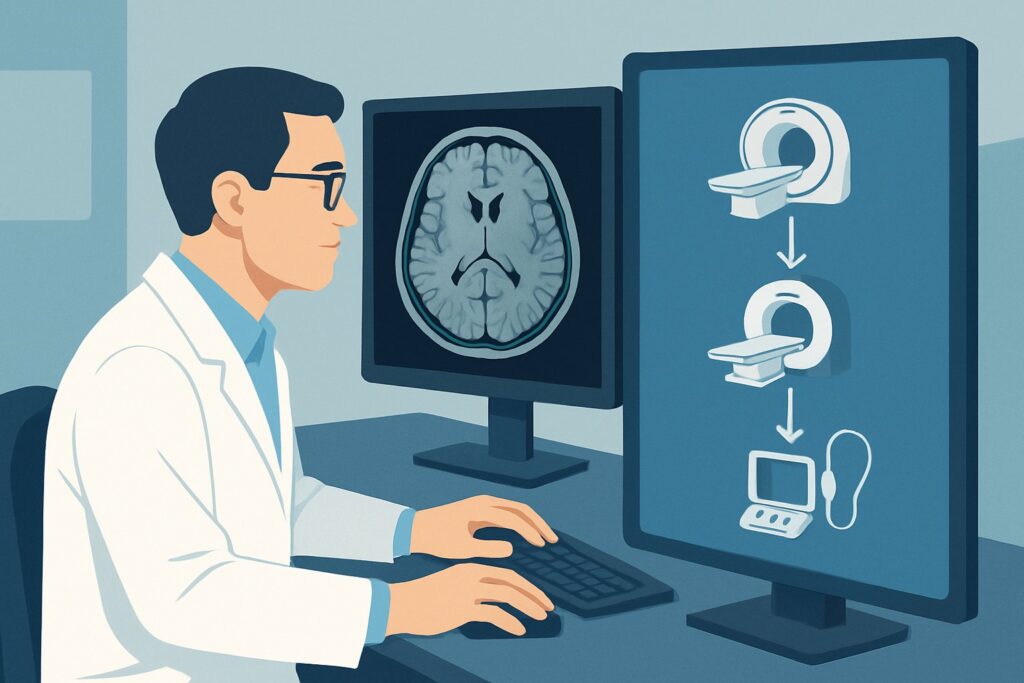
Given the differential diagnosis centered on the brainstem, skull base, and cranial nerves, magnetic resonance imaging (MRI) is the superior modality. The ACR rates MRI head without and with IV contrast as May be appropriate for this scenario. While not the highest rating of Usually appropriate, it is the most logical and informative initial study because the clinical findings strongly point to an intracranial cause that MRI is best suited to detect.
The rationale for this choice involves several key factors:
- Superior Soft-Tissue Contrast: MRI provides unparalleled detail of the brain parenchyma, particularly the brainstem. It can readily distinguish gray and white matter, making it highly sensitive for detecting demyelinating plaques (MS), subtle ischemic changes from a small stroke, or infiltrating tumors that would be invisible on a non-contrast CT.
- Role of Intravenous Contrast: The administration of gadolinium-based contrast is critical. It helps identify enhancement patterns characteristic of tumors, active demyelinating lesions, or inflammation, which is essential for narrowing the differential. A non-contrast study alone could miss these key pathologies.
- Radiation Safety: MRI does not use ionizing radiation (Relative Radiation Level: O), a significant advantage over CT, especially in younger patients or those who may require follow-up imaging.
### How Do Alternative Studies Compare?
- CT head without IV contrast and CTA head and neck with IV contrast: This combined study is also rated May be appropriate. It is a strong alternative if MRI is contraindicated (e.g., incompatible implanted device) or less available. CTA is excellent for evaluating vascular pathology like dissection or aneurysm. However, non-contrast CT has poor sensitivity for non-hemorrhagic brainstem strokes, MS plaques, and many tumors. This study also carries a significant radiation dose (Relative Radiation Level: ☢☢☢☢).
- MRI cervical and thoracic spine without and with IV contrast: This study is rated Usually not appropriate. Ordering an MRI of the spine in this context is a common pitfall. While the sympathetic pathway does travel through the cervical and upper thoracic spine, the presence of cranial nerve or brain signs localizes the suspected lesion intracranially. A spine MRI would fail to visualize the brainstem and would likely miss the causative pathology.
## What Is the Downstream Workflow After the Head MRI?
The results of the head MRI will dictate the subsequent clinical pathway. The goal of the initial imaging is to identify a structural cause and guide further management.
- If the MRI is positive for a specific lesion:
- Tumor: The next step is typically a referral to a neuro-oncology team, which may involve neurosurgery and radiation oncology. Further imaging with specialized MRI sequences or a biopsy may be required to determine the tumor type and grade.
- Stroke: Management will be guided by a stroke neurologist, focusing on secondary prevention and rehabilitation.
- Demyelinating Plaque: A finding suggestive of MS would prompt a referral to a neurologist specializing in multiple sclerosis for further diagnostic workup (e.g., spinal MRI, lumbar puncture) and initiation of disease-modifying therapy.
- If the MRI is negative:
A negative head MRI in a patient with clear localizing signs is a diagnostic challenge. The first step is to re-evaluate the clinical findings and confirm the localization. If suspicion for a brainstem or skull base lesion remains high, consider a repeat MRI with thinner slices or specialized sequences (e.g., FIESTA/CISS) focused on the posterior fossa and cranial nerves. If the localization is less certain, it may be appropriate to proceed to imaging the neck and chest, effectively transitioning to the workup for a nonlocalizable Horner syndrome.
- If the MRI is indeterminate:
An equivocal finding may require discussion with a neuroradiologist to determine the best next imaging step. This could include one of the other May be appropriate studies, such as adding an MRA of the head and neck to evaluate the vasculature or dedicated thin-section imaging of the orbits or skull base.
## Pitfalls to Avoid (and When to Get Help)
Navigating this diagnostic pathway requires careful attention to the clinical details to avoid common errors.
- Pitfall 1: Ordering the wrong regional scan. The most frequent error is ordering a cervical spine or chest study when cranial nerve signs clearly point to an intracranial lesion. Always let the neurological exam guide the imaging location.
- Pitfall 2: Omitting IV contrast. A non-contrast MRI of the head is insufficient for this workup, as it can easily miss enhancing tumors or active demyelinating plaques. Always specify “without and with IV contrast.”
- Pitfall 3: Misinterpreting “nonacute” as “not urgent.” While not an acute stroke presentation, the underlying causes (like a brainstem tumor) can be life-threatening. The diagnostic workup should proceed expeditiously.
If the clinical picture is complex or the imaging results are unclear, consultation with a neurologist or neuroradiologist is essential to ensure the correct diagnostic path is chosen.
## Related ACR Topics and Tools
For a comprehensive overview of all clinical scenarios involving Horner syndrome, further reading and specialized tools can provide additional context and support.
For breadth across all scenarios in Horner Syndrome, see our parent guide: Horner Syndrome: ACR Appropriateness Decoded.
Additionally, the following GigHz resources can help refine imaging orders and facilitate patient discussions:
- ACR Appropriateness Criteria Lookup — To review guidelines for adjacent or alternative clinical presentations.
- Imaging Protocol Library — For detailed technical specifications on MRI and CT protocols.
- Radiation Dose Calculator — To help explain cumulative radiation exposure to patients when considering CT-based studies.
Frequently Asked Questions
Why is MRI rated ‘May be appropriate’ instead of ‘Usually appropriate’ for this scenario?
The ‘May be appropriate’ rating from the ACR likely reflects the clinical nuance and the existence of other reasonable imaging options. While MRI of the head is the most logical first step given the intracranial localizing signs, a combined CT/CTA is also a valid alternative (‘May be appropriate’), especially if MRI is contraindicated. The panel may also have had disagreement on whether to include MRA or dedicated orbit views initially, leading to a more conservative top-line rating.
What if the patient’s only other symptom is headache, not a specific cranial nerve sign?
Headache is a less specific localizing sign. If the headache is new, severe, and associated with neck pain, the workup should pivot to the acute Horner syndrome scenario, prioritizing CTA to rule out a carotid or vertebral artery dissection. If the headache is chronic and nonspecific, the workup may proceed as described here, but the pre-test probability of finding a specific lesion is lower.
Should I order an MRA of the head and neck along with the initial brain MRI?
The ACR panel had disagreement on this point, rating ‘MRI head without and with IV contrast and MRA head and neck’ as ‘May be appropriate (Disagreement)’. Adding an MRA is reasonable if there is any clinical suspicion for a vascular etiology, such as a vertebral artery dissection extending intracranially or an aneurysm. However, for a presentation most suggestive of a tumor or demyelination, a standard brain MRI is a sufficient first step.
If the head MRI is negative, is the workup complete?
Not necessarily. A negative high-quality head MRI makes a structural brainstem or skull base lesion much less likely, but does not entirely exclude it. The next step is to critically re-evaluate the patient’s symptoms and signs. If the clinical localization is certain, a repeat, higher-resolution MRI may be warranted. If the localization is less certain, the investigation may need to be expanded to include the neck and chest to evaluate the second- and third-order sympathetic neurons.
Does this guidance apply to children?
No, this specific workflow is for adults. Horner syndrome in children has a different and often more concerning differential diagnosis, with a high prevalence of neuroblastoma. The imaging workup in a pediatric patient is different and requires specialized pediatric consultation.
Reviewed by Pouyan Golshani, MD, Interventional Radiologist — May 30, 2026