Which Imaging Study Is Best for Nonischemic Visual Loss with Chiasmal Symptoms?
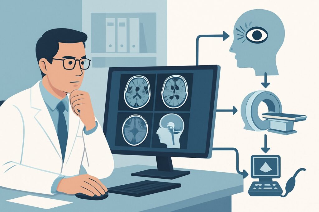
A 45-year-old patient presents to your clinic with a six-month history of “tunnel vision” and bumping into door frames. They deny pain or sudden changes. A formal visual field assessment confirms bitemporal hemianopsia, localizing the deficit to the optic chiasm. You suspect a compressive lesion, but the differential is broad. The immediate clinical question is which imaging study will provide a definitive diagnosis without unnecessary radiation or delay. For this specific presentation, the American College of Radiology (ACR) rates `MRI head without and with IV contrast` as ‘Usually Appropriate’.
Who Fits This Clinical Scenario?
This guidance applies to patients presenting with subacute or chronic, nonischemic visual loss where clinical signs point to a lesion at or behind the optic chiasm. The hallmark symptoms include visual field defects that respect the vertical midline, such as:
- Bitemporal hemianopsia: Loss of the outer half of the visual field in both eyes, strongly suggesting chiasmal compression.
- Homonymous hemianopsia: Loss of the same half (e.g., the right half) of the visual field in both eyes, indicating a lesion in the contralateral optic tract, optic radiation, or visual cortex.
This workflow is for the initial imaging workup in patients without a clear ischemic event (e.g., sudden onset concerning for stroke) or trauma. It is crucial to distinguish this scenario from others that require different imaging approaches:
- Suspected Optic Neuritis: This typically presents with subacute, painful, monocular vision loss and is often associated with multiple sclerosis. It has its own distinct imaging pathway.
- Traumatic Visual Defect: In cases of head or facial trauma, the primary concern is orbital injury, such as fracture or hematoma, which may prioritize different imaging protocols.
- Pre-chiasm Symptoms: If vision loss is monocular and the deficit is localized to the optic nerve or retina by ophthalmologic examination, an `MRI orbits` may be more appropriate than a full head MRI.
What Diagnoses Are You Working Up in This Scenario?
When symptoms localize to the chiasm or retrochiasmal pathways, the differential diagnosis centers on structural lesions that can compress, infiltrate, or displace these neural structures. The goal of imaging is to identify and characterize these potential causes.
Pituitary Macroadenoma: This is the most common cause of chiasmal compression. A benign tumor of the pituitary gland growing superiorly out of the sella turcica can directly impinge on the underside of the optic chiasm, classically causing bitemporal hemianopsia. Imaging is essential to define its size, extent, and relationship to adjacent structures like the cavernous sinuses and carotid arteries.
Meningioma: These are slow-growing, dural-based tumors that can arise from the tuberculum sellae, planum sphenoidale, or clinoid processes. Depending on their origin, they can compress the chiasm from above, below, or anteriorly, leading to a variety of visual field defects.
Craniopharyngioma: While often diagnosed in childhood, these sellar and suprasellar tumors have a second peak incidence in adults. They are notorious for involving the optic chiasm and hypothalamus and often have cystic and calcified components that imaging can delineate.
Aneurysm: Less commonly, a large aneurysm of the internal carotid artery or anterior communicating artery can expand and exert mass effect on the optic chiasm or optic tracts, mimicking a solid tumor. Differentiating this vascular cause is critical for management.
Demyelinating or Inflammatory Disease: While less frequent than compressive lesions in this context, conditions like multiple sclerosis or sarcoidosis can present with plaques or granulomatous inflammation involving the chiasm or optic tracts, causing similar visual symptoms.
Why Is MRI Head without and with IV Contrast the Recommended Study for This Presentation?
The ACR designates `MRI head without and with IV contrast` as ‘Usually Appropriate’ because it offers the highest diagnostic yield for the suspected pathologies in this clinical scenario. Its superior soft-tissue contrast is unmatched for visualizing the delicate structures of the sellar and suprasellar regions, including the pituitary gland, optic chiasm, optic tracts, and hypothalamus.
The non-contrast sequences can identify hemorrhage, calcification, and cystic changes. The addition of intravenous gadolinium-based contrast is critical for several reasons. It helps delineate the margins of tumors like pituitary adenomas and meningiomas, characterizes their vascularity, and can reveal subtle inflammatory or demyelinating lesions that might be invisible on non-contrast images. This detailed characterization is vital for surgical planning and differential diagnosis.
Why are other studies rated lower?
- CT head with IV contrast is rated ‘May be appropriate’. While it can detect large masses and calcifications (common in craniopharyngiomas), its soft-tissue resolution is significantly inferior to MRI for visualizing the chiasm and pituitary. It is primarily a second-line option for patients with contraindications to MRI (e.g., incompatible implants). It also involves ionizing radiation (☢☢☢ 1-10 mSv).
- MRI orbits without and with IV contrast is rated ‘Usually not appropriate’. This study uses high-resolution, thin-slice imaging focused on the globes and intraorbital optic nerves. While excellent for pre-chiasmal pathology, it does not provide adequate coverage or resolution of the chiasm, optic tracts, and brain parenchyma, which are the primary areas of concern in this scenario. Ordering an orbit-specific study would likely miss the causative lesion.
The recommended study, MRI, involves no ionizing radiation (O 0 mSv). When ordering, it is helpful to provide the radiologist with specific clinical details, such as “bitemporal hemianopsia, rule out chiasmal compression,” to ensure the imaging protocol is optimized with thin slices through the sella.
What’s Next After MRI Head without and with IV Contrast? Downstream Workflow
The results of the MRI will guide the subsequent clinical pathway, which almost always involves multidisciplinary consultation.
- If the study is positive for a mass (e.g., pituitary adenoma, meningioma): The next step is referral to neurosurgery and often endocrinology. The neurosurgeon will evaluate for surgical resection, while the endocrinologist will perform a hormonal workup, as many pituitary tumors are hormonally active. The imaging findings—size, invasion of surrounding structures, and relationship to the optic apparatus—are paramount for this planning.
- If the study is negative: A negative high-quality MRI makes a compressive lesion highly unlikely. The workup should be re-evaluated. This may involve referral back to neuro-ophthalmology to reconsider the localization of the deficit. Further investigation for rare inflammatory, paraneoplastic, or hereditary optic neuropathies may be warranted. This could lead to considering a different clinical scenario, such as a workup for an unidentified etiology.
- If the study is indeterminate or shows non-specific findings: If the MRI reveals non-specific white matter changes or subtle enhancement, the differential shifts towards inflammatory or demyelinating causes. This may prompt a lumbar puncture for cerebrospinal fluid analysis (e.g., oligoclonal bands for multiple sclerosis) and referral to a neurologist for further evaluation. In some cases, a follow-up MRI may be recommended to assess for changes over time.
Pitfalls to Avoid (and When to Get Help)
Navigating the workup for chiasmal or post-chiasmal visual loss requires careful attention to detail to avoid common missteps.
- Ordering an MRI of the orbits: As noted, this is a frequent error. Symptoms localizing to the chiasm or beyond require a brain MRI with dedicated views of the sella, not an orbit-focused study.
- Omitting IV contrast: A non-contrast MRI can miss many of the key pathologies, including smaller tumors, meningiomas, and inflammatory lesions. Unless there is a strong contraindication, contrast is essential.
- Misinterpreting the timeline: Attributing chronic, progressive visual loss to an ischemic event can delay the diagnosis of a compressive lesion. The nonischemic nature of the presentation is a key factor guiding the choice of MRI over more acute stroke imaging protocols.
- Failing to provide clinical history: Not specifying the visual field defect on the imaging requisition can lead to a standard brain protocol that may not include the necessary thin-slice sellar sequences, potentially reducing diagnostic sensitivity.
If a large, complex mass is identified or if there is rapid visual deterioration, immediate consultation with neuro-ophthalmology and neurosurgery is warranted.
Related ACR Topics and Tools
This article covers a single, specific clinical scenario. For a broader view of imaging for visual disturbances and to explore related workflows, the following resources are valuable. For breadth across all scenarios in Orbits, Vision, and Visual Loss, see our parent guide: Orbits, Vision, and Visual Loss: ACR Appropriateness Decoded.
- ACR Appropriateness Criteria Lookup — Use this to find guidance for adjacent or alternative clinical presentations.
- Imaging Protocol Library — Review detailed institutional imaging protocols for studies like brain and sellar MRI.
- Radiation Dose Calculator — A useful tool for discussing cumulative radiation exposure with patients, particularly when considering CT as an alternative.
Frequently Asked Questions
Why is MRI preferred over CT for suspected chiasmal lesions?
MRI provides vastly superior soft-tissue contrast, which is essential for visualizing the optic chiasm, pituitary gland, and surrounding neural and vascular structures. CT is limited in this region and is considered a second-line option, typically reserved for when MRI is contraindicated. Additionally, MRI does not use ionizing radiation.
Is an MRA or MRV necessary for the initial workup?
For the initial workup of nonischemic visual loss, a standard `MRI head without and with IV contrast` is sufficient. MRA (Magnetic Resonance Angiography) and MRV (Magnetic Resonance Venography) are rated as ‘May be appropriate’ and are typically reserved as problem-solving tools if the initial MRI suggests a vascular abnormality, such as an aneurysm or dural venous sinus thrombosis.
What if my patient has a pacemaker or other contraindication to MRI?
If a patient has an absolute contraindication to MRI, `CT head with IV contrast` becomes the primary imaging modality. While less sensitive, it can still identify large masses, calcifications, or bony erosion. It is crucial to communicate the clinical suspicion of a chiasmal lesion to the radiologist so they can optimize the CT protocol.
Does the patient’s age change the recommended imaging?
No, the recommended initial study remains `MRI head without and with IV contrast` for both adults and pediatric patients. However, the differential diagnosis may be weighted differently. For example, craniopharyngiomas and optic pathway gliomas are more common in children, while pituitary adenomas and meningiomas are more common in adults.
If the MRI is normal, what is the next step?
A normal, high-quality MRI makes a structural or compressive lesion highly unlikely. The next steps involve a deeper clinical investigation, typically led by a neuro-ophthalmologist. This may include reassessing the visual fields, electrophysiology testing (like VEPs), and laboratory workup for inflammatory, infectious, paraneoplastic, or hereditary optic neuropathies.
Reviewed by Pouyan Golshani, MD, Interventional Radiologist — May 29, 2026