Which Imaging Study Is Best for Staging Intermediate-Risk Prostate Cancer?

A 68-year-old man sits in your urology clinic, having recently received a diagnosis of prostate cancer from a systematic biopsy. His pathology shows a Gleason score of 3+4=7, and his prostate-specific antigen (PSA) is 12 ng/mL. He meets the criteria for intermediate-risk disease, placing him at a crucial decision point between active surveillance and definitive treatment like surgery or radiation. To guide this conversation, you need to accurately stage his cancer, primarily to assess for local extension that would argue against surveillance. What is the next, best imaging step?
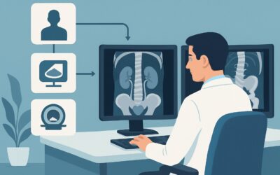
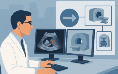
This clinical workflow details the imaging pathway for a patient with clinically established intermediate-risk prostate cancer. For this specific scenario, the American College of Radiology (ACR) Appropriateness Criteria rate multiparametric MRI of the pelvis, often as a precursor to a targeted biopsy, as Usually appropriate, providing the anatomical and functional detail needed to make a well-informed treatment decision.
## Who Fits This Clinical Scenario?
This guidance applies specifically to men with biopsy-proven prostate adenocarcinoma who are classified as having intermediate-risk disease. While definitions can vary slightly, this typically includes patients with at least one of the following features, without any high-risk characteristics:
- Clinical Stage: T2b-T2c (tumor palpable in more than half of one lobe or in both lobes)
- Gleason Score: 7 (either 3+4 or 4+3)
- PSA Level: 10-20 ng/mL
It is critical to distinguish this patient group from others who require different workups:
- Exclusion 1: Biopsy-Naïve Men. This workflow is not for men with an elevated PSA who have not yet had a biopsy. That scenario focuses on initial cancer detection.
- Exclusion 2: Low-Risk Prostate Cancer. Patients with low-risk disease (e.g., Gleason 6, PSA <10 ng/mL, stage T1c-T2a) are often managed with active surveillance, and the role of routine staging imaging is less established.
- Exclusion 3: High-Risk Prostate Cancer. Men with high-risk features (e.g., Gleason score 8-10, PSA >20 ng/mL, clinical stage T3a or higher) have a greater probability of metastatic disease, and their staging workup is often more extensive, frequently including systemic imaging for bone and distant soft tissue metastases.
## What Diagnoses Are You Working Up in This Scenario?
For a patient with intermediate-risk prostate cancer, imaging is not about making the initial diagnosis but about accurately defining the extent of the known cancer. The goal is to differentiate between localized and locally advanced disease, which fundamentally alters prognosis and treatment.
Organ-Confined Disease (Stage T2): This is the most favorable finding. It implies the tumor is entirely contained within the prostatic capsule. Confirming this with high-quality imaging supports the viability of definitive local therapies (prostatectomy, radiation) with a higher likelihood of cure, and may keep active surveillance on the table for select favorable intermediate-risk patients.
Extraprostatic Extension (EPE) (Stage T3a): This refers to the spread of cancer cells through the prostate capsule into the surrounding periprostatic fat. EPE is a key prognostic factor that increases the risk of biochemical recurrence after treatment. Its detection on imaging often leads to modifications in therapy, such as wider surgical margins or the addition of adjuvant radiation therapy.
Seminal Vesicle Invasion (SVI) (Stage T3b): This is the invasion of the tumor into one or both seminal vesicles. SVI is a clear sign of locally advanced disease and is associated with a poorer prognosis than organ-confined cancer. Identifying SVI pre-treatment is critical, as it may prompt a shift toward multimodality therapy, often combining hormonal therapy with radiation.
Regional Lymph Node Metastasis (Stage N1): While the risk of nodal spread is lower in intermediate-risk compared to high-risk disease, it is not zero. Imaging aims to identify suspicious pelvic lymph nodes (typically in the obturator, external iliac, or internal iliac chains). The presence of nodal metastases significantly changes the treatment paradigm, often requiring extended-field radiation and systemic therapy.
## Why Multiparametric MRI Is the Key to Staging and Biopsy in This Scenario
The ACR lists several imaging modalities as Usually appropriate for this scenario, but multiparametric MRI (mpMRI) of the pelvis has emerged as the central tool for local staging. The top-rated procedure, `MRI-targeted biopsy prostate`, presupposes that a high-quality diagnostic mpMRI is performed first to identify and characterize suspicious lesions.
The strength of mpMRI lies in its combination of high-resolution anatomical imaging and functional sequences:
- Anatomical Imaging (T2-weighted): Provides exquisite detail of the prostate’s zonal anatomy, the prostatic capsule, the neurovascular bundles, and adjacent structures like the seminal vesicles and bladder neck. This is the foundation for assessing for visible EPE or SVI.
- Functional Imaging (DWI and DCE): Diffusion-weighted imaging (DWI) assesses the random motion of water molecules, which is restricted in densely packed tumor tissue. Dynamic contrast-enhancement (DCE) evaluates tumor vascularity. Together, these sequences increase the conspicuity of clinically significant cancer and help differentiate it from benign conditions like prostatitis or benign prostatic hyperplasia (BPH).
This combination gives mpMRI superior sensitivity and specificity for detecting and localizing clinically significant cancer compared to other modalities.
### Why Alternatives Are Rated Lower for Local Staging
- TRUS-guided biopsy prostate: Rated May be appropriate. While this is the standard for initial diagnosis, it relies on a systematic (and somewhat blind) sampling of the prostate. For staging and surveillance, it can underestimate tumor grade and volume and may miss aggressive lesions, particularly those in the anterior or apical prostate. An mpMRI provides a “map” to ensure the most suspicious areas are sampled.
- Bone scan whole body: Rated May be appropriate. The pre-test probability of bone metastases in men with intermediate-risk prostate cancer is low. A bone scan is generally not indicated unless the patient has high-risk features (e.g., Gleason score ≥8), a very high PSA, or symptoms like bone pain. It provides no information about local tumor extent.
- CT abdomen and pelvis with IV contrast: Rated Usually appropriate. CT is excellent for detecting enlarged pelvic lymph nodes or distant metastases but offers poor soft-tissue resolution within the prostate itself. It cannot reliably assess for EPE or SVI. It is often used as an adjunct to MRI in higher-risk patients to evaluate nodal basins and abdominal organs, but it is not the primary tool for local staging.
### Radiation and Contrast Considerations
MRI uses no ionizing radiation (0 mSv), a major advantage, especially for patients on active surveillance who may require serial imaging over many years. While `MRI pelvis without and with IV contrast` is rated Usually appropriate, the use of gadolinium-based contrast agents for the DCE sequence is standard for a full diagnostic mpMRI.
Once you’ve decided on multiparametric MRI, our protocol guide covers the technique, contrast, and reading principles: MRI Prostate (Multiparametric).
## What’s Next After MRI? Downstream Workflow
The results of the staging mpMRI directly influence the next steps in management, creating a clear decision tree.
- If the MRI shows organ-confined disease: The findings support that the cancer is localized. This strengthens the case for definitive local therapies like radical prostatectomy or radiation therapy. For patients with “favorable” intermediate-risk disease (e.g., low volume, Gleason 3+4), a reassuring MRI may also support continuing with active surveillance.
- If the MRI shows evidence of EPE or SVI: The patient has locally advanced disease (Stage T3). This finding often prompts a shift in treatment strategy. A surgeon may plan for wider nerve-sparing margins, or the multidisciplinary team may recommend neoadjuvant or adjuvant hormone therapy and/or radiation therapy to improve outcomes. Active surveillance is generally no longer an option.
- If the MRI identifies a suspicious lesion not previously biopsied: The next step is an `MRI-targeted biopsy prostate`. This can be done via cognitive fusion (where the urologist mentally fuses the MRI and ultrasound images), software fusion (which overlays the MRI map onto the live ultrasound), or an in-bore MRI-guided biopsy. This ensures the most aggressive part of the tumor is sampled, potentially leading to a Gleason score upgrade and a change in risk stratification.
- If the MRI shows suspicious pelvic lymph nodes: This finding would typically trigger a biopsy of the node if safely accessible, or it may be sufficient to upstage the patient and recommend systemic therapy (like androgen deprivation therapy) in combination with radiation to the prostate and pelvic nodes.
## Pitfalls to Avoid (and When to Get Help)
- Pitfall 1: Relying on a non-mpMRI protocol. Ordering a generic “MRI Pelvis” without specifying a multiparametric prostate protocol will likely omit the necessary functional sequences (DWI, DCE), rendering the study inadequate for local staging.
- Pitfall 2: Underestimating the importance of radiologist experience. The interpretation of mpMRI of the prostate is complex and has a known learning curve. Ensure the study is read by a radiologist with subspecialty expertise in prostate imaging.
- Pitfall 3: Over-relying on imaging for nodal staging. While MRI and CT can identify enlarged lymph nodes, they have low sensitivity for detecting microscopic metastases in normal-sized nodes. A “negative” scan does not definitively rule out nodal disease.
- Pitfall 4: Not correlating with pathology. The imaging findings must always be interpreted in the context of the patient’s biopsy results, PSA level, and clinical exam.
If imaging reveals unequivocal evidence of metastatic disease (e.g., bone lesions, distant nodal involvement), the patient’s care should be escalated to a multidisciplinary tumor board including medical and radiation oncology to plan for systemic therapy.
## Related ACR Topics and Tools
This article is a deep dive into one specific clinical scenario. For a broader view of imaging across all prostate cancer presentations, from initial detection to surveillance, please see our parent guide.
- For breadth across all scenarios in Pretreatment Detection, Surveillance, and Staging of Prostate Cancer, see our parent guide: Pretreatment Detection, Surveillance, and Staging of Prostate Cancer: ACR Appropriateness Decoded.
For additional decision support and technical guidance, the following GigHz tools are available:
- Imaging Appropriateness Selector — for adjacent scenarios
- Imaging Protocol Library — for technique on the recommended study
- Radiation Dose Calculator — for cumulative dose conversations
Frequently Asked Questions
Is a PSMA PET/CT also appropriate for intermediate-risk prostate cancer staging?
Yes, the ACR rates `PSMA PET/CT skull base to mid-thigh` as Usually appropriate. PSMA PET/CT is highly sensitive for detecting nodal and distant metastatic disease. While its primary role has been in high-risk disease and biochemical recurrence, its use is expanding. For intermediate-risk patients, it may be considered, especially in ‘unfavorable’ cases (e.g., Gleason 4+3, high percentage of positive cores) where the risk of occult metastases is higher.
If a patient has a contraindication to MRI (e.g., an incompatible implanted device), what is the best alternative?
If MRI is contraindicated, `CT abdomen and pelvis with IV contrast` is rated Usually appropriate. While it cannot provide the detailed local staging of an MRI, it is the next best option for assessing pelvic lymph nodes and abdominal organs for metastatic disease. A `Bone scan whole body` (May be appropriate) would also be considered to complete systemic staging in the absence of MRI.
Does every patient with intermediate-risk prostate cancer need staging imaging?
Not necessarily, but it is increasingly common and recommended by many guidelines. For patients with ‘favorable’ intermediate-risk disease (e.g., Gleason 3+4, low tumor volume, PSA just over 10 ng/mL), some clinicians may proceed directly to treatment or active surveillance without advanced imaging. However, an mpMRI provides valuable information that can change management in a significant number of cases, for instance by identifying occult EPE or a higher-grade lesion.
What is the difference between an MRI for staging and an MRI for active surveillance?
The imaging protocol is identical (a standard mpMRI). The difference is the clinical question. A staging MRI is typically a one-time examination at diagnosis to determine the extent of disease and guide initial treatment. An MRI for active surveillance is performed serially (e.g., every 1-2 years) to monitor for changes in tumor size, appearance, or signs of progression that might trigger a recommendation for definitive treatment.
Should I order an endorectal coil with the prostate MRI?
The use of an endorectal coil (ERC) is becoming less common. Modern 3T MRI scanners can often achieve excellent image quality without an ERC, which improves patient comfort and reduces costs. However, on 1.5T scanners or in specific cases (e.g., very large patients), an ERC may still be used to improve the signal-to-noise ratio and image resolution. This decision is typically at the discretion of the radiology department and their specific hardware and protocols.
Reviewed by Pouyan Golshani, MD, Interventional Radiologist — May 26, 2026