Which Imaging Study Is Best for Suspected Appendicitis with Fever and Leukocytosis? ACR-Guided Workflow

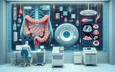
A 34-year-old male presents to the emergency department at 10 PM with sharp, migrating right lower quadrant (RLQ) pain that started this morning. He has a low-grade fever of 101.1°F and his initial labs show a white blood cell count of 16,000/μL. The clinical picture strongly suggests acute appendicitis, but a definitive diagnosis is needed to guide the surgical team. You need to order the most effective initial imaging study to confirm the diagnosis, rule out mimics, and identify any potential complications. This article provides a focused clinical workflow for this exact scenario, based on the American College of Radiology (ACR) Appropriateness Criteria. For this classic presentation, the ACR rates **CT abdomen and pelvis with IV contrast** as *Usually appropriate*.
Who Fits This Clinical Scenario for Suspected Appendicitis?
This guidance is specifically for adult patients who present with a high pre-test probability for acute appendicitis, characterized by the classic triad of:
- Localized right lower quadrant pain
- Fever (typically low-grade)
- Leukocytosis (elevated white blood cell count)
This workflow is intended for the initial diagnostic imaging decision in a non-pregnant adult. It is crucial to recognize presentations that, while similar, fall under different clinical guidelines and require a modified approach. This article does not apply to:
- Pregnant Patients: Suspected appendicitis in pregnancy is a distinct clinical scenario with unique imaging considerations to avoid ionizing radiation to the fetus. Ultrasound and MRI are prioritized.
- Patients with Undifferentiated RLQ Pain: If a patient has RLQ pain but lacks fever and leukocytosis, the differential diagnosis is broader and the urgency may be lower. This represents a different variant in the ACR criteria.
- Most Pediatric Patients: While CT is used in children, ultrasound is often the preferred first-line imaging modality to minimize radiation exposure, a principle known as ALARA (As Low As Reasonably Achievable).
- Patients with a Clear Contraindication to IV Contrast: This includes patients with severe contrast allergies or advanced renal failure, for whom non-contrast CT or MRI may be more appropriate.
What Diagnoses Are You Working Up in This Scenario?
When a patient presents with the triad of RLQ pain, fever, and leukocytosis, the differential diagnosis is focused on acute inflammatory and infectious processes. The choice of imaging must be able to confidently identify or exclude these possibilities.
Acute Appendicitis
This is the leading diagnosis. The imaging study must be able to visualize the appendix directly, identifying signs of inflammation such as luminal distention (>6 mm), wall thickening and enhancement, and surrounding fat stranding. An appendicolith, if present, is a highly specific finding.
Appendiceal Perforation and Abscess
A critical role of imaging is to detect complications that alter surgical management. A perforated appendix can lead to a contained phlegmon or a well-defined abscess. CT with IV contrast excels at identifying fluid collections, defining their extent, and visualizing enhancing abscess walls, which may guide percutaneous drainage in addition to or instead of immediate surgery.
Infectious Ileocolitis
Inflammation of the terminal ileum and cecum, often from infectious agents like Yersinia or Campylobacter, can clinically mimic appendicitis perfectly. Cross-sectional imaging can differentiate by showing wall thickening centered on the ileum and cecum, often with a normal-appearing appendix.
Right-Sided Diverticulitis
While less common than left-sided disease, diverticulitis of the cecum or ascending colon is a key mimic of appendicitis. CT can pinpoint the inflamed diverticulum as the source of the surrounding inflammatory changes, again demonstrating a normal appendix and preventing an unnecessary appendectomy.
Adnexal Pathology (in female patients)
In female patients, ovarian torsion, tubo-ovarian abscess, or a ruptured hemorrhagic cyst can present with identical symptoms. CT can often identify these gynecologic sources of pain, although a dedicated pelvic ultrasound may be required for definitive characterization.
Why Is CT Abdomen and Pelvis with IV Contrast Usually Appropriate for This Presentation?
For an adult patient with a high clinical suspicion of appendicitis, the ACR designates **CT abdomen and pelvis with IV contrast** as *Usually appropriate*. This recommendation is based on its high diagnostic accuracy, speed, and ability to evaluate for alternative diagnoses and complications, which directly impacts clinical management.
The high sensitivity and specificity of contrast-enhanced CT for acute appendicitis often make it the most definitive and efficient test in this setting. Intravenous contrast is crucial because it opacifies blood vessels and enhances inflamed tissues. This allows the radiologist to clearly visualize appendiceal wall enhancement, a key sign of inflammation, and to differentiate a simple phlegmon from a drainable, rim-enhancing abscess.
Let’s consider why other modalities are rated lower for this specific scenario:
- US abdomen / US pelvis: Rated *May be appropriate*. Ultrasound is an excellent modality that involves no ionizing radiation. However, its effectiveness in adults can be limited by patient body habitus and overlying bowel gas, which can obscure the appendix. It is also highly operator-dependent. While often used as a first-line test in children and pregnant women, in the classic adult presentation, CT provides a more consistently definitive answer.
- CT abdomen and pelvis without IV contrast: Rated *May be appropriate*. A non-contrast CT can identify primary and secondary signs of appendicitis, such as an appendicolith or periappendiceal fat stranding. However, it cannot assess for wall enhancement and is significantly less sensitive for detecting complications like ischemia or abscess formation. The addition of IV contrast provides critical information that often changes management.
The primary trade-off with CT is the use of ionizing radiation (Relative Radiation Level ☢☢☢, 1-10 mSv). This exposure is a considered risk, but in the context of a suspected acute surgical abdomen, the clinical benefit of a rapid, accurate diagnosis typically outweighs the small long-term risk. The ordering clinician should also screen for contraindications to iodinated contrast, such as a prior severe allergic reaction or significant renal impairment.
Once you’ve decided on CT abdomen and pelvis with IV contrast, our protocol guide covers the technique, contrast, and reading principles: CT Chest/Abdomen/Pelvis with IV Contrast.
What’s Next After the CT Scan? Downstream Workflow
The results of the CT scan create a clear decision tree for the next steps in patient care. The report should not be the end of the diagnostic process but rather the key that unlocks the appropriate treatment pathway.
Positive for Uncomplicated Appendicitis: If the CT confirms acute appendicitis without evidence of perforation or abscess, the next step is a surgical consultation for appendectomy. The imaging provides the surgeon with a definitive diagnosis, allowing for prompt operative planning.
Positive for Complicated Appendicitis (Abscess/Perforation): If the CT reveals a contained perforation with a well-formed abscess, management may change. The patient will still require a surgical consult, but the team may also involve Interventional Radiology for percutaneous drain placement. In some cases, a course of antibiotics and drainage may be initiated before a delayed (interval) appendectomy.
Negative for Appendicitis, Positive for an Alternative Diagnosis: If the appendix is normal but the CT identifies another cause, such as diverticulitis or ileocolitis, the workflow shifts entirely. A surgical consult may no longer be necessary, and treatment will be directed at the identified pathology (e.g., antibiotics for diverticulitis).
Negative or Indeterminate Study: If the CT is negative and the appendix is clearly visualized and normal, the clinical team should aggressively pursue other causes of the patient’s symptoms. If the appendix is not visualized and the study is indeterminate, further action depends on the patient’s clinical trajectory. If symptoms persist or worsen, a period of observation with serial exams or, in rare cases, diagnostic laparoscopy may be considered.
Pitfalls to Avoid (and When to Get Help)
Navigating this common clinical scenario requires avoiding several potential pitfalls that can delay diagnosis or lead to suboptimal outcomes.
- Ordering Without Contrast: In a patient with normal renal function and no severe allergy, ordering a non-contrast CT for suspected appendicitis significantly reduces the study’s diagnostic power, especially for detecting abscesses.
- Ignoring the “Mimics”: Do not anchor on appendicitis. The radiologist’s report may point to an alternative diagnosis (e.g., cecal diverticulitis, epiploic appendagitis, adnexal torsion) that requires a completely different management plan.
- Delaying the Scan: In a patient with high suspicion for an acute surgical abdomen, delaying imaging for an extended period of observation can increase the risk of perforation and associated morbidity.
- Misinterpreting an Equivocal Report: If the radiology report is equivocal (e.g., “appendix not definitively visualized”), do not discharge the patient without a clear plan. This is the time to re-evaluate the patient, speak directly with the radiologist, and consider a short period of inpatient observation or alternative imaging if clinically warranted.
If the CT findings are complex, such as a large, multiloculated abscess abutting multiple structures, escalate by consulting both General Surgery and Interventional Radiology early to coordinate a multidisciplinary treatment plan.
Related ACR Topics and Tools
This article covers one specific scenario in depth. For a broader view of all clinical variants related to right lower quadrant pain, or to explore the technical details of the recommended imaging studies, the following resources are available.
- For breadth across all scenarios in Right Lower Quadrant Pain, see our parent guide: Right Lower Quadrant Pain: ACR Appropriateness Decoded.
- Explore other scenarios using the ACR Appropriateness Criteria Lookup.
- Review technical specifications for hundreds of studies in the Imaging Protocol Library.
- Discuss radiation exposure with patients using the Radiation Dose Calculator.
Frequently Asked Questions
Why not just order an ultrasound first on every adult with suspected appendicitis?
While ultrasound is the preferred first step in children and pregnant women to avoid radiation, its utility in the general adult population can be limited. Factors like a larger body habitus and overlying bowel gas can prevent clear visualization of the appendix, leading to an indeterminate study. In an adult with classic signs of appendicitis (fever, leukocytosis), CT with IV contrast provides a more rapid and definitive diagnosis, which is why the ACR rates it as ‘Usually appropriate’ for this high-suspicion scenario.
Is oral contrast necessary in addition to IV contrast for suspected appendicitis?
The use of oral contrast for suspected appendicitis is debated and institutional protocols vary. Many institutions have moved away from routine oral contrast because it can delay the scan by 60-90 minutes without significantly improving diagnostic accuracy for appendicitis itself. IV contrast is the critical component for assessing appendiceal wall inflammation and identifying abscesses. The ACR guidelines for this scenario do not specify a requirement for oral contrast.
What if my patient has a mild allergy to IV contrast, like hives from a previous scan?
For patients with a prior mild allergic-like reaction to iodinated contrast, premedication with corticosteroids and antihistamines is a standard and effective strategy. This allows for the safe administration of IV contrast, ensuring the most diagnostically useful study can be performed. A history of a mild reaction is not an absolute contraindication, but it does require this extra preparation step.
If the CT shows a normal appendix but significant right-sided colonic wall thickening, what is the next step?
This finding shifts the diagnosis away from appendicitis and toward an inflammatory colitis, most commonly infectious or, less commonly, an initial presentation of Crohn’s disease or right-sided diverticulitis. The immediate next step is medical management, which may include antibiotics and supportive care. Surgical consultation is typically not required unless there are signs of complications like perforation or abscess related to the colon itself.
Can I use MRI instead of CT for a non-pregnant adult with suspected appendicitis?
Yes, MRI is an option and is rated as ‘May be appropriate’ by the ACR. It offers excellent diagnostic accuracy without using ionizing radiation. However, MRI is often less readily available in an emergency setting, takes longer to perform, and is more expensive than CT. For these logistical reasons, CT remains the more common and recommended initial study for the typical adult patient with high suspicion for appendicitis, while MRI is a primary problem-solving tool, especially in pregnant patients.
Reviewed by Pouyan Golshani, MD, Interventional Radiologist — May 21, 2026