Which Imaging Study Is Best for Suspected Rectosigmoid Endometriosis in Adults?
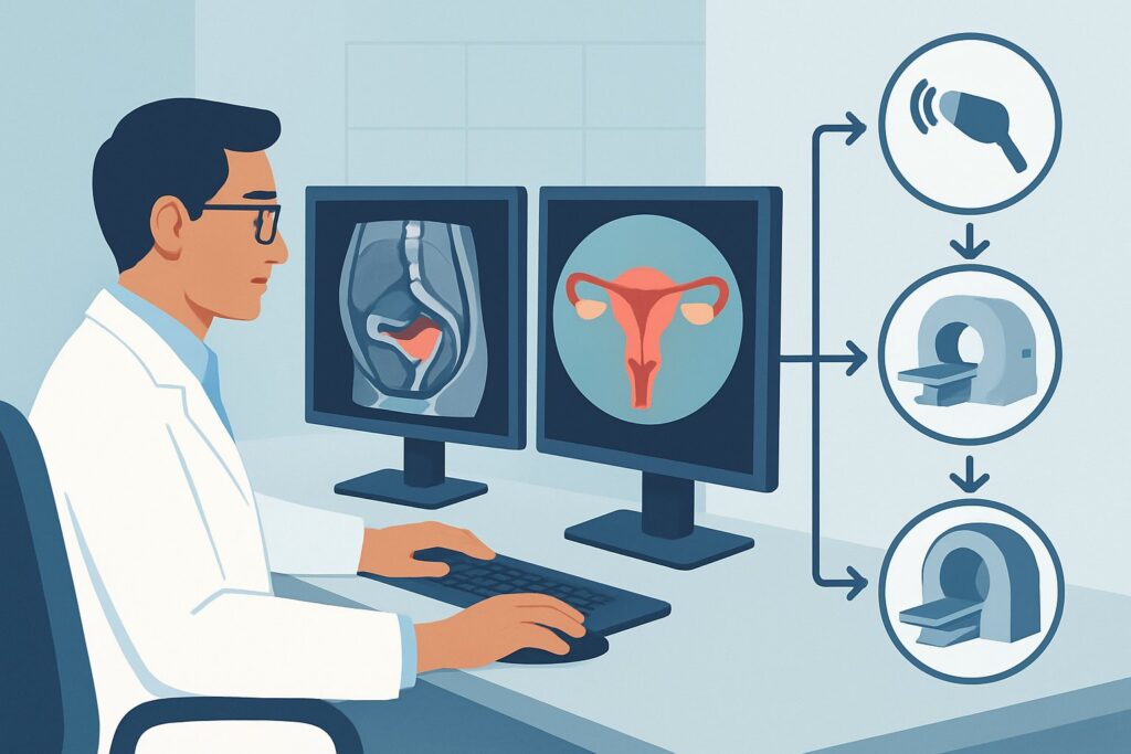
A 34-year-old patient presents to your clinic with a multi-year history of severe dysmenorrhea, but her symptoms have evolved. She now describes cyclical rectal pain, painful defecation (dyschezia), and a sensation of incomplete evacuation, all worsening significantly during her menses. Her gynecologist suspects deep infiltrating endometriosis with rectosigmoid involvement and has sent her for consultation and preoperative planning. You are now faced with the critical initial decision: which imaging study will most accurately stage the disease, guide surgical planning, and rule out mimics? This article provides a focused workflow for this specific scenario. According to the American College of Radiology (ACR), the initial recommended study, `US pelvis transabdominal and US pelvis transvaginal`, is rated Usually Appropriate.
Who Fits This Clinical Scenario?
This guidance applies specifically to adult patients, typically premenopausal, presenting for initial imaging with a clinical suspicion of endometriosis involving the rectosigmoid colon. The key feature is the presence of symptoms that point directly to the posterior pelvic compartment and bowel, such as deep dyspareunia, dyschezia, cyclical hematochezia, or tenesmus, in the setting of known or suspected endometriosis.
This workflow is distinct from other related clinical situations. You should seek different guidance if your patient:
- Presents with generalized, non-specific pelvic pain without clear bowel-related symptoms. This presentation aligns with the broader ACR variant for initial imaging of suspected pelvic endometriosis.
- Has already undergone an ultrasound that was negative or indeterminate for deep infiltrating disease. This patient would proceed to the next ACR variant, which addresses the next imaging study for characterization.
- Has an established, surgically confirmed diagnosis of endometriosis and is presenting with new or recurrent symptoms after surgery. This falls under the follow-up imaging scenario.
Correctly identifying the patient’s specific clinical context is crucial for selecting the most appropriate and highest-yield initial imaging examination.
What Diagnoses Are You Working Up in This Scenario?
When ordering imaging for suspected rectosigmoid endometriosis, you are evaluating a focused differential diagnosis. The goal is not only to confirm the primary suspicion but also to exclude other significant pathologies that can present with similar bowel symptoms.
Deep Infiltrating Endometriosis (DIE) of the Rectosigmoid: This is the primary diagnosis of concern. It represents endometrial glands and stroma invading more than 5 mm deep into the retroperitoneal space, often affecting the bowel wall. This invasion causes a significant fibromuscular reaction, leading to nodules, strictures, and adhesions that are responsible for the severe, localized symptoms.
Inflammatory Bowel Disease (IBD): Conditions like Crohn’s disease or ulcerative colitis can cause rectal pain, bleeding, and altered bowel habits. While the cyclical nature of symptoms points away from IBD, the clinical overlap is significant enough that it must be considered. Imaging can help differentiate the pattern of bowel wall thickening and associated inflammatory changes.
Colorectal Neoplasm: Although less common in the typical age demographic for endometriosis, a primary colorectal cancer must be excluded, especially in patients with rectal bleeding or significant changes in bowel caliber. Imaging is critical to identify focal masses or concerning strictures that would warrant urgent endoscopic evaluation.
Pelvic Adhesions: Scar tissue from prior surgeries, pelvic inflammatory disease, or even prior endometriotic inflammation can cause bowel loops to become fixed and tethered, leading to pain and obstructive symptoms. Imaging can help identify the obliteration of normal tissue planes characteristic of severe adhesions.
Why Is Combined Transabdominal and Transvaginal Ultrasound the Recommended First Step?
The ACR designates `US pelvis transabdominal and US pelvis transvaginal` as Usually Appropriate for the initial evaluation of suspected rectosigmoid endometriosis. This combined approach leverages the strengths of two different techniques to provide a comprehensive, non-invasive assessment without using ionizing radiation (0 mSv).
The initial transabdominal portion provides a global survey of the pelvis. It is essential for assessing the upper abdomen for related findings like hydronephrosis (from ureteral involvement) and evaluating large adnexal masses or endometriomas that might be outside the limited field of view of the transvaginal probe.
The subsequent transvaginal portion is the key to diagnosing deep infiltrating disease. The high-frequency probe provides excellent spatial resolution of the posterior cul-de-sac, uterosacral ligaments, and the rectovaginal septum. An experienced sonographer can directly visualize hypoechoic, spiculated nodules invading the muscularis propria of the anterior rectal wall. Dynamic evaluation, including assessment of the “sliding sign,” can reveal adhesions and fixation of the bowel to the posterior uterus or vagina.
Why are other studies rated lower for this initial workup?
- CT Pelvis with IV Contrast: This study is rated Usually Not Appropriate. While excellent for many abdominopelvic pathologies, CT has poor intrinsic soft-tissue contrast for differentiating small endometriotic implants from the normal bowel wall. It also exposes the patient to ionizing radiation (☢☢☢ 1-10 mSv) without adding significant diagnostic value for this specific clinical question.
- Fluoroscopy Contrast Enema: Rated as May be appropriate with panel disagreement, this study is now rarely used as a primary diagnostic tool. It can demonstrate luminal narrowing or a fixed, tethered appearance of the bowel but cannot visualize the endometriotic nodule itself or determine the depth of invasion. It also involves radiation (☢☢☢ 1-10 mSv).
While MRI is also rated Usually Appropriate, ultrasound is often more accessible, less costly, and, in expert hands, highly accurate for this diagnosis, making it the preferred initial examination. When ordering, be sure to clearly state “suspected rectosigmoid endometriosis” on the requisition to alert the sonographer to perform a dedicated evaluation of the posterior compartment.
What’s Next After Ultrasound? Downstream Workflow
The results of the initial pelvic ultrasound will guide your next steps in management and potential further workup. The decision tree branches based on whether the findings are positive, negative, or indeterminate.
If the study is positive for rectosigmoid endometriosis: A definitive ultrasound report describing the size, location, and depth of invasion of a rectosigmoid nodule provides crucial information for surgical planning. This patient can be referred to a gynecologic surgeon with expertise in advanced endometriosis excision, often in collaboration with a colorectal surgeon. In some centers, a preoperative MRI may still be obtained to map disease in other pelvic compartments (e.g., bladder, ureters) and serve as a visual aid for patient counseling and surgical road-mapping.
If the study is negative: A high-quality negative ultrasound from an experienced operator significantly lowers the likelihood of large, deep-infiltrating nodules. However, if clinical suspicion remains high based on classic symptoms, the next step often involves proceeding to MRI. This aligns with the ACR variant for “Indeterminate or negative ultrasound,” where `MRI pelvis without and with IV contrast` is rated Usually Appropriate to serve as a second-line problem-solving tool.
If the study is indeterminate: Findings such as focal bowel wall thickening without a discrete nodule or severe adhesions obliterating normal anatomical planes may be inconclusive. In this scenario, MRI is the ideal next step to better characterize the tissue and differentiate fibrotic change from active endometriotic infiltration. It can also provide a more global assessment of the pelvis that may have been limited on ultrasound due to patient body habitus or pain.
Pitfalls to Avoid (and When to Get Help)
Navigating the workup for rectosigmoid endometriosis requires careful attention to detail to avoid common diagnostic pitfalls.
- Underestimating Operator Dependence: The accuracy of transvaginal ultrasound for DIE is highly dependent on the skill and experience of the sonographer. If your institution does not have a dedicated protocol or experienced sonographers for this indication, consider referring the patient to a specialized center or proceeding directly to MRI.
- Inadequate Bowel Preparation: Fecal material and gas in the rectosigmoid can create acoustic shadowing that obscures underlying pathology. Some protocols recommend a mild bowel prep before the ultrasound to improve visualization.
- Misinterpreting Bowel Contraction: A transient bowel wall contraction can mimic a focal endometriotic nodule. A careful, real-time dynamic evaluation is necessary to differentiate this normal physiologic activity from a fixed, pathologic lesion.
If the imaging findings are discordant with a high degree of clinical suspicion, or if there is evidence of ureteral obstruction (hydronephrosis) or a large, complex adnexal mass, escalation to a multidisciplinary team including a gynecologic surgeon and radiologist is warranted.
Related ACR Topics and Tools
For a comprehensive overview of imaging recommendations across all clinical presentations of endometriosis, please consult our parent topic hub article. For further exploration of adjacent scenarios or imaging techniques, the following GigHz resources are available:
- For breadth across all scenarios in Endometriosis, see our parent guide: Endometriosis: ACR Appropriateness Decoded.
- To look up appropriateness criteria for other clinical scenarios, use the ACR Appropriateness Criteria Lookup.
- For detailed imaging techniques, explore the Imaging Protocol Library.
- To discuss radiation exposure with patients, consult the Radiation Dose Calculator.
Frequently Asked Questions
Why is MRI also rated ‘Usually Appropriate’ if ultrasound is the recommended first step?
MRI is also an excellent, non-radiation modality for evaluating deep infiltrating endometriosis and is considered ‘Usually Appropriate.’ However, expert-level transvaginal ultrasound is often more accessible, faster, and less expensive, making it the preferred initial test in many clinical settings. MRI is an outstanding problem-solving tool, especially if the ultrasound is negative or indeterminate despite high clinical suspicion, or for comprehensive preoperative surgical mapping.
Is a specific bowel prep required before the ultrasound for suspected rectosigmoid endometriosis?
While not universally mandated, many expert centers recommend a simple bowel preparation, such as a fleet enema administered 1-2 hours before the scan. This helps to clear stool and gas from the rectosigmoid colon, significantly improving visualization of the bowel wall and the detection of small endometriotic nodules that might otherwise be obscured.
What is the ‘sliding sign’ and why is it important in this scenario?
The ‘sliding sign’ is a dynamic sonographic assessment. The sonographer uses gentle pressure with the transvaginal probe to see if the anterior rectum and sigmoid colon glide freely against the posterior uterus and vagina. If they do not, it indicates the presence of adhesions, a key feature of deep infiltrating endometriosis. A ‘negative sliding sign’ (i.e., organs are stuck together) suggests obliteration of the posterior cul-de-sac.
If the patient has an IUD, can she still have a transvaginal ultrasound?
Yes, the presence of an intrauterine device (IUD) is not a contraindication to transvaginal ultrasound. The IUD will be visible within the endometrial cavity, but it does not interfere with the visualization of the myometrium, ovaries, or the posterior compartment structures like the rectosigmoid colon, which are the primary focus of this examination.
Should I order a transrectal ultrasound instead of a transvaginal one?
Transrectal ultrasound is also rated ‘Usually Appropriate’ and can provide excellent images of the rectal wall. However, transvaginal ultrasound is generally preferred as the initial step because it provides a better overall assessment of the uterus and adnexa. Transrectal ultrasound is typically reserved as a problem-solving tool or for patients who are not candidates for a transvaginal exam, such as those who are not yet sexually active.
Reviewed by Pouyan Golshani, MD, Interventional Radiologist — May 30, 2026