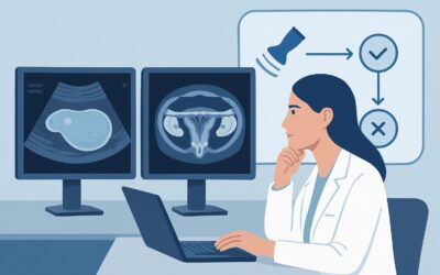
Which Imaging Study Should You Order for Fetal Soft Markers Found on Ultrasound?

A 29-year-old G1P0 patient is at her routine 20-week anatomy scan. The initial transabdominal ultrasound is largely unremarkable, but the sonographer notes a single, bright echogenic intracardiac focus (EICF). The patient’s cell-free DNA screening was low-risk. As the ordering clinician, you must now decide the next step in the diagnostic workflow. Is this an incidental finding, or does it warrant further investigation? This article details the clinical workflow for evaluating fetal soft markers found on a second or third trimester ultrasound. According to the American College of Radiology (ACR) Appropriateness Criteria, the next step in this specific scenario is a US pregnant uterus transabdominal detailed scan, which is rated Usually Appropriate.
Who Fits This Clinical Scenario for Fetal Soft Markers?
This guidance applies to a specific patient population: pregnant patients in the second or third trimester who have already undergone a standard screening ultrasound where one or more “soft markers” were identified. Soft markers are nonspecific sonographic findings that are not anomalies themselves but are associated with an increased statistical risk for aneuploidy or other underlying conditions. Common examples include:
- Echogenic intracardiac focus (EICF)
- Mild ventriculomegaly
- Echogenic bowel
- Increased nuchal fold thickness (in the second trimester)
- Shortened long bones (femur or humerus)
- Pyelectasis (mild renal pelvic dilation)
It is crucial to distinguish this situation from similar but distinct clinical presentations that follow different diagnostic pathways. This article does not apply to:
- Patients with major structural anomalies: If the initial ultrasound reveals a definitive structural defect like a neural tube defect, a large omphalocele, or a complex cardiac anomaly, the workup is different. That scenario falls under the ACR variant for major anomalies found on ultrasound.
- Patients undergoing initial screening: This guidance is for the next step after a finding. The initial choice of imaging for a low-risk or high-risk pregnancy without prior findings is a separate clinical question.
- Patients with abnormal genetic screening and a normal ultrasound: If a patient has a high-risk cell-free DNA (cfDNA) result but the initial ultrasound appears normal, the management pathway focuses on genetic counseling and potential diagnostic testing rather than primarily on repeat imaging.
What Diagnoses Are You Working Up with Fetal Soft Markers?
The discovery of a soft marker prompts a focused evaluation to rule out several potential underlying conditions, though most cases resolve favorably. The differential diagnosis guides the rationale for subsequent imaging and counseling.
Normal Anatomic Variant
This is by far the most common outcome. Many soft markers, particularly when they appear in isolation in an otherwise low-risk pregnancy, represent transient findings or normal variants with no adverse clinical significance. For example, an isolated echogenic intracardiac focus is seen in up to 5% of normal pregnancies. The primary goal of the follow-up detailed scan is to confirm the finding is isolated and that the rest of the fetal anatomy is normal, thereby providing reassurance.
Aneuploidy (e.g., Trisomy 21, Trisomy 18)
This is the most significant concern associated with soft markers. While not diagnostic, these findings act as risk modifiers, slightly increasing the baseline probability of a chromosomal abnormality. The presence of multiple soft markers confers a higher risk than a single isolated marker. The detailed ultrasound aims to meticulously search for any other subtle sonographic signs or structural anomalies that would further increase the suspicion for conditions like Down syndrome (Trisomy 21) or Edwards syndrome (Trisomy 18).
Non-Chromosomal Syndromes or Subtle Structural Anomalies
Less commonly, a soft marker can be the first clue to a non-chromosomal genetic syndrome (e.g., Noonan syndrome) or a subtle structural issue that was not fully characterized on the initial screening exam. For instance, an increased nuchal fold may be associated with certain cardiac defects. The detailed scan provides a higher-resolution assessment of all organ systems to identify these less common but consequential possibilities.
Congenital Infection
Certain soft markers, such as echogenic bowel, intracranial calcifications, or ventriculomegaly, can be associated with congenital infections like cytomegalovirus (CMV), toxoplasmosis, or parvovirus B19. While less common than other etiologies, this possibility is an important part of the differential, as it can have significant implications for fetal prognosis and postnatal management.
Why Is a Detailed Transabdominal Ultrasound the Recommended Next Study for Soft Markers?
When a soft marker is detected on a screening ultrasound, the ACR designates a US pregnant uterus transabdominal detailed scan as Usually Appropriate. This study, often referred to as a level II or targeted ultrasound, provides a more comprehensive evaluation than the initial screening examination.
The rationale for this recommendation is rooted in the study’s ability to clarify the clinical picture safely and effectively. A detailed scan is typically performed using high-resolution equipment by a sonographer with advanced training in fetal imaging, and it is interpreted by a maternal-fetal medicine (MFM) specialist or a radiologist with expertise in fetal anomalies. The examination involves a systematic and meticulous survey of all fetal organ systems, including the brain, face, heart, abdomen, spine, and limbs. This comprehensive approach is critical to determine if the soft marker is an isolated finding—which is often reassuring—or part of a constellation of findings that suggests an underlying syndrome or aneuploidy.
In contrast, other imaging modalities are rated lower for this specific clinical scenario:
- MRI fetal without IV contrast is rated Usually not appropriate. Fetal MRI is a powerful problem-solving tool, particularly for complex anomalies of the central nervous system. However, for the initial evaluation of common soft markers, it does not typically add diagnostic information beyond what a high-quality detailed ultrasound can provide. It is more resource-intensive and is best reserved for cases where ultrasound findings are inconclusive or suggest a complex brain or spine abnormality.
- US echocardiography fetal is rated May be appropriate. A dedicated fetal echocardiogram provides an in-depth evaluation of cardiac structure and function. This study becomes appropriate if the soft marker is cardiac in nature (like an EICF) and there are other risk factors for congenital heart disease, or if the detailed anatomic survey raises suspicion for a cardiac anomaly. However, performing a full detailed scan first is preferred to ensure no other non-cardiac abnormalities are missed.
All recommended and alternative imaging studies for this scenario, including ultrasound and MRI, are non-ionizing and have a relative radiation level of O (0 mSv). The use of IV contrast with fetal MRI is Usually not appropriate due to the theoretical risks of gadolinium-based contrast agents crossing the placenta.
What Is the Downstream Workflow After a Detailed Fetal Ultrasound?
The results of the detailed transabdominal ultrasound will guide the subsequent management, which primarily involves patient counseling and decisions regarding further testing.
If the Detailed Scan is Normal (Isolated Soft Marker)
If the detailed scan confirms the presence of a single soft marker but finds no other structural anomalies, the finding is deemed “isolated.” In this common situation, the next step is comprehensive counseling. The provider will integrate the ultrasound finding with the patient’s prior risk from maternal age and serum or cfDNA screening to generate an adjusted, patient-specific risk for aneuploidy. For many isolated markers in an otherwise low-risk patient, the residual risk is very low, and no further testing may be necessary. The decision to pursue invasive diagnostic testing (amniocentesis) is left to the patient after informed discussion.
If the Detailed Scan Shows Additional Markers or an Anomaly
If the scan reveals multiple soft markers or a definitive structural anomaly, the risk of an underlying genetic or structural condition increases substantially. The patient should be referred for genetic counseling and offered diagnostic testing via amniocentesis to obtain a definitive fetal karyotype or microarray analysis. A referral to a pediatric subspecialist, such as a pediatric cardiologist or surgeon, may also be warranted depending on the specific findings.
If the Detailed Scan is Indeterminate or Limited
Occasionally, the detailed ultrasound may be technically limited due to factors like maternal body habitus, fetal position, or oligohydramnios. In these cases, a follow-up ultrasound may be recommended in 2-4 weeks. If a specific organ system like the brain cannot be adequately visualized and there is high suspicion for an anomaly, a fetal MRI (Usually not appropriate as a first step) may become a reasonable next consideration to resolve the diagnostic uncertainty.
Pitfalls to Avoid (and When to Get Help)
Navigating the workup of fetal soft markers requires careful interpretation and communication. Here are a few common pitfalls to avoid:
- Over-interpreting an isolated finding: Avoid causing undue anxiety. In a patient with low-risk genetic screening, an isolated soft marker often represents a normal variant. Context is key.
- Failing to perform a comprehensive scan: Do not just re-examine the identified marker. The main value of the detailed scan is the meticulous evaluation of all other fetal structures to rule out associated abnormalities.
- Using outdated risk tables: The significance of many soft markers has diminished in the era of cfDNA screening. Ensure that risk counseling is based on current literature that incorporates prior screening results.
- Ignoring parental values: The decision to pursue invasive testing is highly personal. The clinician’s role is to provide accurate risk assessment and support shared decision-making, not to dictate a course of action.
If the detailed ultrasound reveals multiple anomalies or findings suggestive of a complex syndrome, immediate escalation to a maternal-fetal medicine specialist and a genetic counselor is the appropriate next step.
Related ACR Topics and Tools
For a comprehensive overview of all clinical variants related to fetal anomaly screening, please see our parent topic hub article. Additional GigHz tools can help you navigate adjacent scenarios, understand imaging protocols, and discuss radiation safety with patients.
- Parent Topic Hub: For breadth across all scenarios in Second and Third Trimester Screening for Fetal Anomaly, see our parent guide: Second and Third Trimester Screening for Fetal Anomaly: ACR Appropriateness Decoded.
- ACR Criteria Lookup: ACR Appropriateness Criteria Lookup
- Imaging Protocols: Imaging Protocol Library
- Dose Calculator: Radiation Dose Calculator
Frequently Asked Questions
Does an isolated soft marker always require a detailed (level II) ultrasound?
Yes, according to the ACR Appropriateness Criteria, a detailed transabdominal ultrasound is the ‘Usually Appropriate’ next step. While many isolated markers are benign, the detailed scan is crucial to confirm the finding is truly isolated and to meticulously rule out any other subtle structural abnormalities that might have been missed on the initial screening exam.
If a patient had a low-risk cell-free DNA (cfDNA) test, does a soft marker still matter?
Yes, but the context is critical. A low-risk cfDNA result significantly reduces the probability that a soft marker is associated with common aneuploidies like Trisomy 21. However, soft markers can also be associated with other conditions not covered by standard cfDNA panels, such as certain microdeletion syndromes, congenital infections, or structural anomalies. Therefore, a detailed anatomic survey is still recommended to ensure fetal well-being.
When is a fetal echocardiogram indicated for a soft marker?
A dedicated fetal echocardiogram is rated ‘May be appropriate.’ It is most often considered when the soft marker is an echogenic intracardiac focus (EICF), especially if there are other risk factors for congenital heart disease (e.g., family history, maternal diabetes). It may also be ordered if the detailed anatomic survey raises any suspicion about cardiac structure or function. However, the comprehensive detailed scan is the recommended first step to evaluate the entire fetus.
Should I order a fetal MRI after finding a soft marker on ultrasound?
Generally, no. Fetal MRI is rated ‘Usually not appropriate’ as the next imaging study for a soft marker. Its primary role is as a problem-solving tool for complex anomalies, particularly of the fetal brain, or in cases where ultrasound is severely technically limited. For most soft markers, a high-quality detailed ultrasound provides all the necessary diagnostic information.
What is the difference between a ‘follow-up’ and a ‘detailed’ ultrasound in this context?
Both are rated ‘Usually Appropriate.’ A ‘detailed scan’ (or level II) is a comprehensive, systematic evaluation of the entire fetal anatomy. A ‘follow-up scan’ might be ordered to re-evaluate a specific finding after a short interval or to check for resolution. In the context of a newly discovered soft marker, the detailed scan is the more definitive first step to ensure nothing else has been missed.
Reviewed by Pouyan Golshani, MD, Interventional Radiologist — May 29, 2026