Why Is Imaging Not Indicated for Very-Low-Risk Pediatric Minor Head Trauma?

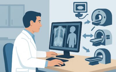
It’s 10 PM in the pediatric emergency department. A worried parent brings in their 18-month-old, who took a tumble off the couch while playing. The child cried immediately, has no visible signs of serious injury apart from a small bump on the forehead, and is now back to their usual playful self. The parent, understandably anxious, asks if a scan is needed to “make sure everything is okay.” This common clinical crossroads—balancing parental concern with evidence-based practice—is the focus of this workflow. For a child with minor acute blunt head trauma who meets the Pediatric Emergency Care Applied Research Network (PECARN) criteria for very low risk of a clinically important brain injury, the American College of Radiology (ACR) rates all forms of initial imaging, including CT, MRI, and even skull radiography, as Usually Not Appropriate. This article explains the robust clinical reasoning behind this recommendation to avoid imaging.
Who Fits This Clinical Scenario?
This guidance applies specifically to children presenting with minor, acute, blunt head trauma who are stratified into the “very low risk” category for a clinically important traumatic brain injury (ciTBI) by the validated PECARN clinical prediction rule. A ciTBI is defined as an injury resulting in death, neurosurgery, intubation for more than 24 hours, or hospital admission for two or more nights.
The PECARN rule divides children into two age groups:
- Children under 2 years: Must have a normal mental status, no scalp hematoma (except frontal), no loss of consciousness (or loss of consciousness <5 seconds), no palpable skull fracture, are acting normally per their parents, and have a non-severe mechanism of injury.
- Children 2 years and older: Must have a normal mental status, no signs of basilar skull fracture, no loss of consciousness, no vomiting, no severe headache, and a non-severe mechanism of injury.
This workflow explicitly excludes children who do not meet these strict criteria. Patients with intermediate-risk features (e.g., a large scalp hematoma, history of brief loss of consciousness, or a severe mechanism of injury) or high-risk features (e.g., altered mental status, palpable skull fracture) fall into different clinical scenarios with distinct imaging recommendations, typically involving a non-contrast head CT.
What Diagnoses Are You Working Up in This Scenario?
In any case of pediatric head trauma, the primary concern is identifying or ruling out a clinically important traumatic brain injury. The decision to not image in this very-low-risk cohort is based on the extremely low pre-test probability of finding such an injury. The differential diagnosis, while broad, is effectively managed through clinical assessment and observation rather than imaging.
Clinically Important Traumatic Brain Injury (ciTBI): This is the most consequential category clinicians aim to rule out. It includes injuries requiring neurosurgical intervention like an epidural or subdural hematoma, as well as cerebral contusions or depressed skull fractures. The PECARN rule was specifically designed to identify a group of children in whom the risk of a ciTBI is less than 0.02%, a risk level far below the potential harms of imaging.
Clinically Unimportant Skull Fracture: A child may sustain a simple, linear, non-displaced skull fracture without any underlying brain injury. While technically a positive finding, these fractures typically heal without intervention and do not alter clinical management. Identifying them with radiography does not improve outcomes and may lead to unnecessary follow-up imaging and anxiety.
Scalp Hematoma or Contusion: This is the most common finding in minor head trauma. It represents localized bleeding in the soft tissues outside the skull. While a large, non-frontal scalp hematoma can be a marker of an underlying fracture (and thus moves a child into a higher risk category), an isolated, small frontal hematoma in an otherwise well child is a benign finding that resolves on its own.
Why Is Imaging Usually Not Appropriate for This Presentation?
For a child meeting the PECARN very-low-risk criteria, the guiding principle is the avoidance of harm. The risk of radiation-induced malignancy from a CT scan, though small on an individual basis, outweighs the vanishingly small probability of detecting a brain injury that requires intervention. The ACR Appropriateness Criteria reflect this consensus by rating all imaging modalities as Usually Not Appropriate.
CT Head without IV contrast, often the go-to study for more significant head trauma, is rated Usually Not Appropriate. The rationale is clear: the radiation dose (pediatric relative radiation level ☢☢☢ 0.3-3 mSv) is not justified when the risk of a ciTBI is less than 1 in 5,000. Children’s developing brains are more radiosensitive than adults’, and minimizing cumulative radiation exposure is a critical public health goal.
Even Radiography skull is rated Usually Not Appropriate. While its radiation dose is lower (pediatric RRL ☢☢ 0.03-0.3 mSv), its clinical utility is extremely limited. A skull radiograph cannot visualize the brain parenchyma, so it cannot rule out an intracranial hemorrhage. A negative result can provide false reassurance, while a positive finding of a simple linear fracture often does not change management and may trigger a subsequent, higher-dose CT scan that was not initially indicated.
Other advanced modalities like MRI head without IV contrast are also Usually Not Appropriate. While MRI avoids ionizing radiation, it is a resource-intensive study that typically requires sedation or general anesthesia in young children to prevent motion artifact. The risks associated with sedation, combined with the cost and time, are not warranted for a patient cohort with such a low pre-test probability of significant injury.
What’s Next? Downstream Workflow After Clinical Assessment
The appropriate workflow for a very-low-risk child does not involve imaging but rather focuses on observation, parental education, and clear return precautions. This approach is both safe and effective.
Observation Period: For many of these children, a period of observation in the emergency department (e.g., 2 to 4 hours) can provide definitive reassurance. If the child continues to eat, play, and interact normally without developing any new symptoms like vomiting or irritability, they can be safely discharged.
Discharge with Head Injury Precautions: The cornerstone of management is empowering parents with clear, written instructions. They should be advised to return immediately if the child develops any “red flag” signs, including worsening headache, repeated vomiting, changes in behavior (e.g., difficult to arouse, inconsolable crying), confusion, seizures, or any focal neurologic deficits like weakness or vision changes.
If Symptoms Evolve: If, during observation or after discharge, the child develops symptoms that move them into a higher-risk category (e.g., begins vomiting, becomes lethargic), the clinical scenario has changed. At that point, the child should be re-evaluated, and the workflow for an intermediate- or high-risk patient should be initiated, which typically involves ordering a non-contrast head CT.
Pitfalls to Avoid (and When to Get Help)
Navigating this scenario requires careful clinical judgment and communication. Several common pitfalls can lead to unnecessary imaging or missed diagnoses.
- Misclassifying Risk: The most critical error is failing to identify a feature that moves a child from the very-low-risk to the intermediate-risk group. Carefully re-assess for mechanism of injury, scalp hematomas (especially non-frontal), and subtle changes in mental status.
- Imaging for Reassurance: Ordering a skull radiograph “just in case” or to appease parental anxiety is a frequent pitfall. This practice goes against evidence-based guidelines, provides minimal useful information, and can lead to a cascade of further unnecessary tests.
- Inadequate Discharge Instructions: Simply telling a parent to “watch the child” is not enough. Provide specific, written instructions on what to look for and when to return to the emergency department.
If at any point the child’s neurologic status deteriorates, they develop signs of a basilar skull fracture, or their GCS drops, escalate care immediately. This represents a shift to a high-risk scenario requiring emergent non-contrast head CT and consultation with neurosurgery or a pediatric trauma service.
Related ACR Topics and Tools
For a comprehensive overview of all clinical variants related to pediatric head trauma, from minor to severe, please consult our parent guide. For tools to assist in applying these criteria and understanding the recommended studies, the following resources are available.
- For breadth across all scenarios in Head Trauma-Child, see our parent guide: Head Trauma-Child: ACR Appropriateness Decoded.
- To look up adjacent scenarios and their ACR ratings, use the ACR Appropriateness Criteria Lookup.
- To review standard imaging techniques, explore the Imaging Protocol Library.
- To facilitate conversations about radiation exposure with families, consult the Radiation Dose Calculator.
Frequently Asked Questions
What are the specific PECARN criteria for ‘very low risk’ in a child with minor head trauma?
For children under 2, they must have a normal mental status, no scalp hematoma (except frontal), no loss of consciousness (or <5 sec), no palpable skull fracture, normal behavior, and a non-severe mechanism of injury. For children 2 and older, they must have a normal mental status, no signs of basilar skull fracture, no loss of consciousness, no vomiting, and no severe headache.
What if the parents are very anxious and insistent on getting an X-ray?
This requires clear communication and shared decision-making. Explain the evidence from the PECARN rule, highlighting that the risk of finding a significant injury is extremely low (less than 0.02%). Contrast this with the risks of radiation, even from a plain film, and explain why a skull X-ray is not a good test to rule out brain injury. Offering a period of observation in the ED can often provide the reassurance parents are seeking.
Is there ever a role for a skull radiograph in this specific scenario?
According to the current ACR Appropriateness Criteria, a skull radiograph is ‘Usually Not Appropriate’ for this very-low-risk group. Its use has been largely replaced by validated clinical decision rules like PECARN, which are more accurate for risk stratification and help avoid the pitfalls of a test that cannot visualize the brain and has poor sensitivity for clinically important injuries.
How long should I observe a very-low-risk child in the ED before discharge?
There is no universally mandated observation time, but a period of 2 to 4 hours is common practice. The goal is to ensure the child remains at their neurologic baseline and does not develop any new or worsening symptoms. If the child is well-appearing, tolerating oral intake, and acting normally after this period, they can typically be discharged safely with clear head injury precautions.
What if the child has a large ‘goose egg’ (scalp hematoma) but seems perfectly fine otherwise?
A large scalp hematoma (especially one that is non-frontal in a child under 2) is an intermediate-risk factor according to PECARN. This finding moves the child out of the ‘very low risk’ category discussed in this article. In that case, the clinical scenario is different, and a discussion about the risks and benefits of a head CT is warranted.
Reviewed by Pouyan Golshani, MD, Interventional Radiologist — May 29, 2026