What Is the Best Initial Imaging for an Adult with Suspected New-Onset Heart Failure?
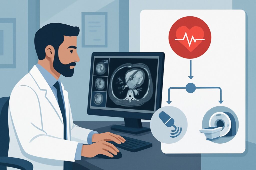
A 68-year-old male with a history of hypertension and type 2 diabetes presents to your outpatient clinic with a two-month history of progressive dyspnea on exertion and new bilateral lower extremity edema. He has no prior cardiac diagnoses and no chest pain. On exam, you note bibasilar crackles and 2+ pitting edema to the mid-shins. You suspect new-onset heart failure and need to confirm the diagnosis, assess cardiac function, and guide initial therapy. The central question is which imaging study to order first to provide the most diagnostic value safely and efficiently. For this specific clinical scenario—an adult with suspected heart failure and no prior history—the American College of Radiology (ACR) Appropriateness Criteria rate a resting transthoracic echocardiogram as Usually Appropriate.
Who Fits This Clinical Scenario?
This guidance applies to the initial imaging workup for an adult patient in whom you have a new clinical suspicion for heart failure. The key inclusion criteria are:
- Adult patient.
- New onset of signs or symptoms suggestive of heart failure, such as dyspnea, orthopnea, paroxysmal nocturnal dyspnea, or peripheral edema.
- No previously established diagnosis of heart failure.
It is crucial to distinguish this presentation from similar but distinct clinical situations that require a different diagnostic approach. This workflow is not intended for:
- Patients with a known history of heart failure. If a patient has an established diagnosis and you are investigating an unknown etiology or performing routine follow-up, the imaging strategy changes. These situations are covered in separate ACR variants, such as Adult. Known heart failure. Unknown etiology. Initial imaging.
- Patients presenting with acute, unstable symptoms. A patient with acute chest pain, hemodynamic instability, or suspected acute coronary syndrome requires an emergent workup focused on ischemia, which may involve different or additional imaging modalities beyond the scope of this initial outpatient evaluation.
This article focuses squarely on the first diagnostic imaging step for a stable patient with a new suspicion of heart failure.
What Diagnoses Are You Working Up in This Scenario?
When ordering initial imaging for suspected heart failure, the primary goal is to confirm the diagnosis and begin to differentiate its underlying cause and phenotype. The differential diagnosis is broad, but the initial imaging is designed to evaluate several key possibilities.
Heart Failure with Reduced Ejection Fraction (HFrEF): This is a critical diagnosis to establish, as it has specific, mortality-reducing therapies. HFrEF is characterized by systolic dysfunction of the left ventricle (LV), leading to an ejection fraction (EF) of 40% or less. Common causes include ischemic heart disease (prior myocardial infarction), non-ischemic dilated cardiomyopathies, or uncontrolled hypertension. Imaging must directly assess LV size and systolic function.
Heart Failure with Preserved Ejection Fraction (HFpEF): Many patients with heart failure symptoms have a normal or near-normal ejection fraction (EF ≥50%). In these cases, the primary abnormality is diastolic dysfunction—the heart muscle is stiff and does not relax properly, leading to high filling pressures. HFpEF is commonly associated with hypertension, diabetes, and obesity. Imaging must evaluate diastolic parameters, left atrial size, and ventricular wall thickness.
Valvular Heart Disease: Significant aortic stenosis or mitral regurgitation can be the primary cause of heart failure symptoms. These conditions can progress insidiously and present as new-onset dyspnea. The initial imaging study must be able to visualize valve structure and quantify the severity of any stenosis or regurgitation, as this directly impacts management and potential surgical or transcatheter intervention.
Pericardial Disease: Less common but important to consider, conditions like a large pericardial effusion causing tamponade physiology or constrictive pericarditis can mimic heart failure symptoms by restricting cardiac filling. Imaging can readily identify pericardial fluid and assess for the hemodynamic consequences of constriction.
Why Is a Resting Transthoracic Echocardiogram the Recommended Initial Study?
For an adult with suspected new-onset heart failure, the ACR designates two studies as Usually Appropriate: resting transthoracic echocardiography (TTE) and chest radiography. While a chest radiograph is invaluable for assessing for cardiomegaly, pulmonary edema, and pleural effusions, the TTE provides the direct structural and functional information needed to diagnose and phenotype heart failure.
A resting TTE is the cornerstone of the initial workup for several key reasons:
- Comprehensive Assessment: It is the single best non-invasive test to answer the most critical initial questions. A TTE directly visualizes and quantifies left and right ventricular size and systolic function (including ejection fraction), regional wall motion abnormalities (suggesting ischemia), ventricular hypertrophy, valvular morphology and function (stenosis/regurgitation), and pericardial abnormalities. It can also estimate pulmonary artery pressures and assess diastolic function.
- Safety and Accessibility: TTE is widely available, relatively inexpensive, and involves no ionizing radiation (Adult RRL=O 0 mSv). It can be performed at the bedside in hospitalized patients or in an outpatient clinic, making it a highly practical first-line tool.
- Diagnostic and Prognostic Value: The findings on the initial TTE—particularly the left ventricular ejection fraction (LVEF)—are fundamental to classifying the type of heart failure (HFrEF, HFmrEF, HFpEF). This classification immediately guides evidence-based medical therapy and provides crucial prognostic information.
The ACR rates other powerful imaging modalities lower for this specific initial workup because they are not the right tool for the first step. For example:
- Cardiac MRI (CMR) is rated May be appropriate. While CMR provides excellent detail on cardiac morphology, function, and tissue characterization (e.g., for scar or infiltration), it is typically reserved as a second-line test to clarify etiology once the diagnosis of heart failure is established by echocardiography. It is less available and more costly than TTE for an initial screen.
- Coronary CTA is rated Usually not appropriate for this indication. This study is designed to evaluate coronary artery anatomy for stenosis. While ischemic disease is a common cause of heart failure, a CTA is not the primary tool to assess cardiac function. It is an anatomic test, not a functional one, and it exposes the patient to significant radiation (Adult RRL=☢☢☢ 1-10 mSv) and iodinated contrast.
What’s Next After a Resting Transthoracic Echocardiogram? Downstream Workflow
The results of the initial TTE create a critical branch point in the patient’s management. The downstream workflow depends directly on the findings.
If the TTE confirms HFrEF (LVEF ≤40%): The diagnosis is confirmed. The immediate next steps involve initiating guideline-directed medical therapy (GDMT), which typically includes a beta-blocker, an ACE inhibitor/ARB/ARNI, and an MRA. Further investigation into the etiology is warranted. If regional wall motion abnormalities suggest coronary artery disease, an ischemic workup (e.g., stress testing or coronary angiography) may be necessary.
If the TTE suggests HFpEF (LVEF ≥50% with diastolic dysfunction): The diagnosis is supported. Management focuses on treating comorbidities like hypertension and diabetes, managing volume status with diuretics, and considering newer therapies approved for this population. The workup may proceed to rule out other causes of dyspnea if the clinical picture is not entirely clear.
If the TTE reveals significant valvular disease: The etiology of the heart failure is likely the valve pathology. The next step is referral to a cardiologist and potentially a cardiothoracic surgeon. The severity of the valvular lesion, patient symptoms, and LV function will determine the timing and type of intervention (e.g., valve repair or replacement).
If the TTE is normal or non-diagnostic: If the echocardiogram shows normal systolic and diastolic function with no significant valvular or pericardial disease, a primary cardiac cause for the patient’s symptoms becomes less likely. The workup should then pivot to investigate non-cardiac causes of dyspnea and edema, such as pulmonary disease (e.g., COPD, pulmonary hypertension), renal disease (nephrotic syndrome), or liver disease.
Pitfalls to Avoid (and When to Get Help)
In the initial workup of suspected heart failure, several common pitfalls can delay diagnosis or lead to inappropriate testing.
- Delaying the Echocardiogram: Do not delay ordering a TTE while awaiting responses to initial diuretic therapy. The echo is a diagnostic test, not just a confirmatory one, and its results are essential for guiding the correct long-term therapy from the outset.
- Misinterpreting a “Normal” EF: Remember that a normal ejection fraction does not rule out heart failure. Be sure to review the report for comments on diastolic function, wall thickness, and left atrial size, which are key indicators of HFpEF.
- Overlooking the Chest Radiograph: While TTE is the primary functional test, a chest X-ray remains Usually Appropriate. It provides complementary information about pulmonary congestion and can reveal alternative diagnoses like pneumonia or interstitial lung disease that can mimic heart failure symptoms.
- Ordering the Wrong Advanced Test: Avoid jumping to advanced imaging like cardiac MRI or CTA as the first step. These are powerful tools but should be used to answer specific questions that arise from the initial clinical and echocardiographic evaluation.
If the patient presents with acute hemodynamic instability, severe respiratory distress, or chest pain concerning for an acute coronary syndrome, escalate care immediately to the emergency department for a more urgent and intensive workup.
Related ACR Topics and Tools
For a comprehensive overview of imaging recommendations across all heart failure presentations, from initial workup to follow-up and advanced etiologic investigation, please consult the parent topic guide. Further resources for selecting appropriate imaging and understanding technical parameters are also available.
- For breadth across all scenarios in Suspected and Known Heart Failure, see our parent guide: Suspected and Known Heart Failure: ACR Appropriateness Decoded.
- ACR Appropriateness Criteria Lookup — for adjacent scenarios
- Imaging Protocol Library — for technique on the recommended study
- Radiation Dose Calculator — for cumulative dose conversations
Frequently Asked Questions
Should I order a chest X-ray along with the echocardiogram for suspected new-onset heart failure?
Yes, in most cases. The ACR rates both a resting transthoracic echocardiogram and a chest radiograph as ‘Usually Appropriate’ for this scenario. The chest X-ray is excellent for assessing pulmonary edema, pleural effusions, and cardiomegaly, and it helps rule out primary pulmonary causes of dyspnea. The two tests provide complementary information for the initial diagnosis.
If the patient has renal insufficiency, is a transthoracic echocardiogram still the best first test?
Yes. A transthoracic echocardiogram does not use iodinated contrast or gadolinium and has no impact on renal function, making it an ideal and safe initial imaging test for patients with any degree of renal insufficiency. This is a significant advantage over CT and MRI studies that may require contrast.
When should I consider a stress echocardiogram instead of a resting one for this patient?
A stress echocardiogram is rated ‘May be appropriate’ for the initial workup. It is most useful when there is a high suspicion of active ischemia as the cause of the new heart failure symptoms. However, for most patients, a resting study is sufficient to establish the diagnosis, assess baseline function, and guide initial therapy. A stress test is often a downstream step to investigate etiology after the initial diagnosis is made.
What if the transthoracic echo provides poor quality images?
If the acoustic windows are poor and the TTE is technically limited or non-diagnostic, other modalities may be necessary. A transesophageal echocardiogram (TEE) can provide clearer images but is more invasive. Cardiac MRI is an excellent non-invasive alternative for assessing function and morphology when TTE is inadequate and is rated ‘May be appropriate’ in the initial workup.
Does this guidance apply to a patient presenting to the emergency department with acute decompensated heart failure?
While the choice of imaging study (echocardiography) is often the same, the clinical context is different. This article focuses on the initial workup for a stable patient with a new suspicion of heart failure. In an acute, unstable setting, a point-of-care ultrasound (POCUS) may be used for rapid assessment at the bedside, followed by a comprehensive TTE once the patient is stabilized. The urgency and ancillary testing will differ significantly from the outpatient setting.
Reviewed by Pouyan Golshani, MD, Interventional Radiologist — May 30, 2026