What Imaging Is Best for Initial Staging of Asymptomatic Hairy Cell Leukemia?

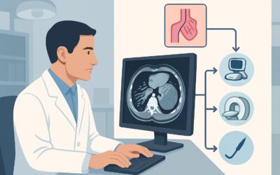
A 58-year-old man presents to your hematology clinic for follow-up on an abnormal complete blood count found during a routine physical. He feels well, reporting no fevers, weight loss, or night sweats. A bone marrow biopsy confirms the diagnosis of hairy cell leukemia (HCL). Now, as you plan his initial management, which is often “watch and wait” in asymptomatic cases, the immediate question arises: what is the appropriate imaging to establish a baseline and complete his initial staging? This article provides a detailed clinical workflow for this specific scenario, guiding you through the American College of Radiology (ACR) Appropriateness Criteria. For an asymptomatic adult with a new diagnosis of hairy cell leukemia, the ACR rates a `CT chest abdomen pelvis with IV contrast` as ‘May be appropriate’ for initial staging.
Who Fits This Clinical Scenario for Hairy Cell Leukemia Staging?
This guidance is specifically for an asymptomatic adult patient with a new, pathologically confirmed diagnosis of hairy cell leukemia. The key inclusion criteria are:
- Adult Patient: The epidemiology and presentation of leukemias differ significantly in pediatric populations.
- Confirmed Hairy Cell Leukemia: The diagnosis has been established, typically through bone marrow aspiration and biopsy with characteristic morphology and flow cytometry results (e.g., positive for CD11c, CD25, CD103, CD123). Imaging in this context is for staging, not diagnosis.
- Asymptomatic: The patient does not report constitutional “B” symptoms such as fevers, drenching night sweats, or unintentional weight loss. They also lack symptoms related to organ infiltration or cytopenias, such as recurrent infections or bleeding.
This workflow does not apply to patients who present differently, even if the underlying disease is a leukemia. Exclusions include:
- Symptomatic Patients: Patients with fever, abdominal pain, or palpable masses may require a different or more urgent imaging workup to investigate these specific signs.
- Different Leukemia Subtypes: This guidance is not for other leukemias. For example, initial staging for acute lymphoblastic leukemia (ALL) or acute myeloid leukemia (AML) follows distinct pathways, often involving different imaging priorities, such as assessing for central nervous system or other extramedullary involvement.
- Post-Therapy Evaluation: Imaging for restaging or assessing treatment response is a separate clinical question with different imaging recommendations.
What Are You Looking for During Initial Staging of Hairy Cell Leukemia?
In an asymptomatic patient with confirmed hairy cell leukemia, staging imaging is not performed to make the diagnosis but to establish a comprehensive baseline of disease extent. The primary goals are to document organ involvement and identify any occult findings that could influence future management decisions. The key findings you are assessing for include:
Splenomegaly: This is the most common and characteristic physical finding in HCL, present in over 80% of patients at diagnosis. While often palpable, cross-sectional imaging provides an objective, reproducible measurement of splenic volume. This baseline is critical for monitoring disease progression or response to therapy, as changes in spleen size are a key clinical indicator.
Lymphadenopathy: Unlike many other B-cell malignancies such as chronic lymphocytic leukemia (CLL), bulky lymphadenopathy is uncommon in HCL. However, mild to moderate abdominal, retroperitoneal, or mesenteric adenopathy can be present. CT is highly sensitive for detecting these nodes, which are often not apparent on physical examination, providing a complete picture of the nodal burden.
Hepatomegaly: Liver enlargement due to leukemic infiltration is the second most common organomegaly in HCL, though less frequent and typically less pronounced than splenomegaly. CT accurately assesses liver size and can rule out other hepatic pathology.
Occult Infections or Other Pathologies: Patients with HCL are immunocompromised due to both the disease and the associated neutropenia. A comprehensive baseline CT of the chest, abdomen, and pelvis can uncover subclinical infections or other incidental findings that may be relevant before initiating potential future therapies that further suppress the immune system.
Why Is CT of the Chest, Abdomen, and Pelvis with IV Contrast Rated ‘May Be Appropriate’ for HCL Staging?
The ACR designates `CT chest abdomen pelvis with IV contrast` as ‘May be appropriate’ for this scenario. This rating reflects that while imaging provides valuable baseline information, its immediate impact on management for an asymptomatic patient may be limited, as treatment is often deferred. However, it remains the most effective non-invasive tool for comprehensively assessing the common sites of extramedullary disease in HCL.
The rationale for this choice over other modalities is rooted in its diagnostic utility for the key staging questions. CT provides excellent spatial resolution to accurately measure the spleen and liver and to detect even small-volume lymphadenopathy in the retroperitoneum and mesentery. The use of intravenous contrast enhances the visibility of solid organs, vasculature, and lymph nodes, improving the detection of subtle infiltrative disease and differentiating it from other pathologies.
Alternative studies are rated lower for specific reasons in this context:
- US abdomen is rated ‘Usually not appropriate’. While ultrasound can easily assess the spleen and liver for organomegaly, it is operator-dependent and provides poor visualization of the retroperitoneum and mesenteric lymph nodes, which are critical areas to evaluate for a complete baseline stage.
- FDG-PET/CT is also rated ‘Usually not appropriate’. Hairy cell leukemia is typically a low-grade, indolent malignancy with low metabolic activity. Consequently, it often demonstrates little to no FDG avidity, leading to a high rate of false-negative results. Its utility is limited in HCL compared to more aggressive lymphomas.
The recommended CT study involves a radiation dose of 10-30 mSv (ACR RRL ☢☢☢☢). This exposure is a key consideration, but in the context of establishing a crucial oncologic baseline in an adult, the diagnostic benefit is generally considered to outweigh the risk. The information gathered provides a definitive reference point for all future clinical decisions.
Once you’ve decided on this study, our protocol guide covers the technical parameters, contrast administration, and interpretation principles in detail: CT Chest/Abdomen/Pelvis with IV Contrast.
What’s Next After CT? Downstream Workflow
The results of the initial staging CT scan will guide the subsequent clinical pathway, primarily by establishing a detailed baseline for future comparison. The downstream workflow depends on the findings:
- If the study confirms expected findings (e.g., isolated splenomegaly): For an asymptomatic patient, this result solidifies the decision for active surveillance or a “watch-and-wait” approach. The CT provides objective measurements (e.g., maximal splenic dimension) that will be used as a benchmark. Follow-up will be guided by symptoms and blood counts, not routine repeat imaging.
- If the study is negative (i.e., no splenomegaly or adenopathy): This is an excellent prognostic indicator. The patient can be confidently managed with active surveillance, with the baseline scan serving as proof of low-volume disease at diagnosis.
- If the study reveals unexpected or extensive disease (e.g., bulky retroperitoneal adenopathy): While atypical for HCL, such a finding might prompt a re-evaluation of the diagnosis or consideration of an earlier-than-planned therapeutic intervention, even in an otherwise asymptomatic patient. It provides critical information about the total disease burden that may influence the choice and timing of therapy when it becomes necessary.
- If the study shows incidental findings (e.g., a suspicious lung nodule or renal mass): The CT serves a dual purpose by identifying unrelated but clinically significant pathologies that require their own distinct workup, completely separate from the management of HCL.
In all cases, the initial staging CT is a foundational data point. It is not typically repeated unless the patient develops new symptoms, their blood counts worsen significantly, or they are being evaluated for response to treatment.
Pitfalls to Avoid (and When to Get Help)
When ordering and interpreting imaging for HCL staging, be mindful of these common pitfalls:
- Omitting IV Contrast: Ordering a non-contrast CT significantly reduces its sensitivity for detecting subtle lymphadenopathy and hepatic or splenic infiltration. Unless there is a strong contraindication (e.g., severe renal failure or significant contrast allergy), IV contrast is essential for a complete staging study.
- Over-reliance on Ultrasound: While tempting for its lack of radiation, relying solely on abdominal ultrasound provides an incomplete picture. It cannot adequately assess the retroperitoneal and mesenteric nodes, which are key staging sites.
- Inappropriate Use of PET/CT: Do not order a PET/CT for routine HCL staging. The low FDG avidity of this indolent disease makes it an unreliable tool that can lead to false reassurance from a negative scan.
- Routine Imaging for Surveillance: Avoid ordering serial CT scans to monitor an asymptomatic, stable patient. Follow-up should be driven by clinical changes (symptoms, physical exam) and laboratory values (blood counts), not a fixed imaging schedule.
If the imaging reveals features atypical for HCL, such as rapidly growing bulky masses or lytic bone lesions, escalate by discussing the case with the reporting radiologist and consider a biopsy of the atypical site to rule out a concurrent or transformed process.
Related ACR Topics and Tools
The ACR Appropriateness Criteria are a powerful resource for ensuring evidence-based imaging decisions. For a broader overview of imaging across all leukemia subtypes and clinical scenarios, please see our parent guide. For tools to help with ordering, protocoling, and discussing radiation dose, see the resources below.
- For breadth across all scenarios in Staging and Follow-Up of Leukemia, see our parent guide: Staging and Follow-Up of Leukemia: ACR Appropriateness Decoded.
- ACR Appropriateness Criteria Lookup — for adjacent scenarios
- Imaging Protocol Library — for technique on the recommended study
- Radiation Dose Calculator — for cumulative dose conversations
Frequently Asked Questions
Is imaging always necessary for an asymptomatic patient with hairy cell leukemia?
Not always, which is why the ACR rates it ‘May be appropriate’ rather than ‘Usually appropriate’. In an asymptomatic patient with a clear diagnosis and non-palpable spleen, some clinicians may opt for observation without baseline imaging. However, a baseline CT provides the most complete assessment of disease burden, which can be valuable for future comparisons if the patient becomes symptomatic or requires treatment.
Why is CT preferred over MRI for staging hairy cell leukemia?
CT is generally faster, more widely available, and provides excellent, high-resolution assessment of the solid organs and lymph nodes in the abdomen and retroperitoneum. While MRI avoids ionizing radiation, it is more susceptible to motion artifact, is more expensive, and offers no clear diagnostic advantage over CT for the specific questions of organ size and adenopathy in this clinical context. The ACR rates various MRI protocols as ‘Usually not appropriate’ for this indication.
If the patient has an allergy to iodinated contrast, what is the next best step?
If a patient has a severe contrast allergy, a non-contrast CT of the chest, abdomen, and pelvis is the next best option. The ACR also rates this study as ‘May be appropriate’. While less sensitive for small lymph nodes and subtle organ infiltration, it still provides excellent assessment of splenomegaly, hepatomegaly, and any gross adenopathy.
How does the imaging workup differ if the patient had symptoms like fever or weight loss?
The presence of constitutional symptoms would increase the clinical suspicion for more extensive disease or an alternative process, such as an occult infection. While a CT of the chest, abdomen, and pelvis would still be the primary imaging modality, the urgency would be higher, and the interpretation would focus more on identifying potential infectious sources in addition to staging the leukemia.
Should a bone scan be ordered to look for bone involvement in hairy cell leukemia?
No. A whole-body bone scan is rated ‘Usually not appropriate’ by the ACR for initial staging of HCL. While bone marrow infiltration is the defining feature of the disease, bone scans (technetium-99m MDP) detect osteoblastic activity, which is not a typical feature of HCL. Bony involvement is rare, and if suspected based on specific symptoms like bone pain, a targeted MRI or CT would be more appropriate.
Reviewed by Pouyan Golshani, MD, Interventional Radiologist — May 30, 2026