ACR Guide: What Initial Imaging Is Best for Sepsis with Cough or Dyspnea?
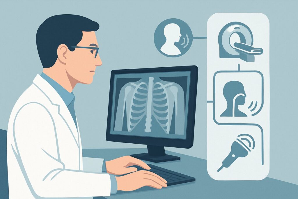
It’s 2 a.m. in the emergency department, and you’re evaluating a 68-year-old patient with a fever, tachycardia, and borderline hypotension. The sepsis protocol is underway with fluids and broad-spectrum antibiotics. The patient is now complaining of a new, productive cough and worsening shortness of breath. You need to identify the source of the infection quickly to tailor therapy, and the chest seems like the most probable culprit. What is the right first imaging study to order in this time-sensitive situation? This article provides a detailed workflow for this specific clinical scenario, grounded in the American College of Radiology (ACR) Appropriateness Criteria. For a patient with suspected sepsis and pulmonary symptoms, the ACR designates Radiography chest as Usually Appropriate.
Who Fits This Clinical Scenario for Sepsis Imaging?
This guidance applies to a specific and common clinical presentation: a patient with a strong clinical suspicion or confirmed diagnosis of sepsis who also presents with localizing signs or symptoms pointing to a thoracic source.
Inclusion criteria for this workflow:
- Systemic Signs: The patient meets clinical criteria for sepsis (e.g., via Sequential Organ Failure Assessment [SOFA] or Systemic Inflammatory Response Syndrome [SIRS] criteria), including findings like fever, hypothermia, tachycardia, tachypnea, hypotension, or altered mental status.
- Localizing Thoracic Symptoms: The patient has a new or worsening cough, dyspnea (shortness of breath), or chest pain, raising suspicion for a pulmonary or mediastinal source of infection.
- Initial Imaging Decision: This is the first imaging study being ordered for this presentation to identify a source.
Exclusion criteria (patients who fit a different workflow):
- Sepsis with Abdominal Pain: If the patient’s primary localizing symptom is abdominal pain, they fit a different ACR variant focused on abdominal imaging.
- Sepsis with No Localizing Signs: If the patient is septic but has no clear symptoms pointing to a specific organ system, the initial imaging strategy is different.
- Post-Radiograph Decision: This guidance does not apply if a chest radiograph has already been performed and was normal, equivocal, or nonspecific. That situation triggers a distinct “next step” ACR scenario.
What Diagnoses Are You Working Up with Chest Imaging in Sepsis?
When a septic patient presents with respiratory symptoms, the primary goal of initial imaging is to rapidly identify or exclude common, life-threatening thoracic pathologies that can serve as a septic focus. The differential diagnosis is broad, but the initial workup is focused on the most probable and actionable causes.
Pneumonia is by far the most common infectious source of sepsis originating from the chest. This can include community-acquired, hospital-acquired, or aspiration pneumonia. Imaging is crucial for confirming the presence of a parenchymal infiltrate or consolidation, which solidifies the diagnosis and guides antibiotic therapy.
Pleural Effusion and Empyema are also key considerations. A parapneumonic effusion can develop secondary to pneumonia, and if it becomes infected (an empyema), it represents a walled-off collection of pus that requires drainage for source control. A simple chest radiograph can often identify moderate to large effusions.
Acute Respiratory Distress Syndrome (ARDS) is a severe inflammatory lung injury that is often a consequence of sepsis, rather than its source. However, its characteristic imaging finding of diffuse, bilateral airspace opacities is a critical diagnosis to make. Identifying ARDS on the initial radiograph signals a higher severity of illness and has significant implications for respiratory management, such as low-tidal-volume ventilation.
Less commonly, imaging may reveal other etiologies. Septic pulmonary emboli, often from right-sided endocarditis or infected intravenous catheters, can appear as multiple, often peripheral, nodular opacities. While rare, mediastinitis from causes like esophageal perforation is a surgical emergency that may be suggested by mediastinal widening or air on a chest radiograph.
Why Is a Chest Radiograph the Recommended Initial Study for Sepsis with Pulmonary Symptoms?
The ACR rates Radiography chest as Usually Appropriate for the initial imaging of a septic patient with cough, dyspnea, or chest pain. This recommendation is based on a careful balance of diagnostic utility, speed, accessibility, and patient safety.
A chest radiograph is highly effective for diagnosing the most common causes in the differential. It can readily detect the lobar or diffuse consolidation of pneumonia, identify significant pleural effusions, and reveal the widespread bilateral opacities characteristic of ARDS. Its ability to answer these primary clinical questions quickly makes it the ideal first-line test.
Perhaps its greatest advantage in the acute setting is its accessibility. A portable chest radiograph can be performed at the patient’s bedside in the emergency department or intensive care unit, avoiding the risks associated with transporting a potentially unstable patient to a scanner. The images are acquired in seconds and are available for review almost immediately. Furthermore, the radiation dose is extremely low (adult relative radiation level ☢ <0.1 mSv), minimizing iatrogenic risk. **Why are other studies rated lower for this initial step?**
- CT chest with or without IV contrast is rated May be appropriate. While CT is more sensitive than radiography for detecting subtle pneumonia, small effusions, and pulmonary emboli, it is not the recommended first test. It requires patient transport, involves a significantly higher radiation dose (☢☢☢ 1-10 mSv), and, if contrast is used, carries a risk of contrast-induced nephropathy in patients who may already have sepsis-related acute kidney injury. CT is an excellent second-line study when the chest radiograph is inconclusive but clinical suspicion remains high.
- MRI chest is rated Usually not appropriate. MRI has a very limited role in evaluating for acute pulmonary infection. It is susceptible to motion artifact from breathing, has long acquisition times, and is not as effective as CT for visualizing the lung parenchyma. Its use is typically reserved for specific indications like evaluating chest wall masses or complex mediastinal pathology, not for an initial sepsis workup.
What’s the Next Step After a Chest Radiograph in a Septic Patient?
The results of the initial chest radiograph create a critical branch point in the patient’s management, dictating subsequent diagnostic and therapeutic steps.
If the radiograph is positive for a clear source (e.g., lobar pneumonia):
The primary goal is achieved. The finding confirms a pulmonary source of sepsis, allowing for focused antibiotic therapy. No further imaging may be necessary unless the patient fails to respond to treatment as expected. If a large pleural effusion is seen, a subsequent ultrasound may be performed to assess for loculations and guide thoracentesis for both diagnosis and source control.
If the radiograph is negative, equivocal, or shows nonspecific findings:
This is a common and important scenario. If your clinical suspicion for a thoracic source of sepsis remains high despite a non-diagnostic radiograph, you have now entered a new clinical situation. This directly corresponds to the sibling ACR scenario: “Suspected or confirmed sepsis. Cough or dyspnea or chest pain. Normal or equivocal or nonspecific chest radiograph. Next.” In this case, proceeding to a CT chest (often with IV contrast) becomes appropriate to look for more subtle disease that the radiograph may have missed.
If the radiograph shows findings suggestive of an alternative diagnosis:
Occasionally, the radiograph may reveal an unexpected finding, such as pneumomediastinum (suggesting esophageal rupture) or a prominent cardiac silhouette with vascular congestion (suggesting cardiogenic pulmonary edema mimicking sepsis). These findings would trigger an entirely different diagnostic and management pathway, potentially involving a CT scan or echocardiogram.
Pitfalls to Avoid (and When to Get Help)
Navigating the initial imaging workup for sepsis requires vigilance to avoid common errors that can delay diagnosis or compromise patient safety.
- Over-reliance on a negative radiograph: A normal chest radiograph does not definitively exclude a pulmonary source of sepsis, especially in patients who are early in their disease course, dehydrated, or severely immunocompromised. Maintain a high index of suspicion if the clinical picture points to the chest.
- Forgetting the portable option: In a hemodynamically unstable patient, do not delay imaging. A portable upright or supine chest radiograph is a valuable tool that avoids the risks of transporting a critically ill patient to the radiology suite.
- Ignoring the kidneys: Before ordering a contrast-enhanced CT as a next step, always assess renal function. Sepsis frequently causes acute kidney injury, which increases the risk of contrast-induced nephropathy.
- Misinterpreting ARDS as simple pneumonia: Diffuse, bilateral opacities in a septic patient should immediately raise concern for ARDS, a condition with high mortality that requires specific ventilator management strategies.
If the imaging findings are complex, unclear, or discordant with the clinical picture, a direct consultation with the interpreting radiologist is invaluable for correlating findings and planning the most appropriate next imaging step.
Related ACR Topics and Tools
For a comprehensive overview of imaging in all sepsis-related scenarios, from abdominal pain to an unknown source, please refer to our parent guide. For tools to help you navigate other clinical questions, the resources below are available.
- For breadth across all scenarios in Sepsis, see our parent guide: Sepsis: ACR Appropriateness Decoded.
- ACR Appropriateness Criteria Lookup — for adjacent scenarios
- Imaging Protocol Library — for technique on the recommended study
- Radiation Dose Calculator — for cumulative dose conversations
Frequently Asked Questions
Why not order a CT scan first to be more sensitive?
While a CT scan is more sensitive, a chest radiograph is rated ‘Usually Appropriate’ as the initial study because it is faster, more accessible (can be done portably), involves significantly less radiation, and can effectively diagnose the most common causes like pneumonia and ARDS. A CT is often the appropriate next step if the radiograph is negative but clinical suspicion remains high.
What if my patient is too unstable to go to the CT scanner?
This is a key reason why the chest radiograph is the recommended initial test. A portable chest X-ray can be performed at the bedside without moving the patient, providing critical diagnostic information while minimizing risk. The decision to proceed to CT must always include an assessment of the patient’s hemodynamic stability.
Does a normal chest radiograph rule out a pulmonary source of sepsis?
No. A normal chest radiograph significantly lowers the likelihood of a major pulmonary process like lobar pneumonia or ARDS, but it cannot definitively rule out all thoracic sources. Early pneumonia, especially in dehydrated or immunocompromised patients, may not be visible. If clinical suspicion persists, further imaging like a CT scan may be warranted.
Should I order a chest radiograph if I strongly suspect a pulmonary embolism (PE) is causing the patient’s symptoms?
If PE is the leading diagnosis, the imaging algorithm changes, and a CT pulmonary angiogram (CTPA) is the test of choice. However, in the undifferentiated septic patient with dyspnea, a chest radiograph is still the appropriate first step. It can identify alternative diagnoses (like pneumonia) that explain the symptoms and is often performed before a CTPA anyway to evaluate for other pathologies.
Is there a role for ultrasound in this scenario?
Yes, point-of-care ultrasound (POCUS) is increasingly used as an adjunct. A lung ultrasound can be performed rapidly at the bedside to look for B-lines (suggesting edema), consolidations, and pleural effusions. While it is highly operator-dependent, it can supplement the findings of a chest radiograph. However, the ACR guidelines formally recommend radiography as the standard initial imaging modality.
Reviewed by Pouyan Golshani, MD, Interventional Radiologist — May 30, 2026