CT TAVR Planning — Dictation, Appropriateness, and Dose for Residents
1. The Pre-TAVR Read: More Than Just a CT Angiogram
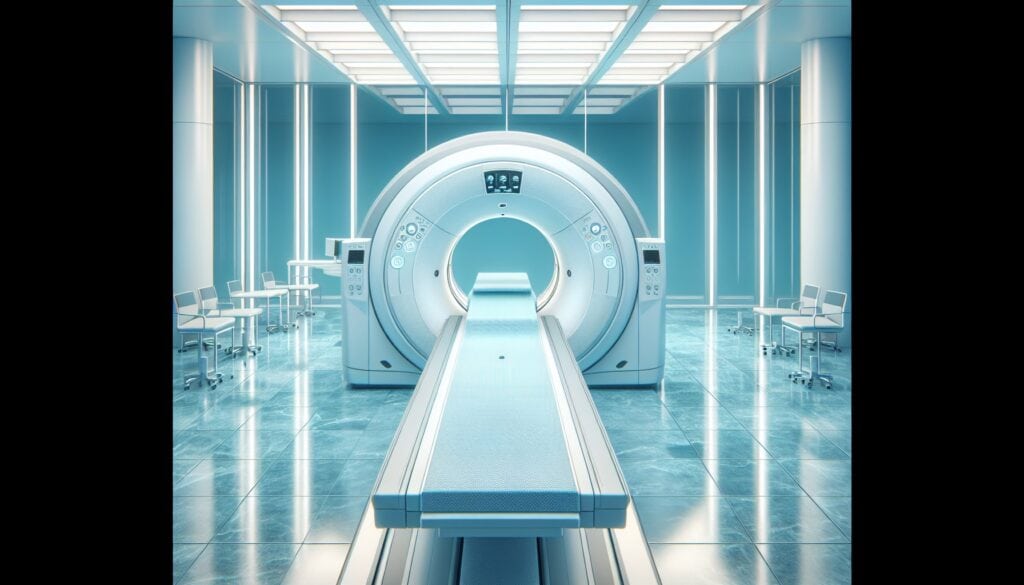
The structural heart team just added a stat CT for Transcatheter Aortic Valve Replacement (TAVR) planning to your list. This isn’t a routine CTA runoff. They need precise annular measurements, coronary heights down to the millimeter, and a definitive call on the femoral access route. Get one number wrong, and you risk paravalvular leak, coronary obstruction, or a vascular complication. The pressure is on, and your attending expects a report that reads like a pre-op checklist for the valve team.
When I was a fellow, these were the reads that made me sweat. You’re juggling multiplanar reformats, centerline analysis, and ECG-gated phases, all while trying to remember the exact cutoff for a “horizontal” aorta. There’s no room for ambiguity. Having a solid template and a systematic approach is the only way to stay sane and deliver the goods. For other high-yield guides and tools, check out the residents free-reference hub we’ve put together.
2. What a CT for Transcatheter Aortic Valve Replacement (TAVR) Planning Covers and What Attendings Look For
A CT TAVR planning study is a comprehensive evaluation designed to answer a very specific set of questions for the interventional cardiologist and cardiothoracic surgeon. It combines a high-resolution, ECG-gated acquisition of the aortic root with a full CTA runoff to the common femoral arteries. Your report needs to be a one-stop shop for every critical measurement.
Your attending and the structural heart team will be looking for these key data points, which should form the core of your report:
- Aortic Annular Sizing: The most critical component. This includes area, perimeter, and both area- and perimeter-derived diameters to guide valve size selection.
- Coronary Ostia Heights: The distance from the annulus to the left and right coronary ostia. Too low, and the native valve leaflets can obstruct the coronaries when the new valve is deployed.
- Aortic Root Anatomy: Diameters of the Sinus of Valsalva and the sinotubular junction (STJ).
- Aortic Angulation: The angle between the left ventricular outflow tract (LVOT) and the ascending aorta, which predicts the difficulty of the transfemoral approach.
- Calcification Burden: Quantification of calcification in the aortic valve leaflets and the LVOT, a key predictor of paravalvular leak.
- Peripheral Access Assessment: A detailed evaluation of the iliofemoral arteries, including minimum vessel diameter, tortuosity, and calcification.
- Bonus Findings: Evaluation for significant coronary artery disease (CAD) on the same dataset.
3. Radiology Report Template for CT TAVR Planning
This template provides a structured format to ensure you hit every required measurement. You can adapt this for your institution’s macros in PowerScribe or other dictation software.
Technique
ECG-gated CT angiography of the chest, abdomen, and pelvis was performed for TAVR planning. A retrospectively gated, helical acquisition of the aortic root was obtained, followed by a continuous helical acquisition from the aortic arch through the common femoral arteries. Multiplanar reformatted images, including reconstructions of the aortic annular plane and curved planar reformats of the aorta and iliofemoral arteries, were reviewed on a dedicated 3D workstation.
Findings
AORTIC VALVE AND ROOT:
Valve Morphology: [Tricuspid / Bicuspid / Other], with [mild/moderate/severe] calcification of the leaflets.
Annular Plane Measurements (Systolic Phase, [XX]% R-R):
Annular Area: [XXX] mm²
Annular Perimeter: [XX.X] mm
Area-Derived Diameter: [XX.X] mm
Perimeter-Derived Diameter: [XX.X] mm
Annular Max Diameter: [XX.X] mm
Annular Min Diameter: [XX.X] mm
Coronary Ostia Heights:
Height of Left Main Ostium: [XX.X] mm from annulus
Height of Right Coronary Ostium: [XX.X] mm from annulus
Aortic Root Dimensions:
Sinus of Valsalva Diameter (average): [XX.X] mm
Sinotubular Junction (STJ) Diameter: [XX.X] mm
Ascending Aorta Diameter (at level of PA): [XX.X] mm
Left Ventricular Outflow Tract (LVOT):
LVOT Calcification: [None / Mild / Moderate / Severe]
LVOT Diameter (5 mm below annulus): [XX.X] mm
Aortic Angulation:
Angle between Annular Plane and Horizontal Plane: [XX] degrees
VASCULAR ACCESS ASSESSMENT:
Right Common Femoral Artery:
Minimal Diameter: [X.X] mm
Calcification: [None / Mild / Moderate / Severe]
Tortuosity: [Minimal / Mild / Moderate]
Left Common Femoral Artery:
Minimal Diameter: [X.X] mm
Calcification: [None / Mild / Moderate / Severe]
Tortuosity: [Minimal / Mild / Moderate]
Other significant findings in the iliac arteries or descending aorta: [Describe any significant stenosis, aneurysm, or dissection].
OTHER FINDINGS:
Coronary Arteries: [No significant stenosis / Mild CAD / Moderate CAD / Severe CAD in LAD/LCx/RCA].
Lungs: [Clear / Other findings].
Other Incidental Findings: [None].
Impression
1. CT evaluation for TAVR planning. Key measurements are provided above.
2. Aortic annulus area is [XXX] mm² and perimeter is [XX.X] mm, suggesting a TAVR valve size of [e.g., 26 mm, 29 mm – often left to the heart team].
3. Coronary ostia heights are adequate at [XX.X] mm (left) and [XX.X] mm (right).
4. The bilateral common femoral arteries appear suitable for large-bore access, with a minimum diameter of [X.X] mm on the right. The right femoral artery is the preferred access site.
5. [Significant LVOT calcification, bicuspid valve, severe aortic angulation, or other key findings that impact the procedure].
4. Where to Find More Free Radiology Templates
Building a personal library of high-quality templates is a career-long project. While you’re building your own, two great free repositories exist that are worth bookmarking. They are maintained by radiologists for radiologists, without a paywall.
- RadReport.org: This is the RSNA-curated library. It’s comprehensive, peer-reviewed, and covers nearly every modality and subspecialty. It’s the gold standard for structured reporting templates.
- Radiology Templates (AU): An excellent resource maintained by Australian radiologists. It offers a slightly different flavor of templates and can be a great place to find alternative phrasing or structures.
5. The Next-Level Move: Free-Form Dictation to Structured Report
The biggest time sink in a complex read like TAVR planning isn’t finding the pathology; it’s organizing the dozens of measurements into a clean, logical report that the clinical team can actually use. You dictate the positive findings—annular area, coronary heights, femoral diameter—in whatever order you find them. Then you spend five minutes cutting, pasting, and reformatting inside the dictation box.
This is where AI-powered tools can streamline your workflow. Instead of wrestling with your dictation software’s macro editor, you can dictate your findings naturally. GigHz Precision AI is designed to take that free-form dictation and automatically generate a perfectly structured draft based on pre-loaded ACR and SIR templates. It helps ensure every key measurement is in the right place, every time. For studies with specific reporting frameworks, it can also surface the appropriate Clinical Decision Support (CDS) to guide classification, though for TAVR planning, the focus is on nailing the structured measurements.
6. When Should You Order a CT for TAVR Planning? ACR Appropriateness Criteria
The decision to order a CT for TAVR planning is typically made by the cardiology or cardiothoracic surgery team, but it’s crucial for radiologists to understand the official guidance. The American College of Radiology (ACR) provides clear recommendations.
Per the ACR Appropriateness Criteria for Preprocedural Planning for Transcatheter Aortic Valve Replacement, CT is the first-line, “Usually Appropriate” imaging modality. Specifically, for the initial pre-intervention planning to assess the aortic root, CT angiography receives the highest rating. Similarly, for assessing the supravalvular aorta and the vascular access route, CT angiography is also rated “Usually Appropriate.”
This high rating reflects CT’s unique ability to provide all the necessary information in a single exam: high-resolution aortic root anatomy, calcification scoring, and a complete map of the vascular access from the arch to the groin. Alternative modalities like transesophageal echocardiography (TEE) are excellent for assessing valve morphology and function but cannot evaluate the peripheral access route. MRI can be used for valve assessment but is less common and often reserved for centers with specific expertise or for patients with contraindications to iodinated contrast.
7. How Much Radiation Does a CT for TAVR Planning Deliver?
A common question from patients and referring providers relates to radiation dose. It’s important to be able to contextualize it accurately. A comprehensive CT TAVR planning study delivers an estimated effective dose of 10-20 mSv.
This places it in the moderate dose tier for diagnostic CT scans. To put it in perspective, this is equivalent to several years of natural background radiation. While not insignificant, the diagnostic yield is extremely high and directly informs a major, life-saving procedure. In patients with chronic kidney disease (CKD), protocols can be modified to use minimal contrast volumes (e.g., 50 mL with a high-flow technique) to reduce the risk of contrast-induced nephropathy.
| Scan Type | Typical Effective Dose (mSv) | Background Radiation Equivalent |
|---|---|---|
| Chest X-ray (PA/LAT) | ~0.1 mSv | ~10 days |
| CT TAVR Planning | 10-20 mSv | ~3-6 years |
| Annual Background Radiation | ~3 mSv | 1 year |
8. CT TAVR Planning Imaging Protocol — Phases, Contrast, and Reconstructions
The success of a TAVR planning study hinges on a meticulously executed protocol. It’s a two-part scan: a gated acquisition for the heart and a helical runoff for the access vessels. The contrast bolus timing must be perfect to opacify the entire arterial tree from the aortic root to the femoral heads.
The key is the ECG-gated cardiac component, which allows for reconstruction at different phases of the cardiac cycle. Annular measurements must be performed in systole (typically 30-40% of the R-R interval) when the annulus is at its largest and most circular.
| Phase / Sequence | Coverage | Key Parameters | Contrast |
|---|---|---|---|
| Topogram | Chest/Abd/Pelvis | kVp: 120 | None |
| ECG-gated Cardiac CT | Aortic root & Ascending Aorta | Retrospective gating; Slice: 0.6-0.8 mm; Rotation: ~0.27s | 100-150 mL @ 4-5 mL/s |
| Continuous Helical Runoff | Aortic Arch to Femoral Arteries | Slice: 1-2 mm | Same bolus as cardiac phase |
| Reconstructions | Workstation | Annular plane, centerline CPRs (aorta, iliofemoral) | N/A |
Common protocol pitfalls: The most common error is motion artifact from arrhythmia or poor ECG gating, which can render annular measurements inaccurate. Another is poor contrast opacification in the distal runoff vessels, which can happen with low cardiac output or a poorly timed scan. Always review the raw data for these issues before starting your measurements.
9. The 3-Months-Free Offer for Radiology Residents and Fellows
3+ months free for radiology residents and fellows
Look like a rockstar on your reports — dictate positive findings in free form, and the AI generates a structured report using ACR + SIR templates with the appropriate clinical decision support firing automatically. All we ask is feedback so we can keep improving the product for trainees.
Signup is simple. No credit card, no long forms. To get set up, just provide these three items:
- Your PGY year (e.g., PGY-2, PGY-4)
- Your training type (radiology residency or specific fellowship — IR, body, MSK, neuro, peds, breast, nucs)
- Your training program / hospital name
To get started, apply for the residents free-access program and reply to the application email with the information above.
10. Frequently Asked Questions
Is GigHz Precision AI HIPAA-compliant?
Yes. The platform is designed for de-identified workflows by default. It operates on the anonymized text of your dictated report and does not require access to protected health information (PHI) or your hospital’s PACS/EMR.
Does this require a complex IT setup?
No. It’s a browser-based tool that works on any modern computer, including the call-room PC or your personal laptop/iPad. There is no software to install and no need to involve your hospital’s IT department.
How does it work with PowerScribe or other dictation software?
It works alongside your existing dictation system. You dictate as you normally would. You can then copy your free-form dictation into the tool, which instantly structures it. You then copy the structured report back into your dictation window for final signature.
Can I use this on my phone or iPad?
Yes, the platform is fully responsive and works on mobile devices, which is great for reviewing templates or checking a quick reference on the go.
Can I customize the templates?
Yes, you can customize the base templates to match your institution’s specific formatting preferences or your attending’s preferred language. The system will learn your modifications over time.
What happens after my residency or fellowship ends?
Trainees who provide valuable feedback during the free-access period are often eligible for significant discounts on post-training plans. The goal is to build a tool that grows with you from training into practice.
Free GigHz Tools That Pair With This Article
Three free tools that complement the material above:
- Imaging Appropriateness Selector — Type an indication or clinical scenario in plain language and get the imaging studies the ACR rates for it, with adult and pediatric radiation levels. Built directly from 297 ACR topics, 1,336 clinical variants, and 15,823 procedure ratings.
- GigHz Imaging Protocol Library — A searchable library of 131 imaging protocols with the physics specs surfaced and the matching ACR Appropriateness Criteria alongside. Plain-English narratives readable in 60 seconds, organized by modality.
- GigHz Radiation Dose Calculator — Pick the imaging studies a patient has had and see total dose in millisieverts (mSv) with comparisons to natural background radiation, transatlantic flights, and chest X-rays. Useful for shared decision-making.
Last reviewed by Pouyan Golshani, MD — 2026-08-04.
Reviewed by Pouyan Golshani, MD, Interventional Radiologist — August 4, 2026