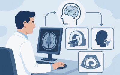
For a New-Onset Seizure Without Trauma, Is MRI or CT the Right First Step?

A 34-year-old architect is brought to the emergency department by coworkers after a witnessed tonic-clonic seizure at his desk. He has no prior history of seizures, no recent illness, and no history of head trauma. On arrival, he is postictal but slowly returning to his neurologic baseline. The initial labs are unremarkable. As the treating physician, you must decide on the most appropriate initial neuroimaging to investigate the underlying cause of this first-time, unprovoked seizure. This is a critical decision point, as the choice of study can directly impact your ability to identify a structural etiology and guide subsequent management.
This article provides a focused workflow for this exact scenario, grounded in the American College of Radiology (ACR) Appropriateness Criteria. For a new-onset, non-traumatic seizure, the ACR rates MRI head without IV contrast as Usually Appropriate, representing the preferred initial imaging modality to evaluate for an underlying structural cause.
Who Fits This Clinical Scenario for a New-Onset Seizure?
This guidance applies to a well-defined patient population: adults or children presenting with their first-ever seizure that is not associated with a recent, significant head injury. The patient is typically evaluated after the seizure has resolved and they are neurologically stable or have returned to their baseline. The primary goal of imaging in this context is to search for a structural lesion that could be an epileptogenic focus.
It is crucial to distinguish this presentation from similar but distinct clinical situations that follow different diagnostic pathways:
- Patients with recent, significant trauma: If the seizure occurs in the setting of a head injury, the imaging workup is guided by trauma protocols, where CT is often the first-line study to assess for acute intracranial hemorrhage, fracture, or contusion. This falls under the ACR variant for New-onset seizure with a history of trauma.
- Patients with a known seizure disorder: An individual with a pre-existing diagnosis of epilepsy who presents with a typical seizure does not routinely require imaging. Imaging is reconsidered only if there is a significant change in seizure type (semiology), a new focal neurologic deficit, or a failure to return to their previous baseline.
- Patients in status epilepticus: While imaging is essential, the immediate priority is medical stabilization. The choice and timing of imaging are dictated by the emergent clinical context, which may favor the speed of CT initially.
This article is for the classic, unprovoked, first-time seizure where the immediate clinical question is: “Is there an underlying structural problem in the brain?”
What Diagnoses Are You Working Up With Initial Imaging?
When ordering imaging for a new-onset seizure, you are primarily investigating for a structural brain abnormality that could lower the seizure threshold. The differential diagnosis guides the choice of imaging modality, favoring a study with high sensitivity for subtle parenchymal changes.
Brain Tumor
A primary brain tumor (like a low-grade glioma) or a metastatic lesion is a leading consideration, particularly in adults over 30. These lesions can irritate the surrounding cortex, leading to focal or generalized seizures. Low-grade tumors may be subtle and isodense on CT, making them difficult to detect without the superior soft-tissue resolution of MRI.
Vascular Malformations
Congenital vascular anomalies such as cavernous malformations (cavernomas) or arteriovenous malformations (AVMs) are important causes of seizures, especially in younger adults. Cavernomas, in particular, can be occult on CT but are readily identified on specific MRI sequences (like gradient echo or susceptibility-weighted imaging) due to chronic microhemorrhages.
Sequelae of Prior Ischemic Stroke
A clinically silent stroke in the past can result in an area of gliosis or encephalomalacia, creating an epileptogenic scar. A seizure may be the first clinical manifestation of this old vascular insult. MRI is far more sensitive than CT for detecting evidence of chronic ischemia and gliosis.
Cortical Dysplasia
Focal cortical dysplasia is a malformation of cortical development and a common cause of epilepsy, particularly in children and young adults. These are often very subtle abnormalities in the cortical gray matter, such as abnormal gyral patterns or blurring of the gray-white matter junction, that are typically only visible on high-resolution MRI.
Cerebral Infection or Inflammation
While less common as an isolated presentation, a brain abscess, herpes encephalitis, or autoimmune encephalitis can present with seizures. These conditions cause focal or diffuse inflammation and edema, which are best characterized by MRI.
Why Is MRI Head Without Contrast Usually Appropriate for a First-Time Seizure?
The ACR designates MRI head without IV contrast as Usually Appropriate because it provides the highest diagnostic yield for the most common structural causes of a new-onset seizure. Its superior soft-tissue contrast is essential for identifying the subtle abnormalities that are often responsible for an epileptogenic focus.
The rationale for this recommendation over other modalities involves a careful balance of diagnostic sensitivity, safety, and accessibility.
- Superior Sensitivity for Key Pathologies: MRI is significantly more sensitive than CT for detecting low-grade tumors, cortical dysplasia, cavernous malformations, and the gliotic changes of a prior stroke. Many of these findings are simply not visible on a non-contrast CT scan. An MRI protocol for seizures typically includes high-resolution, thin-section T1, T2, and FLAIR sequences oriented to the hippocampus, which are specifically designed to detect these subtle epileptogenic lesions.
- Comparison to Alternatives:
- CT head without IV contrast: This study is also rated Usually Appropriate. Its primary advantage is speed and wide availability, making it a reasonable first test in the emergency department to rapidly exclude large, emergent findings like a significant hemorrhage, hydrocephalus, or a large mass with midline shift. However, a negative non-contrast CT does not adequately rule out an underlying structural cause for a seizure. It is often considered a screening tool, with the understanding that an MRI will likely be needed if the CT is unrevealing.
- MRI head without and with IV contrast: This is rated May be appropriate. While gadolinium contrast is crucial for evaluating for active inflammation, infection, or characterizing a known or suspected tumor, it is not always necessary for the initial evaluation. A high-quality non-contrast MRI is often sufficient to identify a lesion. The decision to add contrast can be deferred and guided by the findings of the non-contrast study or a high clinical suspicion for a specific diagnosis like a metastatic lesion or abscess.
- Radiation and Contrast Considerations: A key advantage of MRI is the complete absence of ionizing radiation (0 mSv), which is an important consideration in all patients, especially children and young adults who may require follow-up imaging. In contrast, a head CT delivers a dose of 1-10 mSv (☢☢☢). Opting for a non-contrast MRI as the initial study also avoids the potential risks associated with gadolinium-based contrast agents, such as nephrogenic systemic fibrosis in patients with renal impairment or gadolinium deposition.
What’s Next After MRI Head Without Contrast? Downstream Workflow
The results of the initial MRI will dictate the subsequent clinical pathway. The goal is to move from detecting a potential cause to confirming a diagnosis and initiating appropriate treatment, which often involves a neurology consultation.
- If the MRI is positive for a structural lesion:
- Tumor or Mass: The next step is typically a referral to neurology and neurosurgery. A follow-up MRI head without and with IV contrast (May be appropriate) will likely be ordered to better characterize the lesion, assess for enhancement, and plan for biopsy or resection.
- Vascular Malformation: A referral to neurology and possibly neurosurgery or neurointerventional radiology is warranted. Further characterization with MRA, MRV, or conventional angiography may be required depending on the suspected type of malformation.
- Cortical Dysplasia or Gliosis: This finding confirms a structural basis for the seizure. The patient should be referred to a neurologist or epileptologist for long-term management with anti-epileptic drugs (AEDs) and continued monitoring.
- If the MRI is negative:
A normal, high-quality MRI is a common and reassuring finding. It significantly lowers the likelihood of a structural cause but does not exclude a diagnosis of epilepsy, which is ultimately a clinical diagnosis based on seizure history and EEG findings. The next step is a referral to neurology for an electroencephalogram (EEG) to evaluate for epileptiform discharges and to discuss the risks and benefits of starting AEDs.
- If the MRI is indeterminate:
Occasionally, a finding may be non-specific. In these cases, discussion with the interpreting radiologist is key. Depending on the finding, next steps could include a follow-up MRI to assess for stability, adding IV contrast, or proceeding with an EEG and clinical follow-up.
Pitfalls to Avoid (and When to Get Help)
Navigating the workup for a first-time seizure requires careful consideration to avoid common diagnostic errors.
- Pitfall 1: Accepting a negative CT as a definitive workup. While a non-contrast head CT is useful for emergent triage, it is not sensitive enough to rule out many key causes of seizures. A patient with a first-time, unprovoked seizure and a negative CT still warrants an MRI.
- Pitfall 2: Not ordering a dedicated seizure-protocol MRI. A “routine” brain MRI may not include the specific thin-section coronal sequences through the temporal lobes that are optimized for detecting subtle hippocampal sclerosis or cortical dysplasia. When ordering, specify “MRI brain with seizure protocol.”
- Pitfall 3: Delaying imaging unnecessarily. While the workup is not typically as time-sensitive as a stroke evaluation, prolonged delays can postpone the diagnosis of a serious underlying condition like a brain tumor. Imaging should be arranged in a timely manner, usually on an outpatient basis if the patient is stable.
If the patient presents with focal neurologic deficits that do not resolve, has a change in mental status, or experiences recurrent seizures, escalate care immediately and consider an emergent inpatient evaluation.
Related ACR Topics and Tools
For a comprehensive overview of imaging recommendations across all seizure-related scenarios, from initial presentation to surgical planning, please see our parent guide. You can also use the tools below to explore adjacent ACR criteria, review imaging techniques, and discuss radiation dose with patients.
- For breadth across all scenarios in Seizures and Epilepsy, see our parent guide: Seizures and Epilepsy: ACR Appropriateness Decoded.
- Imaging Appropriateness Selector — for adjacent scenarios
- Imaging Protocol Library — for technique on the recommended study
- Radiation Dose Calculator — for cumulative dose conversations
Frequently Asked Questions
Is a CT scan ever the right first and only test for a new-onset seizure?
Rarely. A non-contrast head CT is rated Usually Appropriate and is excellent for rapid exclusion of acute, life-threatening conditions like a large hemorrhage or mass in the emergency setting. However, a negative CT does not rule out the most common subtle structural causes of seizures (e.g., cortical dysplasia, low-grade tumors). For a comprehensive initial workup, an MRI is the definitive study.
If the patient is a child, does the recommendation for MRI still hold?
Yes, even more so. The ACR guidance applies to both adult and pediatric patients. MRI is strongly preferred in children due to its lack of ionizing radiation and its superior ability to detect congenital or developmental abnormalities, such as cortical dysplasia, which are more common causes of seizures in younger age groups.
When should I add IV contrast to the initial MRI for a first-time seizure?
According to the ACR, MRI without and with contrast is rated May be appropriate. You should consider adding contrast if there is a high clinical suspicion for a brain tumor (e.g., in an older adult or a patient with a history of cancer), or if you suspect an infectious or inflammatory process like an abscess or encephalitis. In many cases, it is reasonable to start with a non-contrast study and add contrast later if an abnormality is detected.
What if the patient has a contraindication to MRI, like a non-compatible pacemaker?
In cases where MRI is contraindicated, a CT head without IV contrast is the best alternative. If clinical suspicion for a tumor or infection is high, a CT head with IV contrast (Usually not appropriate for the initial workup but may become necessary) can provide additional information, though it remains less sensitive than MRI for many epileptogenic pathologies. Consultation with neurology and radiology is recommended in these complex cases.
Does a normal MRI mean the patient does not have epilepsy?
No. Epilepsy is a clinical diagnosis based on the occurrence of unprovoked seizures. A normal MRI is a very common finding and simply means there is no identifiable structural cause for the seizures. These patients are often diagnosed with idiopathic or cryptogenic epilepsy. The next diagnostic step is typically an EEG to look for electrical evidence of seizure activity.
Reviewed by Pouyan Golshani, MD, Interventional Radiologist — June 27, 2026