Which Imaging Is Best for Suspected Arterial Injury After Head Trauma?
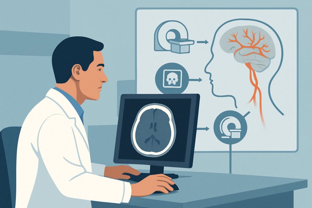
It’s 2 a.m. in the emergency department, and you’re evaluating a patient from a high-speed motor vehicle collision. The initial non-contrast CT of the head, ordered for altered mental status, reveals a complex basilar skull fracture extending through the carotid canal. While there’s no large intracranial hemorrhage, your concern immediately shifts to a potential blunt cerebrovascular injury (BCVI). The patient is hemodynamically stable but remains obtunded. The next decision is critical: which imaging study will definitively evaluate the intracranial and cervical arteries for dissection, pseudoaneurysm, or occlusion? This article provides a focused workflow for this exact scenario—head trauma with suspected intracranial arterial injury. According to the American College of Radiology (ACR) Appropriateness Criteria, the definitive first step is clear: CTA head and neck with IV contrast is rated Usually Appropriate.
Who Fits This Clinical Scenario?
This guidance applies to a specific subset of head trauma patients where the clinical suspicion for an arterial injury is high. This suspicion can arise from two primary pathways: high-risk clinical factors or concerning findings on a prior imaging study (typically an initial non-contrast head CT).
Inclusion criteria for this workflow include patients with:
- Specific fracture patterns known to be high-risk for vascular injury, such as basilar skull fractures involving the petrous bone or carotid canal, Le Fort II or III facial fractures, or cervical spine fractures extending through a transverse foramen.
- Clinical signs suggestive of arterial injury, including a neck hematoma, cervical bruit, neurological deficit inconsistent with the initial CT findings (e.g., signs of a stroke), or a Glasgow Coma Scale (GCS) score of less than 8.
- Findings on a prior non-contrast CT that are indeterminate but raise suspicion for a vascular abnormality.
This workflow is distinct from other head trauma scenarios. It does not apply to the initial workup of mild head trauma (GCS 13-15) where clinical decision rules do not indicate a need for imaging. It also differs from the initial evaluation of moderate to severe head trauma (GCS 3-12) where the primary goal is to identify life-threatening intracranial hemorrhage, in which case a non-contrast CT head is the first step. This scenario is specifically for the next step when the focus shifts to the vessels.
What Diagnoses Are You Working Up in This Scenario?
When ordering a dedicated vascular study after head trauma, you are investigating a spectrum of potentially devastating blunt cerebrovascular injuries (BCVIs). These injuries can be clinically occult initially but may lead to delayed stroke and significant morbidity or mortality if missed. The differential diagnosis is focused and critical.
Carotid or Vertebral Artery Dissection
This is one of the most common and consequential forms of BCVI. The traumatic force creates a tear in the intimal layer of the artery, allowing blood to track into the vessel wall. This can create a flap, a pseudoaneurysm, or an intramural hematoma that narrows (stenosis) or completely blocks (occlusion) the vessel lumen, leading to thromboembolic stroke.
Traumatic Pseudoaneurysm
Unlike a true aneurysm, a pseudoaneurysm (or false aneurysm) is a contained rupture of the vessel wall. A hematoma forms outside the vessel but is contained by the surrounding adventitia or soft tissues. These are highly unstable and carry a significant risk of delayed rupture and hemorrhage.
Arteriovenous Fistula
High-energy trauma, particularly involving skull base fractures, can create an abnormal connection between an artery and a vein. The most classic example is a carotid-cavernous fistula (CCF), where high-pressure arterial blood from the internal carotid artery shunts directly into the low-pressure cavernous sinus, causing orbital congestion, proptosis, and cranial nerve palsies.
Vessel Occlusion
Direct injury or a large intimal flap from a dissection can lead to acute thrombosis and complete occlusion of a major cervical or intracranial artery. This is a direct cause of acute ischemic stroke in the trauma setting and requires immediate identification.
Why Is CTA head and neck with IV contrast the Recommended Study?
The ACR designates CTA head and neck with IV contrast as Usually Appropriate for this scenario because it offers the best combination of speed, availability, and diagnostic accuracy in the acute trauma setting. Its ability to simultaneously visualize bony anatomy and the vascular lumen from the aortic arch through the circle of Willis makes it the ideal first-line modality.
CTA is highly sensitive and specific for detecting dissections, pseudoaneurysms, and occlusions. The rapid acquisition time minimizes motion artifact in potentially uncooperative trauma patients, and its widespread availability in most emergency departments allows for immediate diagnosis and intervention. The inclusion of the neck is non-negotiable; many traumatic carotid and vertebral injuries occur in the cervical region before having intracranial consequences.
Why are other studies rated lower for this initial workup?
- Arteriography (Digital Subtraction Angiography): While considered the diagnostic gold standard, arteriography is rated May be appropriate. It is invasive, carrying risks of stroke, vessel injury, and groin hematoma. Its use is generally reserved for cases where CTA is equivocal or when an endovascular intervention (e.g., stenting, coiling) is planned based on the CTA findings.
- MRA head and neck: Magnetic Resonance Angiography is also rated May be appropriate. It avoids ionizing radiation and iodinated contrast, which is an advantage. However, MRA is significantly slower, more susceptible to patient motion, less available in emergencies, and inferior to CTA for visualizing adjacent bony fractures. It serves as an excellent problem-solving tool or a follow-up modality but is not the preferred primary study in an acute, unstable trauma patient.
The radiation dose for a CTA head and neck is moderate (ACR RRL ☢☢☢, 1-10 mSv for an adult), a necessary trade-off for the critical diagnostic information it provides. When ordering, be specific: request a “CTA of the head and neck” or “cervicocranial CTA” to ensure the protocol covers the entire course of the carotid and vertebral arteries. Once you’ve decided on CTA head and neck, our protocol guide covers the technique, contrast, and reading principles: CTA Head and Neck (Carotid + COW).
What’s Next After CTA head and neck with IV contrast? Downstream Workflow
The results of the CTA will dictate immediate management and consultation. The downstream pathway is typically straightforward, focusing on preventing ischemic complications or hemorrhage.
If the CTA is positive for a BCVI (e.g., dissection, pseudoaneurysm):
This is a critical finding requiring immediate action. The next step is an urgent consultation with the appropriate specialty, which may include neurosurgery, vascular surgery, or interventional neuroradiology, depending on the specific injury and institutional resources. Management often involves initiating antithrombotic therapy (antiplatelet agents like aspirin or anticoagulation with heparin) to prevent stroke from thromboembolism, though this must be balanced against the risk of hemorrhage from other injuries. Some injuries, like pseudoaneurysms or high-flow fistulas, may require endovascular treatment.
If the CTA is negative:
A high-quality negative CTA of the head and neck significantly lowers the likelihood of a clinically significant vascular injury. In most cases, no further vascular imaging is required. The patient’s care can proceed based on their other traumatic injuries. However, if a very high clinical suspicion persists despite the negative scan (e.g., a new, unexplained focal neurologic deficit), a follow-up study or an alternative modality like MRA could be considered in discussion with the radiology and neurology/neurosurgery teams.
If the CTA is indeterminate or equivocal:
Occasionally, findings may be unclear due to motion artifact, vessel tortuosity, or venous contamination. In these cases, the next step is often conventional arteriography (DSA), which remains the gold standard for clarifying ambiguous findings. Alternatively, a short-term follow-up CTA or MRA in 3-5 days may be appropriate for low-grade or uncertain injuries.
Pitfalls to Avoid (and When to Get Help)
In the high-stakes environment of trauma care, several common ordering and interpretation pitfalls can compromise patient care. Avoiding them is key to a timely and accurate diagnosis.
- Ordering the wrong CT: A “CT head with contrast” is not the same as a “CTA head.” The timing of the contrast bolus is completely different, and a standard post-contrast CT will not provide the necessary arterial-phase detail to evaluate for dissection.
- Incomplete coverage: Requesting only a “CTA head” and omitting the neck is a critical error. Many traumatic dissections begin in the cervical portions of the carotid or vertebral arteries. Always order “CTA head and neck.”
- Ignoring contraindications: Before ordering a CTA, assess the patient’s renal function (e.g., creatinine, eGFR) and inquire about severe allergies to iodinated contrast media.
- Delaying the diagnosis: In a patient with high-risk features, vascular imaging should be performed expeditiously. A delay can allow a stroke to evolve from a manageable arterial injury.
If you encounter a positive or equivocal study, or if the clinical picture does not match the imaging findings, escalate immediately. This involves a direct conversation with the interpreting radiologist and the relevant surgical or interventional subspecialist.
Related ACR Topics and Tools
This article focuses on one specific clinical question. For a comprehensive overview of all imaging scenarios in this category, or to explore the technical details of the recommended study, the following resources are essential. For breadth across all scenarios in Head Trauma, see our parent guide: Head Trauma: ACR Appropriateness Decoded.
- Imaging Appropriateness Selector — For exploring adjacent scenarios or different clinical questions.
- Imaging Protocol Library — For technical details on hundreds of imaging protocols.
- Radiation Dose Calculator — For discussing cumulative radiation exposure with patients and families.
Frequently Asked Questions
What are the key clinical signs that should prompt a workup for blunt cerebrovascular injury (BCVI)?
High-risk signs and symptoms include cervical spine fractures (especially C1-C3 or those involving the transverse foramen), basilar skull fractures with carotid canal involvement, Le Fort II or III facial fractures, diffuse axonal injury with a GCS < 6, expanding cervical hematoma, cervical bruit, or a focal neurologic deficit (like hemiparesis or aphasia) that is inconsistent with the findings on an initial non-contrast head CT. These are often formalized in screening criteria like the Denver or Memphis criteria.
Why is a standard ‘CT head with contrast’ not appropriate for this scenario?
A standard ‘CT head with contrast’ uses a delayed-phase imaging protocol designed to detect breakdown of the blood-brain barrier (e.g., in tumors or infection). A CTA (Computed Tomography Angiography) uses a precisely timed bolus of IV contrast to acquire images during peak arterial enhancement. This specific timing is essential to opacify the arteries and visualize subtle abnormalities of the vessel wall like intimal flaps, pseudoaneurysms, or filling defects. A standard contrast-enhanced CT will miss most of these findings.
If my patient has a severe iodine contrast allergy, what is the best alternative to CTA?
For patients with a severe allergy to iodinated contrast or with significant renal impairment, MRA (Magnetic Resonance Angiography) of the head and neck is the best non-invasive alternative. It is rated as ‘May be appropriate’ by the ACR. While it is less ideal in the acute setting due to longer scan times and lower availability, it provides excellent vascular detail without ionizing radiation or iodine-based contrast.
Does a negative CTA completely rule out a vascular injury?
A technically adequate, high-quality negative CTA has a very high negative predictive value and makes a significant cerebrovascular injury highly unlikely. However, no test is perfect. In rare cases with persistent, unexplained neurological symptoms or extremely high-risk injury patterns, a small dissection could be missed. In these situations, discussion with a radiologist about the need for follow-up imaging (e.g., repeat CTA or MRA in several days) or a different modality may be warranted.
Is it necessary to image from the aortic arch upwards for every suspected BCVI?
Yes, it is critical. The origins of the great vessels from the aortic arch are a common site for traumatic injury, as are the cervical portions of the carotid and vertebral arteries. A CTA that only covers the intracranial circulation (a ‘CTA head’) is insufficient and will miss the majority of traumatic dissections. The standard protocol must be a ‘CTA head and neck’ that includes imaging from the aortic arch through the circle of Willis.
Reviewed by Pouyan Golshani, MD, Interventional Radiologist — May 26, 2026