Which Imaging Is Best for a Fixed Neurologic Deficit Lasting Over 6 Hours?
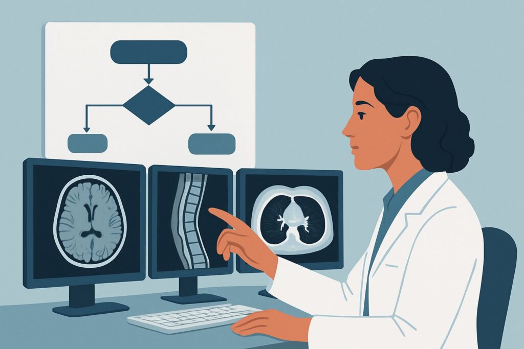
It’s 10 p.m. in the emergency department, and you are evaluating a 68-year-old patient with new, persistent right-sided weakness and aphasia. The symptoms began yesterday evening and have not improved; if anything, they seem slightly worse. The window for thrombolysis has long passed, but the diagnostic question remains urgent: what is the underlying cause, what is the extent of the damage, and how can you best guide secondary prevention? This clinical scenario—a new, fixed or worsening focal neurologic defect lasting longer than six hours—requires a specific imaging strategy to evaluate both the brain parenchyma and the cervicocerebral vasculature. This article details the American College of Radiology (ACR) guided workflow for this presentation. For this specific scenario, the ACR rates MRA head and neck without and with IV contrast as Usually Appropriate.
Who Fits This Clinical Scenario?
This guidance applies to patients presenting with a suspected stroke characterized by a new focal neurologic deficit that is either static or progressing and has been present for more than six hours. The “focal” nature of the deficit (e.g., hemiparesis, aphasia, hemianopsia) points toward a localized process within the brain, and the duration places the patient outside the hyperacute window for interventions like intravenous thrombolysis.
It is critical to distinguish this scenario from similar but distinct clinical presentations that follow different diagnostic pathways:
- Deficit less than 6 hours: If the patient’s symptoms began within the last six hours, they fall into the hyperacute stroke category. This is a time-critical emergency where imaging protocols are optimized to determine eligibility for reperfusion therapies. See the ACR variant for new focal neurologic defect, less than 6 hours.
- Symptoms have resolved: If the patient’s neurologic deficits have completely resolved, the event is classified as a Transient Ischemic Attack (TIA). The imaging workup for TIA focuses on identifying the underlying cause to prevent a future stroke but follows a slightly different algorithm.
- Known hemorrhage: If a non-contrast head CT has already been performed and confirms a parenchymal hemorrhage, the diagnostic question shifts from identifying ischemia to characterizing the hematoma and evaluating for an underlying cause, such as an aneurysm or arteriovenous malformation.
What Diagnoses Are You Working Up in This Scenario?
When a patient presents with a fixed, subacute neurologic deficit, your imaging choice is driven by a differential diagnosis that extends beyond simply confirming an infarct. The goal is to understand the “what” and the “why” to guide treatment.
Completed Ischemic Stroke: This is the primary diagnosis to confirm or exclude. In the subacute phase (beyond 6 hours), imaging aims to identify the location and extent of irreversibly damaged brain tissue (the infarct core). Diffusion-weighted imaging (DWI) on an MRI is exceptionally sensitive for this, showing restricted diffusion within minutes to hours of onset that can persist for days.
Large Vessel Atherosclerosis: A common cause of ischemic stroke is stenosis or occlusion of a major artery in the neck (e.g., internal carotid artery) or head (e.g., middle cerebral artery). Identifying a significant flow-limiting lesion is crucial for secondary prevention, which may include medical management, carotid endarterectomy, or stenting. Vascular imaging is essential to evaluate for this etiology.
Cervical Artery Dissection: While less common than atherosclerosis, dissection is a key consideration, particularly in younger patients or those with a history of neck trauma or connective tissue disease. A tear in the vessel wall can lead to a thrombus that embolizes to the brain or a pseudoaneurysm that compromises flow. Both CT and MR angiography are effective at identifying the characteristic findings of a dissection.
Stroke Mimics: Not all focal neurologic deficits are ischemic strokes. Other pathologies can present identically and must be considered. These include brain tumors (primary or metastatic), brain abscesses, or demyelinating diseases like multiple sclerosis presenting with a large, acute plaque. The superior soft-tissue contrast of MRI is invaluable for differentiating these mimics from a true ischemic event.
Why Is MRA of the Head and Neck the Recommended Study for This Presentation?
For a fixed or worsening neurologic deficit lasting over six hours, the ACR designates MRA head and neck without and with IV contrast as Usually Appropriate. This recommendation is based on the modality’s unique ability to provide a comprehensive, high-resolution assessment of both the brain parenchyma and the full arterial supply from the aortic arch to the circle of Willis in a single examination, all without using ionizing radiation.
The power of the MRI/MRA combination lies in its sequences:
- Diffusion-Weighted Imaging (DWI): This MRI sequence is the most sensitive tool for detecting cytotoxic edema from an acute or subacute ischemic infarct. It provides the definitive answer to “Has a stroke occurred?”
- Angiography (MRA): The MRA component visualizes blood flow within the arteries of the head and neck. It can reliably identify significant stenosis, occlusion, dissection, or aneurysms that may have caused the stroke. Performing this with and without contrast enhances vessel wall imaging and can improve characterization of plaque or dissection flaps.
While MRA is a top choice, other modalities are also rated Usually Appropriate and have important roles. Understanding why they may be chosen or deferred is key:
- CTA head and neck with IV contrast: This is also Usually Appropriate and is an excellent alternative. It is significantly faster than MRA and often more readily available, making it a pragmatic choice in many institutions. It provides superb vascular detail. However, it requires IV iodinated contrast and exposes the patient to radiation (☢☢☢ 1-10 mSv). It also provides less detailed information about the brain parenchyma compared to a full MRI.
- CT head without IV contrast: Also rated Usually Appropriate, a non-contrast CT is typically the very first imaging study performed in any patient with stroke-like symptoms. Its primary role is to rapidly and reliably exclude hemorrhage. In the subacute (>6 hours) timeframe, it may begin to show signs of ischemia (e.g., loss of gray-white differentiation, sulcal effacement), but it is far less sensitive than MRI DWI. It provides no information about the blood vessels.
The ACR rates US duplex Doppler carotid artery as Usually Not Appropriate as a standalone initial test in this scenario. While it can assess the extracranial carotid arteries, it cannot visualize the intracranial vessels or the brain parenchyma, making it an incomplete diagnostic study for this presentation.
Once you’ve decided on MRA of the neck, our protocol guide covers the technique, contrast, and reading principles: MRA Neck With and Without Contrast.
What’s Next After MRA? Downstream Workflow
The results of the head and neck MRA will directly guide the subsequent clinical workflow. The pathway diverges based on whether the findings are positive, negative, or indeterminate.
If the study is positive for an ischemic stroke and a causative vascular lesion (e.g., carotid stenosis): The focus shifts immediately to secondary stroke prevention. This involves initiating or adjusting antiplatelet therapy, high-intensity statins, and blood pressure control. A finding of high-grade, symptomatic carotid stenosis will trigger a consultation with vascular surgery or neuro-intervention to discuss revascularization (e.g., endarterectomy or stenting).
If the study is positive for an ischemic stroke but negative for a large vessel cause: When the MRA confirms an infarct but the head and neck arteries appear normal, the workup must pivot to find an alternative etiology. The next step is often an investigation for a cardioembolic source. This typically includes an echocardiogram (transthoracic or transesophageal) to look for intracardiac thrombus, patent foramen ovale, or valvular disease, as well as prolonged cardiac monitoring (e.g., Holter monitor) to detect paroxysmal atrial fibrillation.
If the study is negative for both stroke and vascular pathology: A completely normal MRI and MRA makes an ischemic stroke highly unlikely. The clinical team must then aggressively pursue stroke mimics. Depending on the specific neurologic deficits and other clinical clues, this may involve further imaging, a lumbar puncture to analyze cerebrospinal fluid (for inflammatory or infectious causes), or electroencephalography (EEG) to rule out seizure with a postictal (Todd’s) paralysis.
Pitfalls to Avoid (and When to Get Help)
Navigating the workup for a subacute stroke requires avoiding several common pitfalls. First, do not mistake a normal non-contrast head CT as ruling out a stroke; its sensitivity for ischemia is low, especially in the first 24 hours. Second, remember to image the entire vascular tree from the aortic arch to the intracranial vessels; ordering only a head MRA or a neck MRA is an incomplete study that can miss the causative lesion. Third, always consider the patient’s renal function before ordering a contrast-enhanced MRA or CTA. If a patient has a contraindication to gadolinium, a non-contrast MRA combined with a CTA or carotid ultrasound may be necessary. If the clinical picture and imaging findings are discordant, or if a complex vascular pathology like a dissection or vasculitis is suspected, it is crucial to escalate care by consulting with a neurologist and neuroradiologist.
Related ACR Topics and Tools
This article covers one specific scenario within the broader topic of cerebrovascular disease. For a comprehensive overview of imaging for all related presentations, from TIA to intracranial hemorrhage, please see our parent guide. For help with other scenarios or technical details, the following resources are available.
- For breadth across all scenarios in Cerebrovascular Disease, see our parent guide: Cerebrovascular Disease: ACR Appropriateness Decoded.
- Imaging Appropriateness Selector — for adjacent scenarios
- Imaging Protocol Library — for technique on the recommended study
- Radiation Dose Calculator — for cumulative dose conversations
Frequently Asked Questions
Why is MRA preferred over CTA when both are ‘Usually Appropriate’ for a fixed deficit over 6 hours?
MRA is often preferred because it provides superior soft-tissue contrast for evaluating the brain parenchyma, particularly with the DWI sequence, which is highly sensitive for subacute infarcts. It also avoids ionizing radiation. However, CTA is much faster and more widely available, making it an excellent and often more practical alternative, especially if MRI access is limited or the patient has contraindications to MRI.
If my initial non-contrast head CT is normal, have I ruled out a stroke?
No. A non-contrast head CT is excellent for ruling out hemorrhage, which is its primary purpose in an acute stroke workup. However, it is not sensitive for detecting ischemic changes in the first 6-24 hours. A normal CT in a patient with persistent focal deficits should prompt further investigation with MRI/MRA or CTA to assess for ischemia and vascular pathology.
Is contrast necessary for the MRA in this scenario?
The ACR recommends MRA ‘without and with IV contrast’ as the top procedure. While a non-contrast Time-of-Flight (TOF) MRA can visualize flow in the major vessels, adding gadolinium-based contrast can improve vessel delineation, help characterize atherosclerotic plaque, and better identify subtle findings like a vessel wall hematoma in a dissection. MRA head and neck without contrast is also rated ‘Usually Appropriate’ and is a valid option if contrast is contraindicated.
What if the patient has a pacemaker or other MRI contraindication?
If a patient cannot undergo an MRI, the best alternative is ‘CTA head and neck with IV contrast,’ which is also rated ‘Usually Appropriate’ by the ACR. This provides excellent vascular detail of the entire cervicocerebral circulation. The parenchymal evaluation would rely on a non-contrast head CT, which is less sensitive for ischemia but can show established infarcts in the subacute phase.
Does this guidance apply to a patient with a ‘wake-up’ stroke?
A ‘wake-up’ stroke (where the time of onset is unknown) is often managed like a hyperacute stroke (<6 hours) if the patient is a potential candidate for reperfusion therapy. In that specific context, advanced imaging like MRI with DWI/FLAIR mismatch or CT perfusion may be used to identify an ischemic penumbra and guide treatment. If the patient is clearly outside any treatment window, their workup would then align with this >6 hour scenario.
Reviewed by Pouyan Golshani, MD, Interventional Radiologist — May 26, 2026