Should You Order MRI for Suspected Alzheimer’s with Typical Memory Deficits?
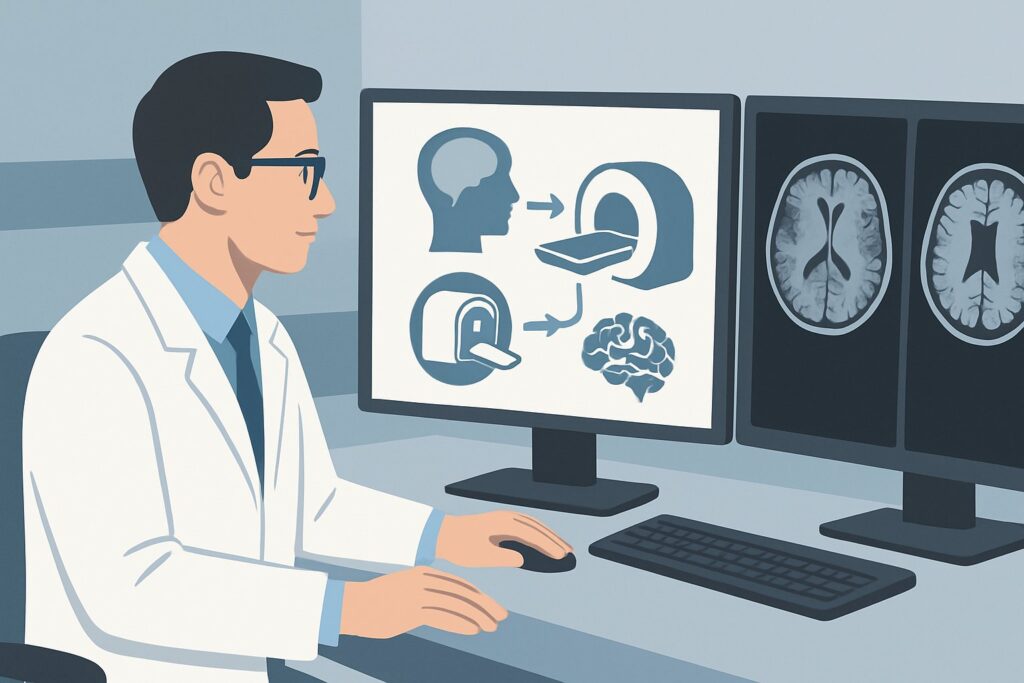
A 68-year-old patient is brought to your clinic by their family, who report a two-year history of insidious, progressive memory loss. The patient repeats questions, frequently misplaces items, and recently got lost driving home from the grocery store. The neurologic exam is otherwise nonfocal, and the clinical picture is highly suggestive of typical Alzheimer disease. You need to initiate a diagnostic workup, starting with neuroimaging. The central question is which initial study will provide the most diagnostic value while excluding critical mimics. According to the American College of Radiology (ACR) Appropriateness Criteria, for this specific presentation, an MRI head without IV contrast is rated Usually appropriate and is the recommended first step.
Who Fits This Clinical Scenario for Suspected Alzheimer’s?
This guidance applies specifically to adult patients presenting with cognitive impairment characterized by a primary and progressive memory deficit. The clinical history should be consistent with a typical, amnestic-predominant Alzheimer disease (AD) phenotype: an insidious onset over months to years and a gradual worsening that interferes with instrumental or basic activities of daily living. The goal of initial imaging in this context is twofold: to identify supportive evidence for AD and, critically, to rule out other structural or vascular causes for the cognitive decline.
This workflow is distinct from several related but different clinical situations:
- Atypical Presentations: If the patient’s primary symptoms are not memory loss but rather early and prominent behavioral changes, executive dysfunction, or progressive language deficits (aphasia), the differential diagnosis shifts towards frontotemporal dementia (FTD). This requires a different diagnostic approach.
- Prominent Parkinsonism or Hallucinations: When parkinsonian motor symptoms or well-formed visual hallucinations are early and core features, the workup should be tailored to suspect dementia with Lewy bodies (DLB).
- Mild Cognitive Impairment (MCI): If the patient has measurable memory deficits but is still functioning independently in their daily life, they may meet the criteria for MCI, not dementia. While the imaging choice may be similar, the clinical context and prognosis are different.
- Rapidly Progressive Dementia: A cognitive decline that unfolds over weeks to months is a neurologic emergency and suggests a different differential, including prion disease or autoimmune encephalitis, which may necessitate different imaging protocols.
What Diagnoses Are You Working Up in This Scenario?
While Alzheimer disease is the leading suspicion, the initial imaging study is crucial for evaluating a broader differential. The goal is to exclude other potentially treatable or co-existing conditions that can mimic or contribute to a dementia syndrome.
Alzheimer Disease (AD): As the most common cause of dementia, this is the primary diagnosis under consideration. Neuroimaging is not used to definitively diagnose AD but to find supportive evidence. The classic finding is a pattern of focal atrophy, particularly in the medial temporal lobes (including the hippocampi and entorhinal cortex), as well as the posterior cingulate gyrus and precuneus.
Vascular Dementia: Cerebrovascular disease is a frequent cause of cognitive impairment, either on its own or as a co-existing pathology with AD (mixed dementia). Imaging is essential to identify evidence of prior cortical or subcortical infarcts, extensive white matter ischemic changes (leukoaraiosis), or cerebral microbleeds that could explain or contribute to the patient’s symptoms.
Normal Pressure Hydrocephalus (NPH): This is a critical, treatable mimic of other dementias. While the full clinical triad includes gait disturbance and urinary incontinence, these may not be prominent early on. Imaging can reveal ventriculomegaly that is out of proportion to the degree of sulcal widening, a key finding that should prompt further investigation and a potential neurosurgical referral.
Structural Lesions: Though less common, it is imperative to rule out structural causes that can present with progressive cognitive decline. These include slow-growing brain tumors (e.g., meningioma, low-grade glioma), chronic subdural hematomas (especially in older adults with a history of falls), and the sequelae of prior head trauma.
Why Is MRI Head without IV Contrast the Recommended Initial Study?
For a patient with suspected typical Alzheimer disease, an MRI of the head without intravenous contrast is the most effective initial imaging test. Its high soft-tissue resolution and lack of ionizing radiation make it superior to other modalities for answering the key clinical questions in this scenario.
The primary strength of MRI is its ability to visualize brain structure in exquisite detail. It is highly sensitive for detecting the specific patterns of regional brain atrophy that support a diagnosis of AD, particularly volume loss in the medial temporal lobes. Standard dementia protocols include high-resolution T1-weighted images acquired in the coronal plane, perpendicular to the hippocampus, to optimize assessment of this key region. Furthermore, sequences like Fluid-Attenuated Inversion Recovery (FLAIR) are excellent for evaluating the burden of white matter ischemic disease, and gradient-echo or susceptibility-weighted imaging (SWI) can detect microhemorrhages associated with amyloid angiopathy or hypertensive vasculopathy.
The ACR rates alternative studies as less appropriate for this initial workup:
- CT head without IV contrast is rated May be appropriate. It serves as a reasonable alternative when MRI is unavailable or contraindicated (e.g., patient with an incompatible implanted device, severe claustrophobia). CT is effective at ruling out major structural causes like large tumors, hydrocephalus, or significant strokes. However, its inferior soft-tissue contrast makes it much less sensitive for detecting subtle atrophy patterns and the full extent of small vessel ischemic disease.
- MRI head without and with IV contrast is rated Usually not appropriate. In the routine evaluation of a typical dementia presentation, gadolinium-based contrast agents add cost, scan time, and a small risk of adverse reaction without providing diagnostically useful information. Contrast is not needed to assess for atrophy or chronic vascular changes. Its use should be reserved for cases where the clinical picture or initial non-contrast findings raise suspicion for an active inflammatory, infectious, or neoplastic process.
From a safety perspective, MRI is the preferred modality as it involves no ionizing radiation (0 mSv). This compares favorably to CT, which involves a radiation dose of 1-10 mSv.
What’s Next After the MRI? Downstream Workflow
The results of the non-contrast head MRI will guide the subsequent steps in the patient’s diagnostic journey and management plan. The report should be interpreted in the context of the patient’s age and clinical presentation.
- Findings Supportive of Alzheimer Disease: If the MRI shows medial temporal lobe atrophy and a pattern of posterior-predominant cortical volume loss, with minimal vascular disease, this evidence supports the clinical diagnosis of AD. The next steps are primarily clinical: counseling the patient and family, initiating symptomatic therapies (e.g., cholinesterase inhibitors), and addressing safety and long-term care planning.
- Findings Suggestive of Vascular Dementia: If the MRI reveals a significant burden of cerebrovascular disease (e.g., multiple cortical infarcts, severe white matter disease) sufficient to explain the cognitive deficits, the diagnosis may shift to vascular dementia. Management will then focus on aggressive secondary stroke prevention, including management of hypertension, diabetes, and hyperlipidemia.
- Findings Suggestive of Other Pathologies: If the MRI identifies evidence of normal pressure hydrocephalus, a tumor, or a chronic subdural hematoma, the workflow shifts dramatically. This requires an urgent referral to neurosurgery for further evaluation and potential intervention.
- A “Normal” or Nonspecific MRI: An MRI that is normal for the patient’s age or shows only mild, nonspecific global atrophy does not rule out a neurodegenerative process like early AD. In such cases, or if the diagnosis remains uncertain, more advanced imaging may be considered. FDG-PET/CT brain or Amyloid PET/CT brain, both rated Usually appropriate, can provide functional or molecular information to increase diagnostic certainty, though access and coverage for these studies can be variable.
Pitfalls to Avoid (and When to Get Help)
Navigating the initial dementia workup requires careful attention to the specific clinical context. Here are a few common pitfalls to avoid:
- Over-reliance on a “Normal” Report: A radiology report describing “age-appropriate atrophy” does not exclude a neurodegenerative disease. The imaging findings must be correlated with the clinical severity of the patient’s symptoms.
- Ordering Contrast by Default: Unnecessarily adding IV contrast to the initial MRI for a typical AD presentation adds cost and potential risk without benefit. Reserve it for atypical cases or specific concerns.
- Dismissing Vascular Changes: Do not underestimate the cognitive impact of small vessel ischemic disease. Even in a patient with suspected AD, a high burden of white matter disease may indicate a mixed dementia, which has implications for management.
- Not Specifying the Indication: When ordering the MRI, clearly state “evaluation for cognitive impairment” or “dementia workup.” This helps the radiology department select the most appropriate imaging protocol, including the crucial high-resolution coronal sequences of the temporal lobes.
If the clinical picture and imaging results are discordant, or if the presentation is atypical, consider a referral to a neurologist or a specialized memory disorders clinic for a more comprehensive evaluation.
Related ACR Topics and Tools
This article focuses on a single, common clinical scenario. For a broader overview of imaging for all types of dementia and related cognitive disorders, please see our parent guide. For additional resources to help select and understand imaging studies, the following tools are available.
- For breadth across all scenarios in Dementia, see our parent guide: Dementia: ACR Appropriateness Decoded.
- ACR Appropriateness Criteria Lookup — for adjacent scenarios
- Imaging Protocol Library — for technique on the recommended study
- Radiation Dose Calculator — for cumulative dose conversations
Frequently Asked Questions
Why not start with a CT scan, which is faster and more accessible?
A CT scan is rated ‘May be appropriate’ and is a valid alternative if MRI is contraindicated or unavailable. It can effectively rule out major structural problems like tumors, hydrocephalus, or large strokes. However, MRI is superior for detecting the subtle patterns of brain atrophy characteristic of Alzheimer disease and for visualizing the full extent of small vessel ischemic disease, providing more diagnostic information for the same clinical question.
Does a normal MRI rule out Alzheimer disease?
No. In the early stages of Alzheimer disease, the brain MRI may appear normal or show only mild, nonspecific changes often described as ‘age-appropriate atrophy.’ A normal MRI is valuable for excluding other causes of dementia, but the diagnosis of Alzheimer disease remains a clinical one, supported by imaging and other biomarkers.
When should I consider a PET scan for a patient with suspected Alzheimer’s?
FDG-PET or Amyloid PET scans are rated ‘Usually appropriate’ but are typically considered second-line tests. They are most useful when the diagnosis remains uncertain after a thorough clinical evaluation and structural MRI. For example, if a patient has an atypical presentation or if it’s difficult to distinguish between Alzheimer disease and frontotemporal dementia, a PET scan showing patterns of hypometabolism (FDG) or amyloid plaque burden can increase diagnostic certainty.
Is intravenous contrast ever needed in a dementia workup?
For the initial evaluation of a typical, slowly progressive dementia like suspected Alzheimer disease, IV contrast is rated ‘Usually not appropriate.’ However, contrast should be used if the clinical scenario suggests an inflammatory process (e.g., autoimmune encephalitis), infection, or if there is a concern for a primary or metastatic brain tumor, especially in a patient with a known history of cancer or a rapidly progressing course.
What specific sequences should I ensure are included in the MRI protocol?
While you don’t need to specify every sequence, it’s helpful to provide the clinical indication ‘dementia evaluation.’ This prompts the radiology team to use a dedicated protocol that typically includes: 1) High-resolution T1-weighted images, including thin coronal slices through the temporal lobes to assess hippocampal volume; 2) FLAIR sequences to evaluate white matter disease; and 3) A susceptibility-weighted or gradient-echo sequence to screen for microhemorrhages.
Reviewed by Pouyan Golshani, MD, Interventional Radiologist — May 30, 2026