Is Imaging Needed for Primary Hypothyroidism? An ACR-Guided Workflow
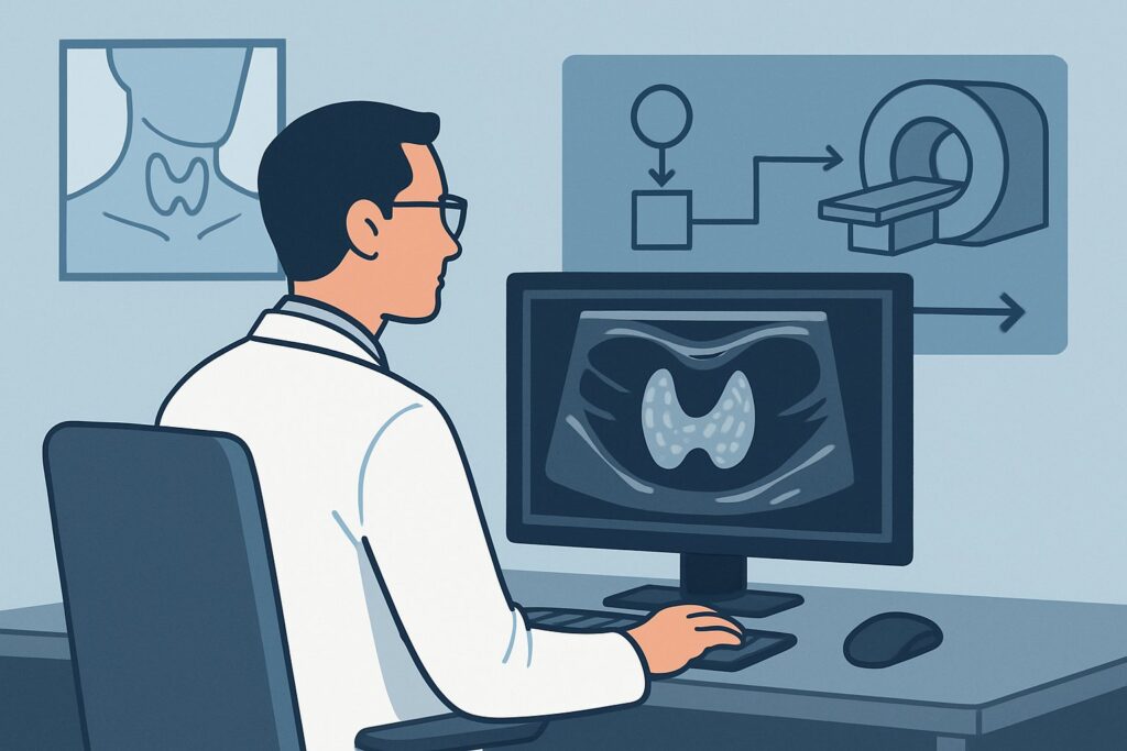
A 48-year-old woman presents to your primary care clinic with several months of fatigue, weight gain, and cold intolerance. Her physical exam is unremarkable, with no palpable thyroid nodules or visible goiter. You suspect hypothyroidism and order labs, which return showing an elevated Thyroid-Stimulating Hormone (TSH) and a low free thyroxine (T4), confirming primary hypothyroidism. As you prepare to start treatment, you pause: should you order an ultrasound of her thyroid to establish a baseline or look for an underlying cause? For this specific clinical scenario—initial imaging for primary hypothyroidism—the American College of Radiology (ACR) finds that imaging studies, including thyroid ultrasound, are Usually not appropriate. This article explains the evidence-based rationale for this recommendation and outlines the correct, non-imaging-based workflow.
Who Fits This Clinical Scenario?
This guidance applies to adult and pediatric patients with a new, biochemically confirmed diagnosis of primary hypothyroidism. The key inclusion criteria are:
- Laboratory evidence of primary hypothyroidism (e.g., elevated TSH with low or normal free T4).
- The clinical question is the initial evaluation of this lab finding.
- The patient has no specific physical exam findings that would trigger a separate imaging workup.
It is crucial to distinguish this scenario from related but distinct clinical presentations where imaging is, in fact, indicated. This guidance does not apply if the patient also presents with:
- A palpable thyroid nodule: This presentation routes to a different ACR variant where ultrasound is the primary recommended study.
- A suspected or visible goiter: A clinically apparent goiter also warrants an initial ultrasound to assess its size, structure, and the presence of any dominant nodules.
- Symptoms of thyrotoxicosis: Patients with low TSH and high thyroid hormone levels require a different workup, often involving a radionuclide uptake scan.
This article is exclusively for the common situation where primary hypothyroidism is discovered via lab testing in a patient without concerning neck exam findings.
What Diagnoses Are You Working Up in This Scenario?
When a patient has primary hypothyroidism, the diagnostic question is not if they have it—the labs have already confirmed that—but why. The differential diagnosis for the underlying cause is addressed through history, physical exam, and further serologic testing, not imaging. The goal of the workup is to identify the etiology to guide long-term management and counseling.
Hashimoto’s Thyroiditis: This is the most common cause of hypothyroidism in iodine-sufficient regions of the world. It is an autoimmune disorder where the body’s immune system attacks the thyroid gland, leading to chronic inflammation and a gradual decline in function. The diagnosis is typically confirmed by the presence of anti-thyroid peroxidase (TPO) antibodies in the blood.
Iatrogenic Hypothyroidism: This category includes hypothyroidism resulting from medical treatment. Common causes are prior radioactive iodine therapy for Graves’ disease, external beam radiation to the neck for cancers like lymphoma, or surgical removal of the thyroid gland (thyroidectomy).
Drug-Induced Hypothyroidism: Certain medications can interfere with thyroid hormone production or metabolism. Amiodarone, lithium, and newer checkpoint inhibitor cancer therapies are well-known culprits. A thorough medication history is key to identifying this cause.
Congenital Hypothyroidism: While typically identified through newborn screening, this remains a consideration, especially in pediatric populations. It results from abnormal thyroid gland development (dysgenesis) or a defect in hormone synthesis (dyshormonogenesis).
Why Is Imaging Usually Not Appropriate for This Presentation?
For the initial evaluation of uncomplicated primary hypothyroidism, the ACR rates all imaging modalities as Usually not appropriate. The diagnosis is biochemical, and imaging does not alter the initial management, which is thyroid hormone replacement. Introducing imaging at this stage can lead to a cascade of unnecessary tests and procedures without benefiting the patient.
The core rationale is that the findings on an ultrasound, while they might show features consistent with Hashimoto’s (e.g., diffuse heterogeneity, micronodulation), do not change the treatment plan. The presence of anti-TPO antibodies is a more direct and cost-effective way to confirm autoimmune thyroiditis. Furthermore, routine ultrasound risks the discovery of incidental, non-palpable thyroid nodules. These “incidentalomas” are extremely common in the general population, overwhelmingly benign, and their discovery often triggers patient anxiety and a workup involving further imaging and potentially fine-needle aspiration (FNA) biopsy, which would not have been indicated otherwise.
Alternative cross-sectional imaging modalities are even less appropriate and carry additional risks:
- CT neck with IV contrast is rated Usually not appropriate. It provides poor detail of the thyroid parenchyma compared to ultrasound and exposes the patient to ionizing radiation (1-10 mSv) and iodinated contrast, which can itself interfere with thyroid function.
- Nuclear medicine scans, such as an I-123 uptake scan, are also rated Usually not appropriate. These studies are used to evaluate thyroid function (hyperthyroidism vs. thyroiditis) and are not indicated in a clear case of primary hypothyroidism. They also involve radiation exposure.
While imaging is not indicated for the initial diagnosis, if a thyroid ultrasound is performed for a separate, valid indication (such as a newly palpated nodule), understanding the proper technique is crucial. Once you’ve decided on US thyroid for an appropriate reason, our protocol guide covers the technique, reporting standards, and reading principles: US Thyroid.
What’s Next? The Downstream Workflow
The correct downstream workflow after a new diagnosis of primary hypothyroidism is clinical and biochemical, not radiological. The steps are focused on treatment and monitoring.
If the diagnosis is confirmed by labs (high TSH, low free T4):
The next step is to initiate treatment with levothyroxine. The starting dose is based on patient weight, age, and comorbidities. You may also order anti-TPO antibody testing to confirm or rule out Hashimoto’s thyroiditis as the underlying cause, which can be useful for patient counseling and prognosis.
After starting treatment:
The primary follow-up is to recheck the TSH level in 6-8 weeks to assess the adequacy of the hormone replacement dose and titrate as needed. The goal is to normalize the TSH level and resolve the patient’s symptoms.
If a new physical finding develops:
If, during follow-up, the patient develops a palpable neck mass or a distinct nodule, the clinical scenario changes. At that point, you would pivot to the workflow for a palpable thyroid nodule, for which a thyroid ultrasound becomes Usually appropriate.
Pitfalls to Avoid (and When to Get Help)
In managing primary hypothyroidism, the most significant pitfall is ordering reflexive, low-yield imaging. Here are key errors to avoid:
- Ordering a “baseline” ultrasound: This is not supported by evidence and initiates the risk of the incidentaloma cascade.
- Using imaging to diagnose Hashimoto’s: Serologic testing (anti-TPO antibodies) is the more direct and appropriate method.
- Ignoring a palpable finding: Do not let the “no imaging for hypothyroidism” rule prevent you from imaging a palpable nodule or goiter, which are distinct and valid indications.
- Misinterpreting lab results: Ensure the diagnosis is truly primary hypothyroidism. Subclinical hypothyroidism or central hypothyroidism have different management pathways.
If the clinical picture is complex, such as a patient with a rapidly enlarging or painful thyroid gland, or if they fail to respond biochemically to standard levothyroxine therapy, escalation to an endocrinologist is the appropriate next step.
Related ACR Topics and Tools
This article focuses on a single clinical scenario. For a comprehensive overview of imaging across all common thyroid-related presentations, from nodules to cancer surveillance, please see our parent guide. It provides a hub-and-spoke model to help you find the specific variant that matches your patient.
- For breadth across all scenarios in Thyroid Disease, see our parent guide: Thyroid Disease: ACR Appropriateness Decoded.
- To explore other clinical scenarios and their corresponding ACR ratings, use the ACR Appropriateness Criteria Lookup.
- To review technical details for hundreds of imaging studies, visit the Imaging Protocol Library.
- For discussions about radiation exposure with your patients, consult the Radiation Dose Calculator.
Frequently Asked Questions
My patient with new hypothyroidism has a palpable goiter. Should I still not order imaging?
No, in that case you should order imaging. A palpable goiter is a distinct clinical finding that warrants evaluation. That presentation falls under the ACR scenario ‘Suspected goiter,’ for which a thyroid ultrasound is ‘Usually appropriate’ to assess the size and character of the gland and to look for any underlying nodules.
What if the patient’s anti-TPO antibodies are negative? Does that mean I need an ultrasound?
Not for the initial diagnosis. The diagnosis of primary hypothyroidism remains biochemical (high TSH, low free T4). While most cases of Hashimoto’s are seropositive, seronegative autoimmune thyroiditis exists. Other causes, like post-viral thyroiditis or drug-induced hypothyroidism, are also not diagnosed via imaging. The management—thyroid hormone replacement—remains the same regardless of the ultrasound findings.
Is there any role for CT or MRI in evaluating primary hypothyroidism?
No, not for the initial diagnosis. The ACR rates both CT and MRI of the neck as ‘Usually not appropriate’ for this specific scenario. They offer less detail of the thyroid tissue than ultrasound, are more expensive, and in the case of CT, expose the patient to unnecessary ionizing radiation. Their use is reserved for complex cases, such as evaluating a large goiter with substernal extension or suspicion of invasive cancer, which are entirely different clinical questions.
Why is it a pitfall to order an ultrasound ‘just to be safe’?
Ordering an ultrasound without a clear indication like a palpable nodule can trigger a ‘diagnostic cascade.’ Thyroid incidentalomas are found in up to 68% of adults on ultrasound. While the vast majority are benign, their discovery often leads to patient anxiety, follow-up imaging, and potentially unnecessary biopsies, all of which carry their own costs and risks without improving the outcome for the initial diagnosis of hypothyroidism.
If I don’t order imaging, how do I diagnose the cause, like Hashimoto’s?
The cause of primary hypothyroidism is determined primarily through the patient’s history and serologic (blood) tests. A history of neck radiation or certain medication use can point to iatrogenic causes. For diagnosing Hashimoto’s thyroiditis, the key test is measuring anti-thyroid peroxidase (TPO) antibodies. A high level of these antibodies is highly specific for autoimmune thyroid disease.
Reviewed by Pouyan Golshani, MD, Interventional Radiologist — May 29, 2026