What Imaging Should You Order for Neck Pain After Cervical Spine Surgery?
A 58-year-old patient, two years post-anterior cervical discectomy and fusion (ACDF) at C5-C6, presents to your clinic. He was doing well until a month ago and now reports increasing, achy neck pain with new tingling radiating down his right arm, mimicking his pre-operative symptoms. He denies any recent trauma. You suspect a hardware issue or adjacent segment disease, but where do you start the workup? This scenario—recurrent or new symptoms after cervical surgery without red flags—requires a specific imaging strategy to safely and effectively evaluate both the surgical hardware and the surrounding anatomy. According to the American College of Radiology (ACR) Appropriateness Criteria, the initial investigation of choice is Radiography cervical spine, a modality rated as Usually Appropriate.
Who Fits This Clinical Scenario for Post-Surgical Cervical Pain?
This clinical workflow is designed for a specific patient population: adults with a history of cervical spine surgery who now present with new, increasing, or changing mechanical neck pain or radiculopathy. The prior surgery could be an ACDF, posterior fusion, laminectomy, or laminoplasty. The key is the presence of surgical hardware or altered anatomy from a previous intervention.
This guidance applies only when there are no “red flag” symptoms. It is crucial to distinguish this scenario from others that require a different, often more urgent, workup. This workflow does not apply if the patient presents with:
- Significant recent trauma: A fall or motor vehicle collision in a post-surgical patient warrants a different imaging protocol, often starting with CT to rule out acute fracture.
- Signs of infection: If the patient has fever, chills, night sweats, or localized erythema and warmth over the surgical site, the primary concern shifts to abscess or osteomyelitis. This routes to a different ACR variant, where MRI with contrast is often the first-line study.
- Suspicion of malignancy: A known history of cancer, unexplained weight loss, or pain that is worse at night should raise concern for metastatic disease, which also requires a distinct imaging pathway.
- Surgery-naïve presentation: Patients experiencing their first episode of cervical pain or radiculopathy without a history of surgery follow a separate, less complex diagnostic algorithm.
What Diagnoses Are You Working Up in a Patient with Prior Cervical Surgery?
In the post-operative cervical spine, the differential diagnosis for recurrent pain or radiculopathy is broader than in a surgery-naïve patient. Your initial imaging choice is aimed at evaluating several key possibilities.
The most immediate concern is often a hardware-related complication. This includes screw loosening (which may appear as a subtle lucency around the implant), hardware fracture, or migration of the cage or plate. These issues can lead to instability and direct neural compression, requiring prompt surgical evaluation.
A highly common long-term issue is adjacent segment disease (ASD). When one or more cervical levels are fused, the biomechanical stress is transferred to the vertebral levels immediately above and below the fusion. This can accelerate degenerative changes, leading to new disc herniations, spinal stenosis, or spondylolisthesis at these adjacent levels, causing a recurrence of symptoms years after a successful surgery.
Another important consideration is pseudoarthrosis, or failed fusion. This occurs when the intended bony bridge fails to form between the vertebral bodies, resulting in persistent micromotion at the surgical level. This instability is a common cause of chronic, persistent mechanical neck pain after fusion procedures.
Finally, you must consider recurrent or residual stenosis. The original pathology may not have been fully decompressed, or scar tissue (epidural fibrosis) may have formed, tethering or compressing the nerve roots. While less common, these remain important considerations in the diagnostic workup.
Why Are Cervical Spine Radiographs the Recommended First Step After Surgery?
For this specific scenario, the ACR panel designates four studies as Usually Appropriate: Radiography, Radiography with flexion-extension views, MRI without contrast, and CT without contrast. However, standard radiographs serve as the ideal starting point due to their unique strengths in the post-operative setting.
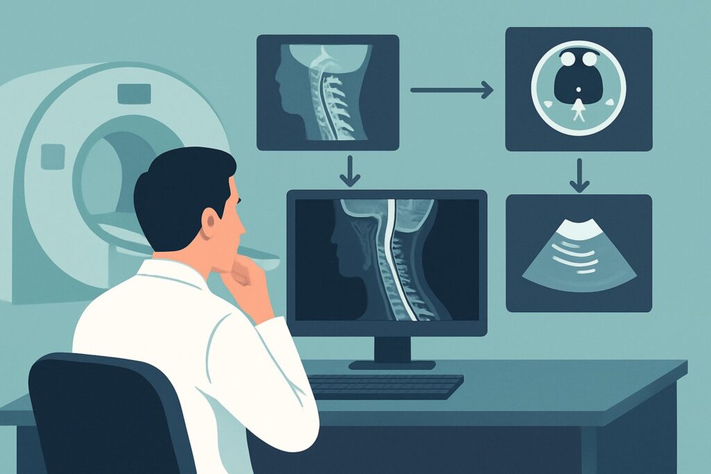
The primary rationale is that Radiography cervical spine is the most effective initial tool for assessing hardware integrity and spinal alignment. Anteroposterior (AP) and lateral views can quickly identify gross hardware failure like a broken screw, plate displacement, or significant subsidence of an interbody cage. It provides a clear overview of the construct’s relationship to the native spine. Adding Radiography cervical spine flexion extension lateral views is also Usually Appropriate and provides critical functional information, revealing instability that may indicate pseudoarthrosis or adjacent segment ligamentous laxity. This modality is fast, widely available, and delivers a low radiation dose (Relative Radiation Level ☢☢, 0.1-1 mSv).
While also highly rated, other modalities are typically used as second-line studies based on the radiographic findings or persistent clinical suspicion:
- CT cervical spine without IV contrast: This study is also Usually Appropriate and is superior to radiographs for definitively assessing bony fusion. If radiographs are suspicious for pseudoarthrosis, a non-contrast CT with thin cuts is the gold standard for confirming whether a solid bony bridge has formed. However, it imparts a higher radiation dose (RRL ☢☢☢, 1-10 mSv) and is best reserved for answering this specific question.
- MRI cervical spine without IV contrast: Also rated Usually Appropriate, MRI is the best modality for evaluating soft tissues: the spinal cord, nerve roots, and intervertebral discs. It is the test of choice for diagnosing recurrent disc herniation or stenosis, particularly at adjacent segments. However, the metallic hardware creates significant susceptibility artifact that can obscure the anatomy at the surgical level itself, making it less reliable than radiographs or CT for evaluating the hardware-bone interface. It is often the logical next step if radiographs are normal but radicular symptoms persist.
- MRI cervical spine without and with IV contrast: This is rated May be appropriate. The addition of gadolinium contrast is not necessary for evaluating mechanical or degenerative causes of pain. Its use is reserved for cases where there is a specific concern for infection, inflammation, or tumor—conditions explicitly excluded from this clinical scenario.
What Is the Downstream Workflow After Initial Cervical Radiographs?
The results of the initial cervical spine radiographs will guide your next steps in a branching decision tree.
- If radiographs show definitive hardware failure: In cases of a fractured screw, plate migration, or obvious loosening, the diagnosis is clear. The immediate next step is a referral back to the operating surgeon or a spine specialist for evaluation and potential revision surgery. Pre-operative CT is often obtained for surgical planning but the clinical path is set.
- If radiographs are negative but symptoms persist: A normal radiograph does not rule out significant pathology. The next study depends on the dominant symptom.
- For persistent radiculopathy (arm pain, numbness, tingling), the primary concern is neural compression. The next step is an MRI cervical spine without IV contrast to directly visualize the nerve roots, spinal cord, and discs, with a focus on the adjacent segments.
- For persistent mechanical neck pain, especially if pseudoarthrosis is suspected, the next step is a CT cervical spine without IV contrast. This will provide a definitive assessment of the bony fusion.
- If flexion-extension radiographs show instability: Dynamic views revealing abnormal motion at the surgical site or an adjacent level indicate instability. This finding, even with otherwise normal static films, warrants a surgical consultation.
Pitfalls to Avoid (and When to Get Help)
Navigating the workup for post-surgical neck pain requires careful attention to detail to avoid common missteps.
A primary pitfall is proceeding directly to MRI without initial radiographs. While MRI is excellent for soft tissues, the artifact from hardware can obscure the very region of interest, and you may miss a clear hardware failure that would be obvious on a simple X-ray.
Another error is underutilizing flexion-extension views. In a patient with mechanical pain and normal static radiographs, these dynamic views are essential for unmasking instability from pseudoarthrosis or adjacent segment disease, which is a common cause of persistent symptoms.
Finally, be cautious not to attribute all symptoms to the prior surgery. New-onset pain could be unrelated, and a thorough history and physical exam are critical to avoid anchoring bias. If any red flag symptoms emerge during the workup, such as progressive myelopathy or signs of infection, escalate care immediately with an urgent surgical or specialty consultation.
Related ACR Topics and Tools
For a comprehensive overview of imaging for all cervical pain scenarios, from acute trauma to chronic pain in surgery-naïve patients, please see our parent guide. For tools to assist in ordering and interpreting these studies, the following resources are available.
- Parent Topic Hub Article: Cervical Pain or Cervical Radiculopathy: ACR Appropriateness Decoded
- ACR Appropriateness Criteria Lookup: ACR Appropriateness Criteria Lookup
- Imaging Protocol Library: Imaging Protocol Library
- Radiation Dose Calculator: Radiation Dose Calculator
Frequently Asked Questions
Why not start with an MRI for every post-surgical patient with radiculopathy?
While MRI is excellent for visualizing nerves, metallic hardware from the surgery creates significant signal artifact that can obscure the anatomy at and immediately around the surgical site. A simple radiograph is better for the initial assessment of hardware position and integrity. If the radiograph is normal, an MRI is often the appropriate next step to evaluate for soft tissue causes of radiculopathy, like a new disc herniation at an adjacent level.
What if the patient has a contraindication to MRI, like a non-compatible device?
If an MRI is contraindicated, a CT myelogram is rated as ‘May be appropriate’ by the ACR. This involves injecting intrathecal contrast followed by a CT scan. It provides excellent visualization of the spinal canal and nerve roots and can effectively diagnose stenosis or nerve compression, serving as a powerful alternative to MRI for evaluating the neural elements.
How soon after surgery can flexion-extension radiographs be safely performed?
The timing for obtaining flexion-extension views depends on the surgeon’s protocol and the type of surgery performed. Generally, they are deferred until there is radiographic evidence that the fusion is maturing, typically at least 3 to 6 months post-operatively. Ordering them too early risks disrupting the healing fusion. It is best to consult with the operating surgeon if there is any uncertainty.
Does the type of prior surgery (e.g., ACDF vs. posterior fusion) change the initial imaging choice?
No, for this specific clinical scenario, the initial imaging choice of cervical spine radiography remains the same regardless of whether the surgical approach was anterior or posterior. Radiographs are effective at evaluating alignment and the integrity of plates, screws, rods, and cages used in both types of procedures.
Is a CT scan always necessary to diagnose pseudoarthrosis (failed fusion)?
While CT is the gold standard for assessing bony fusion, it is not always necessary. Dynamic flexion-extension radiographs showing clear motion at the surgical level can be sufficient to diagnose an unstable pseudoarthrosis. However, if the dynamic films are equivocal or if a surgeon requires definitive confirmation of the lack of a bony bridge for pre-operative planning, a non-contrast CT is the most accurate test.
Reviewed by Pouyan Golshani, MD, Interventional Radiologist — May 30, 2026