Which Imaging Study Is Best for Isolated Accessory Nerve (CN XI) Palsy?

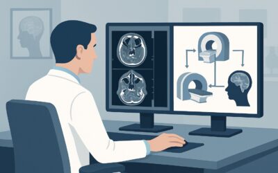
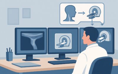
A 62-year-old patient presents to your clinic with a two-month history of a painless “drooping” of his right shoulder and new difficulty turning his head against resistance to the left. On examination, you note atrophy and weakness of the right trapezius and sternocleidomastoid (SCM) muscles, with no other neurologic deficits. The presentation is classic for an isolated right accessory nerve (cranial nerve XI) palsy. The immediate clinical question is how to investigate the cause, which requires visualizing the nerve’s entire, lengthy course from the brainstem to the neck. According to the American College of Radiology (ACR) Appropriateness Criteria, the initial imaging study for this scenario, MRI head without and with IV contrast, is rated Usually Appropriate.
Who Fits This Clinical Scenario for an Accessory Nerve Palsy Workup?
This diagnostic workflow is specifically for patients presenting with new, unilateral, and isolated weakness or paralysis of the sternocleidomastoid and trapezius muscles. The key term is “isolated.” The patient’s history and physical exam should not suggest involvement of other cranial nerves or neurologic systems. This focused presentation points to a lesion affecting the spinal accessory nerve (CN XI) somewhere along its path.
This guidance does not apply if the patient presents with additional symptoms, which would suggest a different localization and require a different imaging strategy. Key exclusion criteria include:
- Multiple Cranial Neuropathies: If the patient also has facial weakness (CN VII), hearing loss (CN VIII), or difficulty swallowing (CN IX, X), the pathology is more likely at the skull base (e.g., jugular foramen syndrome) or within the brainstem, and a broader imaging protocol is needed.
- Long Tract Signs: The presence of hemiparesis, sensory loss below the neck, or hyperreflexia indicates a central nervous system lesion (e.g., brainstem stroke, spinal cord process) rather than an isolated peripheral nerve palsy.
- Known Recent Trauma or Surgery: While imaging is still often performed, a clear history of a radical neck dissection, cervical lymph node biopsy, or penetrating neck trauma makes iatrogenic or traumatic injury the leading diagnosis, which can influence the imaging interpretation and subsequent management.
What Diagnoses Are You Working Up in This Scenario?
The accessory nerve has a long and complex path, originating from both the brainstem (cranial root) and the upper spinal cord (spinal root), ascending into the skull through the foramen magnum, and then exiting through the jugular foramen to innervate the SCM and trapezius muscles in the neck. A lesion anywhere along this course can cause a palsy. Imaging is crucial for identifying a structural cause.
The differential diagnosis includes:
Skull Base and Neck Tumors: This is a primary concern. Benign tumors like schwannomas (specifically jugular foramen schwannomas) or meningiomas can compress the nerve as it exits the skull. Malignant processes are also a key consideration, particularly metastatic disease from head and neck squamous cell carcinoma, lung cancer, or breast cancer, which can manifest as skull base metastases or pathologic lymph nodes in the neck that invade or compress the nerve. Glomus jugulare tumors (paragangliomas) are a less common but important cause of jugular foramen syndromes.
Iatrogenic Injury: The accessory nerve’s superficial course in the posterior cervical triangle makes it highly vulnerable to injury during surgical procedures. It is one of the most commonly iatrogenically injured nerves. Procedures like cervical lymph node biopsy, radical neck dissection for cancer, and even carotid endarterectomy can lead to stretching, transection, or entrapment of the nerve. While the history is often clear, imaging may be used to rule out other concurrent pathology like tumor recurrence.
Trauma: Penetrating injuries to the neck (e.g., stab wounds) can directly sever the nerve. Blunt trauma or traction injuries, sometimes seen in sports or accidents, can also cause damage.
Idiopathic or Inflammatory Causes: In the absence of a structural lesion, an inflammatory process may be the cause. Neuralgic amyotrophy (Parsonage-Turner syndrome) can affect the accessory nerve, though it more commonly involves the brachial plexus. This is a diagnosis of exclusion made after imaging has ruled out a compressive lesion.
Why Is MRI Without and With IV Contrast the Recommended Study for This Presentation?
The ACR panel rates both MRI head without and with IV contrast and MRI orbits face neck without and with IV contrast as Usually Appropriate for an isolated CN XI palsy. The core rationale is MRI’s superior ability to visualize soft tissues, allowing for direct and indirect assessment of the nerve and the structures surrounding it.
The key advantages of an MRI-based approach for this specific scenario include:
- Comprehensive Anatomic Coverage: A dedicated MRI protocol can visualize the entire course of the accessory nerve, from its origins in the medulla and upper cervical spine, through the cisternal space, its exit at the jugular foramen, and its path through the posterior cervical triangle in the neck.
- Superior Soft-Tissue Resolution: MRI is unmatched in its ability to differentiate the nerve from adjacent muscles, vessels, and fat. It can detect subtle abnormalities like nerve thickening or intrinsic signal change, as well as small, compressive tumors that a CT scan might miss.
- Importance of IV Contrast: The administration of gadolinium-based contrast is critical. It helps identify pathologic enhancement within the nerve itself (suggesting inflammation or neoplastic infiltration) and is essential for characterizing tumors like schwannomas, meningiomas, and metastatic deposits, which typically enhance avidly. A non-contrast MRI is rated lower (May be appropriate) because it can easily miss these key diagnoses.
- Detection of Denervation: MRI can show secondary signs of nerve injury. Subacute-to-chronic denervation of the SCM and trapezius muscles will appear as T2-hyperintensity (edema) and later as fatty atrophy and volume loss. Seeing these changes confirms the clinical finding and helps localize the chronicity of the injury.
How do alternative studies compare?
- CT neck with IV contrast is also rated Usually Appropriate. It is an excellent alternative when MRI is contraindicated (e.g., incompatible implanted device, severe claustrophobia) or less available. CT provides superior visualization of the bony margins of the jugular foramen. However, its soft-tissue contrast is significantly lower than MRI, making it less sensitive for small tumors or direct nerve visualization. It also involves ionizing radiation (ACR Relative Radiation Level ☢☢☢), a consideration particularly in younger patients.
- US neck is rated Usually not appropriate. While ultrasound is useful for evaluating superficial neck masses or guiding a biopsy of a known lymph node, it cannot visualize the intracranial or skull base portion of the nerve. It provides an incomplete evaluation and cannot be used as a primary screening tool for this indication.
What’s Next After MRI? Downstream Workflow
The results of the MRI will guide the subsequent clinical pathway. The goal of imaging is to find a treatable structural cause.
- If the MRI identifies a tumor (e.g., schwannoma, meningioma, metastasis): The next step is a referral to the appropriate specialist. For benign tumors, this is typically a neurosurgeon or head and neck surgeon (otolaryngology) to discuss options like observation, stereotactic radiosurgery, or surgical resection. For suspected metastases, a referral to oncology is paramount, and a biopsy may be required to confirm the diagnosis and guide systemic therapy.
- If the MRI is negative for a structural lesion: The focus shifts away from a compressive cause. The next steps often include:
1. Revisiting the history for a missed traumatic or iatrogenic injury.
2. Electrodiagnostic testing (EMG/NCS) to confirm the presence and severity of the axonopathy, which can help determine prognosis for recovery.
3. Referral to physical and occupational therapy to manage the functional deficits, such as shoulder droop and scapular winging, and to prevent secondary complications like frozen shoulder.
4. Considering an idiopathic or inflammatory cause, such as Parsonage-Turner syndrome, which is managed supportively.
- If the MRI is indeterminate: An equivocal finding, such as subtle, non-specific enhancement, may warrant a short-interval follow-up MRI to assess for change. Consultation with a neuroradiologist can help determine if the findings are significant or if an alternative imaging modality, like a high-resolution CT of the skull base, could add value.
Pitfalls to Avoid (and When to Get Help)
When working up an isolated accessory nerve palsy, several common pitfalls can delay diagnosis or lead to incomplete evaluation.
- Ordering an incomplete study: Requesting an “MRI brain” without specifying the need to evaluate the skull base and neck is a frequent error. This may fail to visualize the extracranial course of the nerve, where much of the pathology lies. Be explicit in the order: “Evaluate for CN XI palsy, include skull base and full course of the nerve through the neck.”
- Omitting IV contrast: A non-contrast study is insufficient for this indication, as it will miss most of the enhancing lesions (tumors, inflammation) that are the primary targets of the investigation.
- Misattributing symptoms to musculoskeletal causes: Weakness of the trapezius can be mistaken for a primary shoulder problem (e.g., rotator cuff injury), delaying a neurologic workup. A careful neurologic exam demonstrating weakness in both the trapezius and SCM is key to making the correct diagnosis.
- Ignoring red flags: The new onset of a cranial neuropathy, especially in a patient with a known history of cancer, should be treated with high suspicion for metastatic disease until proven otherwise.
If the clinical picture is complex, involves multiple cranial nerves, or if imaging results are unclear, consultation with a neurologist or neuroradiologist is the appropriate next step.
Related ACR Topics and Tools
This article focuses on a single, specific clinical scenario. For a broader overview of imaging for all cranial neuropathies, from CN I to CN XII, please see our parent guide. For additional resources to help refine your imaging orders, the following tools are available.
- For breadth across all scenarios in Cranial Neuropathy, see our parent guide: Cranial Neuropathy: ACR Appropriateness Decoded.
- ACR Appropriateness Criteria Lookup — for adjacent and alternative clinical scenarios.
- Imaging Protocol Library — for detailed guidance on imaging techniques.
- Radiation Dose Calculator — for discussing cumulative radiation exposure with patients when considering CT.
Frequently Asked Questions
Why is an MRI of the head and neck recommended over just an MRI of the brain?
The accessory nerve (CN XI) has a long course that extends from the brainstem down into the neck. Pathology can occur anywhere along this path, including the skull base (jugular foramen) and the posterior cervical triangle. An MRI of the brain alone would miss extracranial causes like tumors or lymph nodes in the neck, which are common causes of an isolated CN XI palsy. A comprehensive study of the head and neck is required.
Is a CT scan an acceptable first choice for working up an accessory nerve palsy?
Yes, a CT of the neck with IV contrast is also rated as ‘Usually Appropriate’ by the ACR and is a good alternative, especially if MRI is contraindicated or unavailable. CT is excellent for evaluating the bony anatomy of the skull base but is less sensitive than MRI for detecting small soft-tissue tumors, nerve enhancement, or early muscle denervation changes. MRI is generally preferred for its superior soft-tissue contrast.
What if the MRI is completely normal?
A normal MRI is a crucial finding, as it effectively rules out a compressive structural lesion like a tumor. In this case, the focus should shift to non-structural causes. This includes a thorough review of the patient’s history for iatrogenic injury (prior surgery) or trauma. If none is found, the palsy may be idiopathic or inflammatory (e.g., Parsonage-Turner syndrome). Electrodiagnostic studies (EMG/NCS) are often the next step to confirm the diagnosis and assess prognosis.
Do I need to order an MRA or CTA for an isolated CN XI palsy?
No, for an isolated accessory nerve palsy without other symptoms, a standard MRA or CTA is rated ‘Usually not appropriate’ by the ACR. These studies are optimized for visualizing blood vessels to look for conditions like aneurysm or dissection, which are not typical causes of an isolated CN XI palsy. The recommended study is a contrast-enhanced MRI or CT focused on nerve and soft-tissue anatomy.
My patient has shoulder droop but can turn their head normally. Is this still a CN XI palsy?
It could be. The sternocleidomastoid (SCM) and trapezius muscles can be affected to different degrees. Isolated trapezius weakness is a common presentation of CN XI injury, as the branch to the trapezius is particularly vulnerable in the posterior neck. However, it is important to perform a full neurologic exam to ensure the SCM is truly spared and to rule out other causes of shoulder weakness, such as a brachial plexus or long thoracic nerve injury.
Reviewed by Pouyan Golshani, MD, Interventional Radiologist — May 29, 2026