Which Imaging Study Is Best for the Initial Workup of Parkinsonian Syndromes?
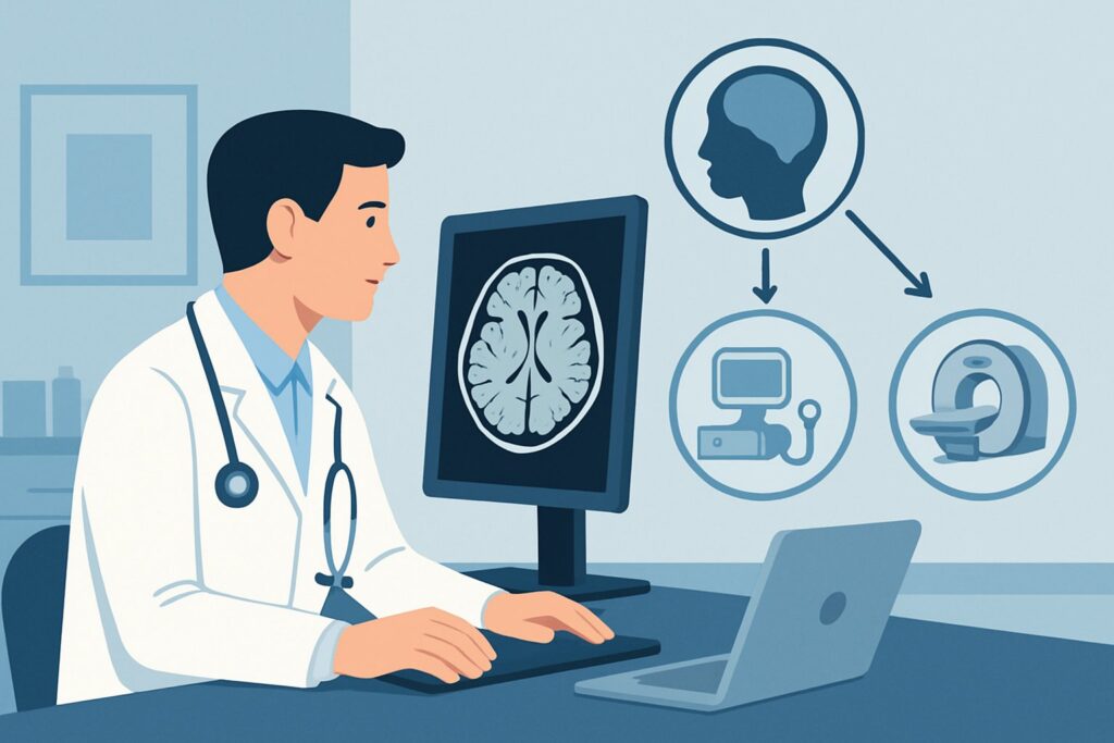
A 68-year-old patient presents to your clinic with a six-month history of a subtle resting tremor in his right hand, a general sense of slowness when getting up from a chair, and a feeling of stiffness in his limbs. His family notes his facial expression has become less animated. You suspect a parkinsonian syndrome and are planning the initial diagnostic workup. The central question is which imaging study, if any, is the most appropriate first step to clarify the diagnosis and rule out mimics. This article provides a detailed clinical workflow for this exact scenario, guided by the American College of Radiology (ACR) Appropriateness Criteria, which rate an MRI head without IV contrast as Usually Appropriate.
Who Fits This Clinical Scenario?
This guidance applies to patients presenting for an initial evaluation of a parkinsonian syndrome. The core clinical features include bradykinesia (slowness of movement) combined with either a resting tremor, rigidity, or both. While many of these patients will ultimately be diagnosed with Idiopathic Parkinson’s Disease (IPD), this workflow is particularly crucial for those with atypical features that raise suspicion for an alternative diagnosis.
Inclusion criteria for this workflow:
- Patients with newly diagnosed or suspected parkinsonism.
- Individuals with atypical features such as early-onset postural instability and falls, symmetric symptoms at onset, rapid progression, poor response to levodopa therapy, or early autonomic dysfunction.
- Cases where the clinical picture is ambiguous and structural brain pathology must be excluded.
It is critical to distinguish this presentation from nearby clinical scenarios that require a different diagnostic approach. This guidance does not apply to:
- Rapidly progressive dementia with myoclonus: This presentation is highly suspicious for Creutzfeldt-Jakob disease and follows a distinct imaging pathway.
- A primary complaint of chorea with a family history: This suggests Huntington disease, which has its own specific imaging considerations.
- Isolated upper or lower motor neuron signs: This clinical picture points toward motor neuron disease, where imaging focuses on the spinal cord and motor cortex.
What Diagnoses Are You Working Up in This Scenario?
The primary goal of initial imaging in parkinsonism is not to “diagnose” Idiopathic Parkinson’s Disease—which remains a clinical diagnosis—but to exclude other conditions and identify features that suggest an alternative cause. The differential diagnosis is broad.
Idiopathic Parkinson’s Disease (IPD) is the most common cause of parkinsonism. In early IPD, structural brain imaging like MRI is typically normal or may show age-appropriate cerebral atrophy. A normal MRI in the setting of a classic clinical presentation and a good response to dopaminergic therapy supports the diagnosis of IPD by excluding mimics.
Atypical Parkinsonian Syndromes (Parkinson-plus) are a group of neurodegenerative disorders that share features with IPD but have additional signs, a different prognosis, and often a poor response to standard therapies. Imaging can reveal characteristic patterns of atrophy. Key considerations include Multiple System Atrophy (MSA), which may show atrophy of the putamen, middle cerebellar peduncles, or pons (the “hot cross bun” sign); Progressive Supranuclear Palsy (PSP), classically associated with midbrain atrophy (the “hummingbird” sign on sagittal views); and Corticobasal Degeneration (CBD), which can cause marked asymmetric cortical atrophy.
Secondary Parkinsonism refers to parkinsonian symptoms caused by another identifiable condition. Imaging is essential for identifying these mimics. Vascular parkinsonism may be suggested by extensive subcortical white matter ischemic changes. Normal Pressure Hydrocephalus (NPH) presents with the classic triad of gait disturbance, cognitive impairment, and urinary incontinence, and imaging shows ventriculomegaly out of proportion to sulcal widening. Other less common structural causes include brain tumors, sequelae of trauma, or toxic exposures.
Why Is MRI Head without IV Contrast the Recommended Study for Parkinsonian Syndromes?
The ACR designates MRI head without IV contrast as Usually Appropriate for the initial imaging of parkinsonian syndromes because it provides the best balance of diagnostic utility, safety, and efficiency for answering the key clinical questions in this scenario.
The primary strength of MRI is its superior soft-tissue contrast, which is unmatched for evaluating brain parenchyma. This allows for the detailed assessment of regional brain volumes, which is critical for detecting the specific patterns of atrophy associated with atypical parkinsonian syndromes like PSP and MSA. Furthermore, MRI is highly sensitive for identifying potential secondary causes, including strokes, tumors, inflammation, and the characteristic ventricular enlargement of NPH.
Why are alternative studies rated lower for this initial workup?
- CT head without IV contrast is rated May be appropriate. While it can be useful for excluding gross pathology like a large tumor, hydrocephalus, or significant hemorrhage, its utility is limited. CT has poor sensitivity for subtle atrophy, posterior fossa structures (where key findings for MSA and PSP are located), and small vessel ischemic disease. It involves ionizing radiation (ACR Relative Radiation Level ☢☢☢, 1-10 mSv) and should be reserved for situations where MRI is contraindicated or unavailable.
- SPECT or SPECT/CT brain striatal (e.g., DaTscan) is also rated May be appropriate. This is a functional study that visualizes the dopamine transporter in the striatum. It is highly effective at differentiating neurodegenerative parkinsonism (like IPD, MSA, or PSP) from conditions with intact dopaminergic systems, such as essential tremor or drug-induced parkinsonism. However, it does not provide anatomical detail and cannot distinguish between the different types of neurodegenerative parkinsonism. Therefore, it is often considered a second-line or problem-solving tool rather than the ideal initial imaging test. This study also involves radiation (ACR RRL ☢☢☢, 1-10 mSv).
An MRI without contrast avoids both ionizing radiation (ACR RRL O, 0 mSv) and the risks associated with IV contrast agents. Contrast is generally not required because the primary differential diagnoses (neurodegeneration, ischemia, NPH) are not typically enhancing processes. An MRI head without and with IV contrast is rated May be appropriate and should be considered only if there is a specific clinical suspicion for a condition like a primary or metastatic tumor or an inflammatory/demyelinating disease.
What’s Next After MRI Head without IV Contrast? Downstream Workflow
The results of the initial MRI will guide the subsequent clinical pathway. The workflow branches based on whether the findings are normal, suggestive of a specific mimic, or indicative of an atypical syndrome.
- If the MRI is normal or shows only non-specific, age-related changes: This is the expected finding in early Idiopathic Parkinson’s Disease. The diagnosis remains clinical, and the next step is typically a therapeutic trial of levodopa. A positive response strongly supports the diagnosis of IPD. The absence of structural abnormalities on MRI provides confidence in proceeding with this clinical management plan.
- If the MRI reveals a structural mimic: Findings such as extensive vascular disease (suggesting vascular parkinsonism), disproportionate ventriculomegaly (NPH), or a mass lesion require a completely different management pathway. For NPH, this would lead to a referral to neurosurgery for consideration of a lumbar drain trial or shunting. For a tumor, it would prompt neuro-oncology consultation.
- If the MRI shows a pattern suggestive of an atypical parkinsonian syndrome: Findings like midbrain atrophy (PSP) or putaminal signal changes (MSA) are significant. While not perfectly specific, they strongly suggest a Parkinson-plus syndrome. This finding shifts the focus of management from simple dopamine replacement to multidisciplinary supportive care, as these conditions have a poorer prognosis and do not respond well to standard IPD therapies. Further functional imaging, such as an FDG-PET scan (rated May be appropriate), may sometimes be used in complex cases to help differentiate these syndromes.
Pitfalls to Avoid (and When to Get Help)
When ordering and interpreting imaging for parkinsonian syndromes, several common pitfalls can lead to diagnostic delays or errors.
- Over-reliance on a “normal” report: In early IPD, a normal MRI is the expected outcome. This result does not rule out the disease but rather helps rule out mimics.
- Accepting a CT when an MRI is needed: Unless MRI is absolutely contraindicated, it should be the first-line study. CT lacks the sensitivity to detect the subtle structural clues that differentiate atypical parkinsonian syndromes.
- Misinterpreting age-related changes: It is common to see some degree of cerebral atrophy and white matter T2 hyperintensities in older adults. The key is to distinguish these non-specific findings from the specific, regional patterns of atrophy seen in PSP or MSA.
- Not providing sufficient clinical history: The radiologist’s ability to identify subtle, relevant findings is greatly enhanced by a clear clinical history. Always specify that the indication is “evaluation of parkinsonism” and mention any atypical features.
If the clinical picture and imaging findings are discordant or the diagnosis remains uncertain, escalation to a movement disorders neurologist is the appropriate next step.
Related ACR Topics and Tools
For a comprehensive overview of imaging in neurodegenerative conditions and access to helpful clinical tools, please see the following resources. For breadth across all scenarios in Movement Disorders and Neurodegenerative Diseases, see our parent guide: Movement Disorders and Neurodegenerative Diseases: ACR Appropriateness Decoded.
- ACR Appropriateness Criteria Lookup — for adjacent scenarios
- Imaging Protocol Library — for technique on the recommended study
- Radiation Dose Calculator — for cumulative dose conversations
Frequently Asked Questions
Does a normal brain MRI rule out Parkinson’s disease?
No. In fact, a normal brain MRI is the most common and expected finding in early Idiopathic Parkinson’s Disease (IPD). The primary purpose of the initial MRI is not to ‘see’ Parkinson’s disease itself, but to exclude other conditions that can mimic its symptoms, such as strokes, tumors, Normal Pressure Hydrocephalus, or the structural changes seen in atypical parkinsonian syndromes.
When should I order a DaTscan (SPECT brain striatal imaging) instead of an MRI?
A DaTscan is a functional study that assesses the integrity of the dopamine system. According to the ACR, it ‘May be appropriate’ but is not the recommended initial study. Its main role is to differentiate neurodegenerative parkinsonism from non-degenerative causes of tremor, like essential tremor or drug-induced parkinsonism. It is best used as a problem-solving tool when the clinical diagnosis is uncertain, rather than as the first-line imaging test, because it provides no anatomical information.
Is intravenous contrast necessary for the initial MRI in a patient with parkinsonism?
Generally, no. The ACR rates ‘MRI head without IV contrast’ as ‘Usually Appropriate.’ The key diagnostic features you are looking for—regional atrophy, ischemic changes, hydrocephalus—are well-visualized without contrast. IV contrast is typically reserved for cases where there is a specific suspicion of a brain tumor, active inflammation, or infection, which are not common causes of a classic parkinsonian syndrome.
What specific MRI sequences are most important for evaluating parkinsonian syndromes?
A standard brain MRI protocol is usually sufficient, but certain sequences are particularly valuable. High-resolution T1-weighted images, especially in the sagittal plane, are crucial for assessing midbrain atrophy (the ‘hummingbird sign’ in PSP). Axial T2-weighted and FLAIR images are essential for evaluating the putamen, pons, and cerebellum for changes seen in MSA, as well as for assessing the burden of any vascular disease. Susceptibility-weighted imaging (SWI) can also be helpful for detecting abnormal iron deposition.
If the MRI suggests an atypical parkinsonian syndrome like PSP or MSA, what is the next step?
An MRI finding suggestive of an atypical parkinsonian syndrome is a critical result that changes the patient’s prognosis and management. The next step is to correlate this finding with the detailed clinical examination. This diagnosis often requires confirmation by a movement disorders specialist. Management will shift from primarily dopaminergic therapy to a multidisciplinary approach focusing on physical therapy, speech and swallow therapy, and management of autonomic symptoms.
Reviewed by Pouyan Golshani, MD, Interventional Radiologist — May 29, 2026