Should You Order CT or MRI for a First-Time Seizure After Head Trauma?
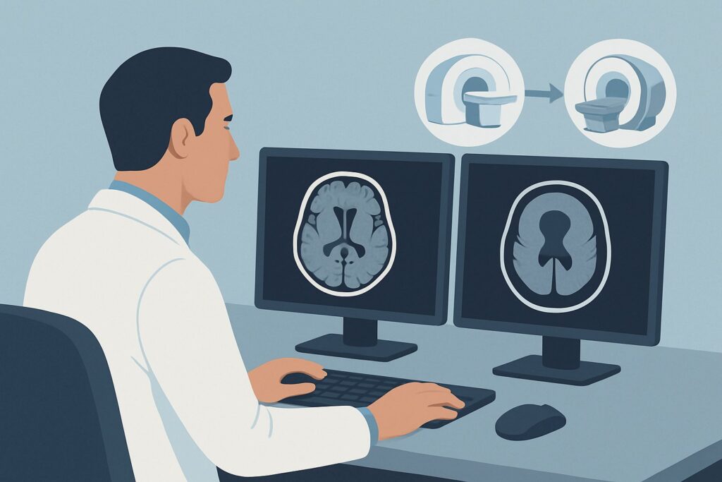
It’s 2 AM in the emergency department, and you’re evaluating a 34-year-old patient who fell, struck their head, and then had a witnessed generalized tonic-clonic seizure. They are now postictal but arousable. The immediate, critical question is whether an intracranial injury from the trauma triggered the seizure. You need to choose the right initial imaging study to quickly and accurately assess for life-threatening pathology. This article provides a focused workflow for this exact scenario: a new-onset seizure with a history of trauma, guiding you through the differential, study rationale, and downstream decisions. Based on the American College of Radiology (ACR) Appropriateness Criteria, the initial imaging study of choice is a CT head without IV contrast, which is rated Usually Appropriate.
Who Fits This Clinical Scenario?
This guidance applies specifically to patients presenting with their first-ever seizure in the context of recent, known trauma. The key inclusion criteria are a clear history linking a traumatic event (e.g., motor vehicle collision, fall, assault) to the subsequent onset of seizure activity. The patient has no prior diagnosis of epilepsy or a known seizure disorder.
It is crucial to distinguish this presentation from similar but distinct clinical situations that follow different diagnostic pathways. This workflow does not apply if:
- There is no history of trauma. A new-onset seizure without a traumatic precipitant requires a different workup, often prioritizing MRI to evaluate for underlying tumors, vascular malformations, or inflammatory conditions. This falls under the New-onset seizure, Unrelated to trauma ACR variant.
- The patient has a known seizure disorder. If a patient with diagnosed epilepsy has a breakthrough seizure after minor trauma, the clinical question is different. The focus may be on whether the trauma was a result of the seizure rather than its cause. Imaging decisions in this group depend on changes in seizure type or neurologic baseline.
- The seizure occurred first, causing the trauma. If a patient has a seizure and then falls, the primary workup is for an unprovoked seizure, though imaging is still required to assess for injury from the fall itself. The clinical history is paramount for differentiation.
What Diagnoses Are You Working Up in This Scenario?
In a post-traumatic seizure, the imaging workup is focused on identifying acute, structurally significant injuries that can cause neuronal irritation and lead to seizures. The differential is heavily weighted toward traumatic brain injury (TBI) and its immediate sequelae.
Intracranial Hemorrhage: This is the most critical and common cause to exclude. It includes several types. Epidural hematomas (EDH) are often arterial and can expand rapidly. Subdural hematomas (SDH) are typically venous and can be acute or subacute. Traumatic subarachnoid hemorrhage (SAH) and intraparenchymal hemorrhage (IPH), or contusions, are also primary concerns. Blood products are highly epileptogenic and are a direct cause of acute symptomatic seizures.
Skull Fracture: A fracture itself is not the cause of the seizure, but its presence, particularly if depressed, indicates a significant force of impact. Depressed skull fractures can directly irritate or injure the underlying cortex, serving as a seizure focus. The presence of a fracture also significantly increases the likelihood of an associated intracranial hemorrhage.
Cerebral Contusion: This is essentially a “bruise” on the brain parenchyma, often occurring at the site of impact (coup injury) or on the opposite side (contrecoup injury). These can be hemorrhagic and are a well-established cause of early post-traumatic seizures. While some non-hemorrhagic contusions are better seen on MRI, significant ones are readily apparent on CT.
Why Is CT Head without IV Contrast the Recommended Study for This Presentation?
The ACR designates CT head without IV contrast as Usually Appropriate for the initial imaging of a new-onset, post-traumatic seizure because it optimally balances speed, accessibility, and diagnostic yield for the most urgent clinical questions.
The primary goal is to rapidly identify or exclude acute intracranial hemorrhage and significant fractures. Non-contrast CT is exceptionally sensitive for detecting acute blood, which appears hyperdense (bright white) against the brain parenchyma. It is also the gold standard for evaluating the bony structures of the skull. In a potentially unstable trauma patient, a non-contrast head CT can be completed in minutes, providing actionable information for immediate neurosurgical consultation.
Alternative studies are rated lower for specific reasons in this initial context:
- MRI head without and with IV contrast and MRI head without IV contrast are both rated May be appropriate. While MRI offers superior soft-tissue detail and is more sensitive for subtle injuries like non-hemorrhagic contusions or diffuse axonal injury, it is not the preferred initial test. MRI scans take significantly longer, are less available in many emergency settings, and require a cooperative or sedated patient who is stable enough to be in the scanner for an extended period. These factors make it impractical for the initial, time-sensitive evaluation of a trauma patient.
- CT head with IV contrast is rated Usually not appropriate. Intravenous contrast is not necessary to diagnose acute hemorrhage or skull fractures. In fact, contrast can sometimes obscure a small subarachnoid hemorrhage or be confused with calcifications. It adds time to the scan, carries a risk of allergic reaction or contrast-induced nephropathy, and increases the radiation dose without providing additional crucial information for the initial decision.
The radiation dose for a non-contrast head CT is moderate (adult relative radiation level ☢☢☢, 1-10 mSv), a necessary trade-off for the speed and diagnostic clarity it provides in this high-stakes scenario. Once you’ve decided on the study, our protocol guide covers the essential technical details. For a deeper dive into the technique, appropriateness, and reading principles, see our complete guide: CT Brain Without Contrast.
What’s Next After CT Head without IV Contrast? Downstream Workflow
The results of the initial non-contrast head CT will dictate the immediate next steps in patient management, creating a clear decision-making fork.
If the CT is positive for acute injury: A finding of epidural or subdural hematoma, significant subarachnoid or intraparenchymal hemorrhage, or a depressed skull fracture is a critical result. The immediate next step is an urgent consultation with neurosurgery. The patient will require admission, likely to an intensive care unit, for close neurologic monitoring, management of intracranial pressure, and potential surgical intervention (e.g., craniotomy for hematoma evacuation).
If the CT is negative: A normal initial head CT is reassuring, as it effectively rules out a large, life-threatening hemorrhage or fracture that would require emergent surgery. However, it does not rule out the possibility of a seizure. The patient still requires a neurology consultation for management of the seizure itself. The downstream workflow typically involves admission for observation, initiation of anti-epileptic drug (AED) therapy based on recurrence risk, and an electroencephalogram (EEG) to assess for epileptiform activity. An MRI may be considered later during the hospital stay to evaluate for more subtle, non-hemorrhagic injuries that could be a seizure focus, especially if the patient’s mental status does not return to baseline.
If the CT is indeterminate: In rare cases, findings may be ambiguous (e.g., concern for a subtle isodense subdural hematoma). An immediate discussion with the on-call radiologist is essential. This may lead to a repeat CT in a few hours, a CT with contrast to look for membrane enhancement, or proceeding to an MRI for definitive characterization.
Pitfalls to Avoid (and When to Get Help)
Navigating this scenario requires vigilance to avoid common diagnostic and management errors. Be mindful of these potential pitfalls:
- Delaying the Scan: In a patient with a new seizure after head trauma, time is critical. Do not delay ordering the non-contrast head CT while awaiting other tests or consultations.
- Forgetting the C-Spine: A mechanism of injury sufficient to cause a TBI and seizure is often sufficient to cause a cervical spine injury. Maintain C-spine precautions until it has been clinically or radiographically cleared.
- Anchoring on Trauma: While trauma is the likely trigger, a negative CT scan should prompt consideration of other underlying causes. The seizure may have been unprovoked, with the fall being a consequence, not the cause.
- Misinterpreting the History: Carefully distinguish between a seizure causing a fall versus a fall causing a seizure. This distinction fundamentally changes the diagnostic pathway and pre-test probability.
If the CT scan shows any acute intracranial pathology, the immediate escalation is to neurosurgery. If the CT is negative but the patient’s neurologic status is not improving or seizures recur, escalate to neurology and consider ICU-level care.
Related ACR Topics and Tools
This article covers one specific variant within the broader topic of Seizures and Epilepsy. For a comprehensive overview of imaging recommendations across all related clinical scenarios, from febrile seizures to surgical planning for refractory epilepsy, please see our parent guide. It provides the breadth that complements this article’s depth.
- For breadth across all scenarios in Seizures and Epilepsy, see our parent guide: Seizures and Epilepsy: ACR Appropriateness Decoded.
- To look up other clinical variants, use the Imaging Appropriateness Selector tool.
- To review technical specifications for this or other studies, visit the Imaging Protocol Library.
- To discuss radiation exposure with patients, our Radiation Dose Calculator can help frame the conversation.
Frequently Asked Questions
Why not order an MRI as the first imaging study after a traumatic seizure?
While MRI provides more detailed images of the brain parenchyma, a non-contrast CT is much faster, more widely available, and superior for detecting the most immediate life-threatening conditions in this scenario: acute hemorrhage and skull fractures. In a potentially unstable trauma patient, the speed and diagnostic focus of CT make it the appropriate initial choice.
If the trauma was minor, like a simple fall from standing, is a CT scan still necessary?
Yes. A new-onset seizure is a significant neurologic event. Even seemingly minor trauma can cause serious intracranial injury, such as a subdural hematoma, particularly in older patients or those on anticoagulants. The presence of a seizure after any head trauma lowers the threshold for imaging and makes a non-contrast head CT a necessary part of the initial evaluation.
Does a normal non-contrast head CT mean the patient is safe to be discharged from the emergency department?
Not necessarily. A normal CT is excellent for ruling out an immediate surgical emergency, but a first-time seizure requires further evaluation. The patient typically requires admission for neurologic observation, consultation with neurology, consideration for an EEG to assess seizure recurrence risk, and initiation of anti-epileptic medication if indicated.
Is there any situation where IV contrast would be used for a post-traumatic seizure?
In the initial evaluation, IV contrast is rated ‘Usually not appropriate.’ However, if the non-contrast CT is negative but there is a high clinical suspicion for an underlying vascular injury (e.g., dissection, fistula) or if a subsequent MRI is ordered to investigate for tumor or infection as an alternative cause, contrast would then be appropriate for that specific indication.
What if the patient is a child? Does the recommendation for a non-contrast CT still hold?
Yes, the principle is the same, but with a heightened emphasis on radiation dose reduction (ALARA – As Low As Reasonably Achievable). Non-contrast CT remains the primary modality for ruling out acute, life-threatening traumatic injury. Pediatric-specific CT protocols must be used to minimize radiation exposure. MRI may be considered more readily as a follow-up study in children once they are stable.
Reviewed by Pouyan Golshani, MD, Interventional Radiologist — May 26, 2026