Should You Image Mild Head Trauma if Clinical Decision Rules Are Negative?
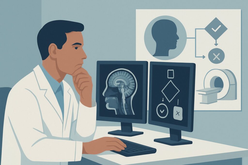
A 24-year-old male presents to the emergency department after tripping and hitting his head on a curb. He had no loss of consciousness, his Glasgow Coma Scale (GCS) is 15, and he has a small scalp hematoma but no other signs of trauma. He is neurologically intact. You apply the Canadian CT Head Rule, and he meets none of the high-risk or medium-risk criteria for brain injury. The patient is anxious and asks if he needs a “scan to be safe.” This scenario—acute, mild head trauma in a patient who does not meet criteria for imaging based on a validated clinical decision rule—is common. This article details the American College of Radiology (ACR) guidance for this specific presentation, where the evidence strongly supports forgoing imaging. For this low-risk cohort, modalities including CT head, MRI head, and even skull radiography are all considered Usually not appropriate.
Who Fits This Clinical Scenario?
This guidance applies to a very specific and well-defined patient population: adults with acute, mild traumatic brain injury (TBI). The inclusion criteria are precise:
- Glasgow Coma Scale (GCS) of 13–15: The patient is conscious and ranges from fully alert to mildly altered.
- Acute Trauma: The injury occurred recently, typically within the last 24 hours.
- Imaging Not Indicated by a Clinical Decision Rule (CDR): This is the critical qualifier. The patient has been risk-stratified using a validated tool, such as the Canadian CT Head Rule or the New Orleans Criteria, and has been found to be at very low risk for a clinically important brain injury.
It is crucial to distinguish this scenario from similar but higher-risk presentations. This guidance does not apply if:
- The patient meets criteria for imaging on a CDR: A patient with mild TBI (GCS 13–15) who has a high-risk feature (e.g., suspected open or depressed skull fracture, GCS <15 at 2 hours post-injury, or age ≥65) falls into a different ACR variant where CT is indicated.
- The trauma is moderate or severe: Patients with a GCS of 3–12 are managed under a separate, more aggressive imaging pathway.
- The patient is on anticoagulation: Many decision rules have specific criteria or are not validated for patients on anticoagulants or with bleeding disorders, who are at higher risk for intracranial hemorrhage even with minor trauma.
Correctly identifying that a patient fits this low-risk scenario is the key to avoiding unnecessary diagnostic testing.
What Diagnoses Are You Working Up in This Scenario?
When a patient presents with head trauma, the primary goal is to identify or exclude life-threatening intracranial injuries that may require neurosurgical intervention or intensive monitoring. Even in this low-risk scenario, the theoretical differential diagnosis remains the same, but the pre-test probability is exceedingly low.
The main concern is intracranial hemorrhage. This includes epidural hematoma (EDH), often from a tear in the middle meningeal artery associated with a temporal bone fracture; subdural hematoma (SDH), from tearing of bridging veins; subarachnoid hemorrhage (SAH), from trauma to cortical vessels; and intraparenchymal hemorrhage or contusion. In patients who are negative by a validated CDR, the risk of any of these injuries requiring intervention is exceptionally small.
Another key diagnosis to consider is a skull fracture. While a simple, linear, non-displaced skull fracture may not require specific intervention, depressed or basilar skull fractures are clinically significant. Depressed fractures may require surgical elevation, while basilar skull fractures increase the risk of cerebrospinal fluid (CSF) leaks, meningitis, and cranial nerve injury. The clinical decision rules are designed with high sensitivity to detect patients at risk for these significant injuries. The absence of high-risk clinical signs (e.g., hemotympanum, “raccoon eyes,” Battle’s sign) makes a basilar skull fracture highly unlikely.
Why Imaging Is Usually Not Appropriate for This Presentation
For patients with mild head trauma who are negative by a clinical decision rule, the ACR Appropriateness Criteria rate all initial imaging modalities as Usually not appropriate. This strong consensus is based on the high negative predictive value of the decision rules and the principle of avoiding low-yield, potentially harmful testing.
The rationale is rooted in risk-benefit analysis. Validated CDRs like the Canadian CT Head Rule were specifically developed to identify nearly all patients with clinically important brain injuries, allowing clinicians to safely forgo imaging in the vast majority of low-risk individuals.
- CT Head (without or with contrast): This is the workhorse for acute TBI when imaging is indicated. However, in this scenario, it is Usually not appropriate. The diagnostic yield is extremely low, and it exposes the patient to ionizing radiation (adult relative radiation level ☢☢☢ 1-10 mSv). The risk of radiation-induced malignancy, while small for a single scan, is cumulative and not justified when the probability of finding a clinically significant injury is negligible.
- Radiography skull: Once a mainstay, the skull radiograph is now Usually not appropriate. Its primary limitation is that it can only identify a skull fracture; it provides no information about the brain parenchyma or potential intracranial hemorrhage, which is the most critical concern. A patient can have a lethal epidural hematoma with a normal skull X-ray, or a benign linear fracture with no underlying brain injury. Because it does not reliably rule out the most dangerous pathologies and rarely alters management, it has been superseded by clinical evaluation and, when needed, CT.
- MRI Head: While it provides excellent anatomic detail without ionizing radiation (adult relative radiation level O 0 mSv), MRI is also Usually not appropriate for initial evaluation. It is more time-consuming, less available in many emergency settings, more costly, and highly sensitive to patient motion. It offers no advantage over CT in the acute setting for detecting the life-threatening bleeds that drive immediate management and is not indicated in this low-risk population.
The core principle is that the clinical examination, combined with a validated decision rule, provides sufficient evidence to rule out a significant injury, making the risks, costs, and time associated with any imaging modality outweigh the minimal potential benefit.
What’s Next? Downstream Workflow After No Imaging
When imaging is appropriately withheld, the clinical workflow shifts from diagnosis to observation, patient education, and safe discharge planning. The “next step” is not a test but a set of clinical actions.
- If the patient remains clinically stable: After a period of observation in the emergency department (if deemed necessary), the patient can be safely discharged. The cornerstone of this step is providing clear, documented head injury precautions. The patient and a responsible family member or friend should be instructed to monitor for “red flag” symptoms, including worsening headache, repeated vomiting, increasing confusion or drowsiness, seizures, or any new focal neurologic symptoms like weakness or numbness. They should be told to return to the emergency department immediately if any of these occur.
- If the patient’s condition changes: If, during observation or after discharge, the patient develops new or worsening symptoms (e.g., a drop in GCS, a new focal deficit, a seizure), they no longer fit this low-risk scenario. This clinical change represents a failure of the initial risk stratification. The patient should be immediately re-evaluated, and this presentation would now align with a different ACR scenario, such as Acute head trauma with new or progressive neurologic deficit(s), for which emergent CT head imaging is appropriate.
The downstream plan relies on a strong safety net built on patient education and clear instructions for when to seek further care.
Pitfalls to Avoid (and When to Get Help)
Managing this scenario effectively requires adherence to evidence-based guidelines and avoiding common pressures.
- Misapplying the Decision Rule: Ensure the patient truly fits the inclusion criteria for the CDR you are using. For example, the Canadian CT Head Rule has specific exclusions (e.g., age <16, anticoagulant use, seizure after injury) that, if ignored, invalidate the rule's predictive power.
- Imaging Due to External Pressure: Avoid ordering a CT scan “just in case” or due to patient or family anxiety when it is not clinically indicated. Engaging in shared decision-making, explaining the rationale for forgoing imaging, and highlighting the risks of radiation can help manage these situations.
- Inadequate Discharge Instructions: Simply telling a patient to “take it easy” is insufficient. Provide written, easy-to-understand instructions detailing specific warning signs and the importance of having a responsible adult observe them for a period after discharge.
- Ignoring a Change in Status: Do not attribute a subtle decline in mental status to the initial injury or intoxication without a thorough re-evaluation. Any worsening of the neurologic exam moves the patient into a higher-risk category that warrants urgent imaging.
If a patient’s clinical status deteriorates or if there is any uncertainty about the application of a decision rule, escalate care by re-assessing the patient, consulting a senior colleague, or proceeding with emergent imaging.
Related ACR Topics and Tools
For further exploration of imaging guidelines and related clinical scenarios, the following resources are available:
- For breadth across all scenarios in Head Trauma, see our parent guide: Head Trauma: ACR Appropriateness Decoded.
- To search for other clinical presentations, use the Imaging Appropriateness Selector.
- To review technical details for various imaging studies, consult the Imaging Protocol Library.
- To discuss radiation exposure with patients, the Radiation Dose Calculator can help frame the conversation.
Frequently Asked Questions
What if a patient with mild head trauma is on an anticoagulant like warfarin or a DOAC?
Patients on anticoagulants are at significantly higher risk for intracranial hemorrhage, even from minor trauma. Most clinical decision rules, including the Canadian CT Head Rule, exclude these patients. Therefore, they do not fit this ‘no imaging’ scenario. A non-contrast CT of the head is generally indicated for this higher-risk group, even with a GCS of 15 and a normal neurologic exam.
Are clinical decision rules 100% effective at ruling out significant injury?
No test or rule is 100% perfect. However, validated rules like the Canadian CT Head Rule have been shown in large studies to have very high sensitivity (approaching 100%) for detecting clinically important brain injuries that require neurosurgical intervention. This means the risk of missing such an injury by following the rule is extremely low and is considered an acceptable trade-off to avoid unnecessary radiation exposure in the vast majority of low-risk patients.
Why not just order a skull X-ray if I’m worried about a fracture? The radiation is very low.
A skull radiograph is rated ‘Usually not appropriate’ because it does not answer the most important clinical question: is there an intracranial bleed? A patient can have a fatal brain injury with a normal X-ray. Furthermore, finding a simple, non-depressed linear skull fracture often does not change clinical management. Because it has low utility for guiding treatment and can provide false reassurance, it has been replaced by clinical risk stratification and, when necessary, head CT.
What specific ‘head injury precautions’ should I provide on discharge?
Patients should be given clear, written instructions. Key points include: have a responsible adult stay with you for the next 24 hours; abstain from alcohol; do not take medications that can cause drowsiness without consulting a physician. Instruct them to return to the emergency department immediately for any of the following: worsening or severe headache, repeated vomiting, confusion or unusual behavior, extreme drowsiness or difficulty waking up, a seizure, or new weakness/numbness in the arms or legs.
Does this guidance apply to children?
No. This guidance and the commonly cited clinical decision rules (Canadian CT Head Rule, New Orleans Criteria) are for adults. Pediatric head trauma has its own set of risks and is evaluated using different, age-specific clinical decision rules, such as the PECARN (Pediatric Emergency Care Applied Research Network) head injury rule.
Reviewed by Pouyan Golshani, MD, Interventional Radiologist — May 26, 2026