Which Imaging Study Is Best for a Suspected Midface Fracture After Trauma?
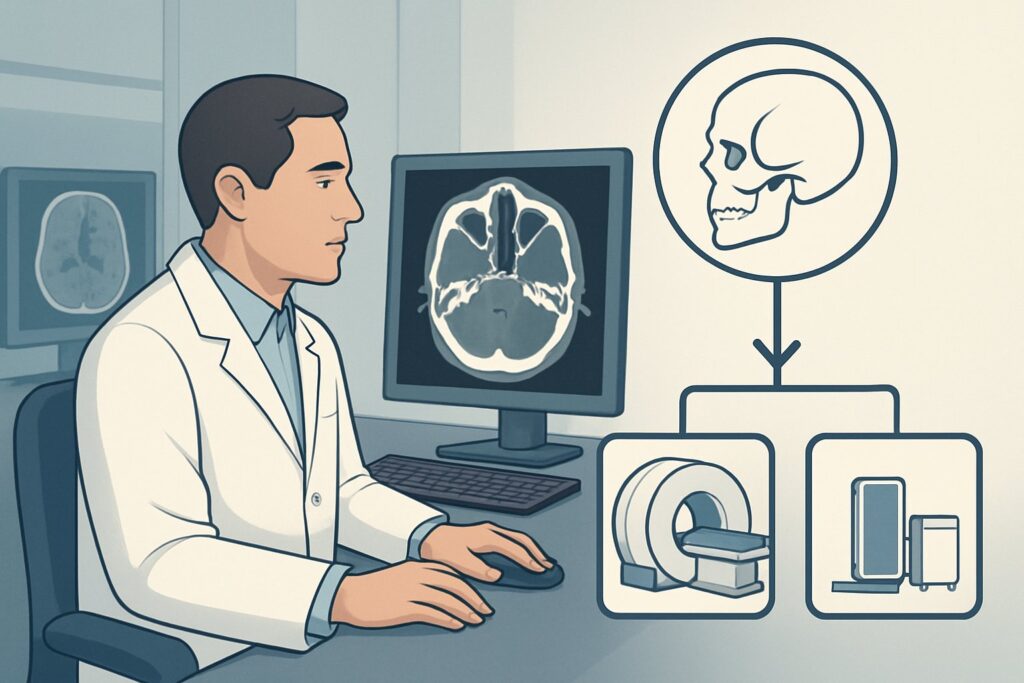
A 34-year-old man presents to the emergency department after an assault, complaining of severe facial pain and the sensation that his “teeth don’t fit together right.” On examination, you note swelling and tenderness over his right cheekbone, and gentle manipulation of his upper jaw elicits significant pain and slight movement. His right cheek feels numb. You suspect a complex midface fracture, but the next step—choosing the right initial imaging study—is critical for accurate diagnosis and guiding surgical consultation. This article provides a detailed workflow for this specific clinical scenario, grounded in the American College of Radiology (ACR) Appropriateness Criteria. For a patient with suspected midface injury, the ACR rates CT maxillofacial without IV contrast as Usually appropriate.
Who Fits This Clinical Scenario?
This guidance is for patients who have undergone a primary trauma survey and present with signs and symptoms suggestive of a midface or zygomaticomaxillary complex (ZMC) injury. The key clinical indicators include:
- Pain with upper jaw manipulation: Suggests instability of the maxilla (Le Fort fractures).
- Pain, deformity, or tenderness over the zygoma: Points toward a ZMC or isolated zygomatic arch fracture.
- Facial elongation: A classic, though less common, sign of severe, inferiorly displaced midface fractures.
- Malocclusion: The patient reports their bite feels “off,” a strong indicator of a fracture involving the dental arches.
- Infraorbital nerve paresthesia: Numbness or tingling in the cheek, upper lip, or upper gingiva on the affected side, suggesting injury to the nerve as it passes through the orbital floor or maxilla.
This workflow is not intended for patients with:
- Isolated nasal trauma: If the injury is confined to the nasal bridge with no signs of broader midface instability (e.g., no malocclusion, normal jaw mobility), the workup follows a different pathway. See the ACR variant for suspected nasal injury.
- Suspected mandibular injury: If the patient presents with trismus, pain localized to the lower jaw, or gingival hemorrhage near the mandible, the focus shifts to evaluating for a mandibular fracture.
- Frontal bone or sinus concerns: Tenderness isolated to the forehead suggests a frontal bone injury, which has its own specific imaging considerations.
What Diagnoses Are You Working Up in This Scenario?
When a patient presents with these findings, your differential diagnosis centers on complex fractures that disrupt the structural integrity of the midface. The primary goal of imaging is to identify, classify, and determine the displacement of these injuries to guide management, which often involves surgical consultation.
Le Fort Fractures: These are complex fracture patterns that involve the separation of all or part of the midface from the skull base. They are classified into three types based on the plane of injury. Le Fort I is a horizontal maxillary fracture separating the teeth from the upper face. Le Fort II is a pyramidal fracture involving the nasal bridge and maxilla. Le Fort III, or craniofacial disjunction, is the most severe, involving a transverse fracture line that passes through the nasofrontal suture, maxillo-frontal suture, orbital wall, and zygomatic arch.
Zygomaticomaxillary Complex (ZMC) Fracture: Often called a “tripod fracture,” this is one of the most common midface injuries. It involves fractures of the zygomatic arch, the inferior orbital rim and orbital floor, and the lateral wall of the maxillary sinus. These injuries often result in facial flattening and can cause entrapment of extraocular muscles or impingement on the infraorbital nerve.
Orbital Floor “Blowout” Fracture: While this can occur in isolation, it is frequently a component of ZMC or Le Fort injuries. A direct blow to the globe increases intraorbital pressure, causing the thin orbital floor to fracture. This can lead to herniation of orbital fat and entrapment of the inferior rectus muscle, causing diplopia on upward gaze.
Naso-orbito-ethmoid (NOE) Complex Fracture: This severe injury involves fractures of the nasal bones, ethmoid sinuses, and medial orbital walls. It can disrupt the medial canthal tendon, leading to traumatic telecanthus (widening of the distance between the eyes), and carries a high risk of associated cerebrospinal fluid (CSF) leak and intracranial injury.
Why Is CT Maxillofacial without IV Contrast the Recommended Study?
The ACR designates CT maxillofacial without IV contrast as Usually appropriate for this presentation because it provides the necessary detail to diagnose and characterize the suspected injuries with optimal efficiency and safety.
The primary strength of CT is its exceptional spatial resolution and ability to visualize fine bony anatomy without the superimposition of structures that plagues plain radiography. It can precisely delineate fracture lines, quantify fragment displacement, and assess for involvement of critical adjacent structures like the orbital apex, optic canal, and skull base. This level of detail is essential for surgical planning.
Why are alternative studies rated lower?
- Radiography (paranasal sinuses): Rated Usually not appropriate. While historically used, plain films like a Waters view have very low sensitivity for complex midface fractures. The intricate, overlapping bones of the face make it nearly impossible to accurately identify and characterize Le Fort, ZMC, or NOE fractures. A negative radiograph does not rule out a clinically significant injury, often leading to delayed diagnosis or the need for subsequent CT anyway.
- CT maxillofacial with IV contrast: Also rated Usually not appropriate for the initial evaluation of bony trauma. Intravenous contrast does not improve the visualization of bone. It adds potential risks (allergic reaction, contrast-induced nephropathy), increases cost, and delivers a slightly higher radiation dose without providing additional diagnostic information for the primary question of a fracture. Contrast is reserved for specific situations where a vascular injury (e.g., carotid-cavernous fistula, dissection) or post-traumatic infection is suspected.
The radiation dose for a non-contrast maxillofacial CT is relatively low, with a typical adult effective dose of 0.1-1 mSv (ACR RRL ☢☢). While dose reduction techniques are always paramount, especially in pediatric patients (pediatric RRL ☢☢☢, 0.3-3 mSv), the diagnostic benefit in identifying potentially vision- or function-threatening injuries far outweighs the risk.
Once you’ve decided on the appropriate study, understanding the technical aspects is key. While the specific protocol for maxillofacial CT may vary by institution, our guide on CT Brain Without Contrast covers foundational principles of non-contrast head and neck CT technique, dictation, and dose considerations relevant to trauma imaging.
What’s Next After CT Maxillofacial without IV Contrast? Downstream Workflow
The results of the CT scan will directly guide your next steps and consultations. The workflow typically branches based on the findings:
- Positive for Displaced or Complex Fracture: If the CT confirms a displaced ZMC, Le Fort, NOE, or orbital floor fracture with muscle entrapment, the immediate next step is an urgent consultation with the relevant surgical service. This is typically Oral and Maxillofacial Surgery (OMFS), Plastic and Reconstructive Surgery, or Otolaryngology (ENT), depending on institutional practice. The 3D reformatted images from the CT are invaluable for these specialists to plan their surgical approach.
- Positive for Nondisplaced Fracture: For a simple, nondisplaced fracture (e.g., an isolated zygomatic arch fracture without functional impairment), management may be non-operative. The patient can often be discharged with close outpatient follow-up with the appropriate surgical service. Instructions on a soft diet and activity restrictions are crucial.
- Negative for Fracture: If the CT is negative for a fracture but the patient remains symptomatic (e.g., persistent malocclusion or diplopia), further evaluation is warranted. This could involve a more detailed dental evaluation to rule out a dental or alveolar ridge injury not well-visualized on CT, or an ophthalmology consultation to assess for soft-tissue orbital injury or cranial nerve palsy. A repeat clinical exam after swelling has subsided can also be revealing.
Pitfalls to Avoid (and When to Get Help)
When managing a suspected midface injury, several common pitfalls can lead to delayed or missed diagnoses.
- Incomplete Examination: Failing to test for infraorbital nerve sensation, extraocular movements, and visual acuity can cause you to miss critical signs of orbital floor involvement or optic nerve injury.
- Missing Associated Injuries: Midface trauma often occurs in the context of high-energy mechanisms. Always maintain a high index of suspicion for concomitant cervical spine injuries, intracranial hemorrhage, or mandibular fractures. Ensure the CT scan coverage is adequate to evaluate the skull base and intracranial contents if clinically indicated.
- Ignoring the Soft Tissues: Even on a non-contrast “bone” scan, evaluate the soft tissues. Look for signs of a retrobulbar hematoma, which can cause orbital compartment syndrome and is a true ophthalmologic emergency requiring immediate intervention.
- Underestimating Pediatric Injuries: In children, facial bones are more malleable, and suture lines can be mistaken for fractures (or vice versa). If there is any uncertainty, consultation with a pediatric radiologist is recommended.
If you identify signs of a retrobulbar hematoma (proptosis, decreased visual acuity, increased intraocular pressure) or a potential CSF leak (clear rhinorrhea), escalate immediately to ophthalmology and neurosurgery, respectively.
Related ACR Topics and Tools
This article focuses on a single, common scenario. For a comprehensive overview of other presentations or to explore the evidence in more detail, the following resources are essential. For breadth across all scenarios in Imaging of Facial Trauma Following Primary Survey, see our parent guide: Imaging of Facial Trauma Following Primary Survey: ACR Appropriateness Decoded.
- ACR Appropriateness Criteria Lookup: Look up other clinical variants or review the full ACR document.
- Imaging Protocol Library: Access detailed technical protocols for various imaging studies.
- Radiation Dose Calculator: Help facilitate informed conversations with patients about cumulative radiation exposure.
Frequently Asked Questions
Why shouldn’t I order plain X-rays first to screen for a midface fracture?
Plain radiographs are rated ‘Usually not appropriate’ by the ACR for this scenario because the complex, overlapping anatomy of the facial skeleton makes them highly insensitive for detecting and characterizing significant midface fractures like Le Fort or ZMC injuries. A negative X-ray does not reliably rule out a fracture, and a positive finding would still require a CT for definitive characterization and surgical planning.
In what situation would I add IV contrast to the maxillofacial CT scan?
IV contrast is generally not needed for the initial assessment of bony facial trauma. However, you should order a CT with contrast (or a CTA) if you suspect an associated vascular injury. Clinical signs suggesting this include pulsatile exophthalmos, a cranial bruit (suggesting a carotid-cavernous fistula), or evidence of a penetrating injury with a high-risk trajectory.
What is ‘facial elongation’ and why is it a significant sign?
Facial elongation, sometimes called ‘long face syndrome,’ is a clinical sign where the midface appears lengthened and flattened. It is a classic indicator of a severe, bilateral Le Fort II or Le Fort III fracture, where the entire midfacial skeleton is displaced inferiorly due to the force of the trauma. It signifies a high-energy injury with significant instability.
Does numbness in the cheek specifically point to one type of fracture?
Yes, paresthesia in the distribution of the infraorbital nerve (cheek, upper lip, upper gums) is highly suggestive of a fracture involving the orbital floor or zygomaticomaxillary complex (ZMC). The nerve passes through the infraorbital canal and foramen in this region and is easily contused, compressed, or transected by fracture fragments.
Are 3D reconstructions from the CT scan always necessary?
While the diagnosis can typically be made from the standard 2D axial, coronal, and sagittal images, 3D reconstructions are extremely valuable for surgical planning. They provide an intuitive, three-dimensional view of the fracture pattern and fragment displacement, which helps surgeons from OMFS, Plastics, or ENT to plan their operative approach and select the appropriate hardware for fixation. Most modern CT scanners can generate these images automatically.
Reviewed by Pouyan Golshani, MD, Interventional Radiologist — May 29, 2026