Why Is Imaging Usually Not Appropriate for Headache Without Red Flags?

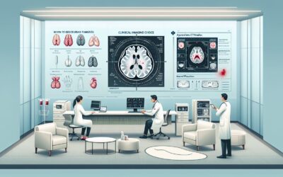
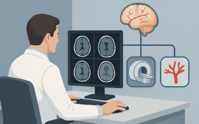
A 34-year-old patient with a multi-year history of episodic headaches presents for a follow-up. The headaches are unilateral, throbbing, and associated with photophobia, consistent with their established diagnosis of migraine without aura. The neurologic exam is, as always, completely normal. Today, they are anxious, having read an article online, and ask, “Shouldn’t I get an MRI just to be sure there’s nothing serious going on?” This common clinical question places you at a decision point: to image or not to image. This article provides a detailed workflow for this specific scenario—a headache without any “red flags”—and explains the clinical reasoning behind the American College of Radiology (ACR) Appropriateness Criteria. For this presentation, neuroimaging modalities like MRI, CT, and arteriography are all rated as Usually not appropriate.
Who Fits This Clinical Scenario?
This guidance applies to a well-defined patient population: individuals presenting for initial imaging of a headache that lacks any concerning features, often referred to as “red flags.” The clinical picture is typically one of a primary headache disorder, such as migraine or tension-type headache, in a patient with a normal neurologic examination.
Inclusion criteria for this workflow:
- The headache pattern is stable or consistent with a known primary headache disorder.
- The neurologic examination is entirely normal.
- The patient is under 50 years of age at the onset of their headache pattern.
Exclusion criteria (patients who require a different workup):
- Sudden, severe “thunderclap” headache: This presentation is a medical emergency and requires a different imaging pathway to evaluate for subarachnoid hemorrhage. It is covered in a separate ACR variant.
- Headache with neurologic deficits: Any new, focal neurologic finding (e.g., weakness, sensory loss, diplopia, ataxia) or altered mental status immediately moves the patient into a higher-risk category where imaging is indicated.
- New-onset headache in a patient over 50: This is a red flag for conditions like giant cell arteritis or intracranial mass and warrants imaging.
- Other red flags: This workflow does not apply if the headache is associated with fever, a history of cancer or immunocompromise, features of high or low intracranial pressure, or recent trauma.
What Diagnoses Are You Working Up in This Scenario?
In a patient with a headache and no red flags, the clinical focus is on confirming a primary headache diagnosis and, just as importantly, confidently excluding serious secondary causes based on the low pre-test probability. The decision not to image is an active clinical judgment.
The most common diagnoses in this category are primary headache disorders. This includes migraine (with or without aura) and tension-type headache. These conditions are diagnosed based on clinical criteria established by the International Classification of Headache Disorders (ICHD-3). Their pathophysiology is not related to a structural brain abnormality that would be visible on standard neuroimaging.
The purpose of considering imaging is to rule out less common but consequential secondary causes. However, in the absence of red flags, the likelihood of finding a clinically significant structural lesion is exceedingly low. The differential for secondary headache includes intracranial tumors, vascular malformations (like aneurysms or arteriovenous malformations), and chronic subdural hematomas. The key insight from decades of research is that these serious conditions almost invariably present with one or more of the red flag signs or symptoms that specifically exclude a patient from this low-risk scenario.
Why Neuroimaging Is Usually Not Appropriate for This Presentation
For a patient with a headache lacking red flags, the ACR rates all initial imaging modalities—including MRI, MRA, CT, and CTA—as Usually not appropriate. This recommendation is based on a careful balance of the very low diagnostic yield against the potential for patient harm, unnecessary cost, and downstream testing.
The core rationale is the low pre-test probability of significant pathology. In patients with stable primary headache patterns and normal neurologic exams, the incidence of finding a causative lesion on neuroimaging is similar to the incidence of finding asymptomatic, incidental abnormalities in the general population. Ordering a scan is far more likely to reveal an incidentaloma—an unrelated finding like a small meningioma, a developmental venous anomaly, or an arachnoid cyst—than it is to find the cause of the headaches. Such findings often lead to patient anxiety, further costly and sometimes invasive testing, and do not change the management of the primary headache disorder.
Let’s consider the specific modalities and why they are not recommended here:
- MRI Head (without or with contrast): While MRI offers excellent soft tissue detail without using ionizing radiation, its high sensitivity becomes a liability in this low-risk population. It is rated Usually not appropriate because of the high likelihood of detecting incidental findings that complicate, rather than clarify, the clinical picture.
- CT Head (without or with contrast): A non-contrast CT is a fast and accessible study, but it is less sensitive than MRI for many potential pathologies and exposes the patient to ionizing radiation (Relative Radiation Level ☢☢☢ 1-10 mSv). Given the benign nature of the underlying diagnosis, this radiation exposure is not justified. It is also rated Usually not appropriate.
- Arteriography cervicocerebral: This invasive procedure carries significant risks (e.g., stroke, vessel dissection) and a high radiation dose. It has no role in the initial evaluation of a non-suspicious headache and is firmly rated Usually not appropriate.
The consensus is that the clinical history and neurologic exam are the most powerful tools for diagnosis in this scenario. Imaging should be reserved for when those tools suggest a secondary cause.
What’s Next? The Downstream Workflow Without Imaging
When you correctly identify a patient who fits this scenario and decide against imaging, the workflow shifts from diagnostic testing to clinical management and patient education.
- If the clinical picture is clear for a primary headache: The next step is to initiate or optimize therapy. This involves selecting appropriate abortive and, if necessary, prophylactic medications. Providing patient education on trigger identification, lifestyle modifications, and the benign nature of their condition is paramount. Reassuring the patient that imaging is not needed because their presentation does not suggest a dangerous underlying cause is a key part of the conversation.
- If the diagnosis is uncertain but still low-risk: A headache diary can be an invaluable tool. Having the patient track the frequency, severity, duration, and associated symptoms of their headaches over a few weeks can help clarify the pattern and confirm a primary headache diagnosis.
- If the headache pattern changes: This is a critical point. If a patient with a previously stable headache pattern develops new, concerning features (e.g., it becomes “the worst headache of my life,” is associated with a new neurologic deficit, or changes in character), they no longer fit this scenario. At that point, the patient should be re-evaluated, and imaging becomes appropriate under a different ACR variant, such as “Sudden onset severe headache” or “Headache with neurologic deficit.”
The primary downstream pathway is one of reassurance, education, and effective medical management of the primary headache disorder.
Pitfalls to Avoid (and When to Get Help)
Navigating the decision to forego imaging requires clinical confidence and clear communication. Here are common pitfalls to avoid in this specific scenario:
- Ordering imaging for patient reassurance alone: While tempting, this can lead to the cascade of incidental findings, increased anxiety, and unnecessary costs without improving outcomes.
- Missing a subtle red flag: A thorough history and a meticulous neurologic exam are non-negotiable. Don’t dismiss a subtle historical clue (e.g., “the headache is worse when I stand up”) that might point to a different scenario like intracranial hypotension.
- Failing to provide a “safety net” plan: Always instruct the patient on exactly what symptoms should prompt them to seek immediate medical attention (e.g., a sudden, explosive onset; new weakness or numbness; fever with stiff neck).
- Underestimating the power of education: Clearly explaining why a scan isn’t needed is often more reassuring to an anxious patient than simply ordering the test.
If the headache pattern evolves or any red flags emerge, the patient’s risk profile has changed. Escalate by proceeding with urgent neuroimaging (typically a non-contrast head CT for a thunderclap headache or an MRI of the brain for a new focal deficit) and consider consultation with neurology.
Related ACR Topics and Tools
This article focuses on a single, low-risk headache scenario. For a comprehensive overview of all headache variants and for tools to help with ordering decisions, the following resources are available:
- For breadth across all scenarios in Headache, see our parent guide: Headache: ACR Appropriateness Decoded.
- ACR Appropriateness Criteria Lookup — for exploring adjacent clinical scenarios.
- Imaging Protocol Library — for technical details on imaging studies when they are indicated.
- Radiation Dose Calculator — for discussing cumulative radiation dose with patients when considering CT.
Frequently Asked Questions
My patient has a family history of a brain aneurysm but their own headaches are typical for migraine. Should I still order an MRA?
In general, screening for unruptured intracranial aneurysms is not recommended unless the patient has two or more first-degree relatives with aneurysms or a specific genetic syndrome. For a patient with a typical primary headache pattern and a more distant family history, the ACR guidance to not perform imaging (‘Usually not appropriate’) still holds. The decision to screen for familial aneurysms is a separate clinical question from the headache workup.
What is the risk of missing a brain tumor if I don’t image a patient with a stable headache and normal exam?
The risk is exceptionally low. Large studies have consistently shown that brain tumors almost always present with other signs or symptoms, such as a new or progressive neurologic deficit, seizures, altered mental status, or signs of increased intracranial pressure (papilledema). A long-standing, stable headache pattern with a normal exam is not a typical presentation for a brain tumor.
If I decide not to image, how should I document my reasoning?
Clear documentation is key. Your note should explicitly state that the patient’s history and physical exam were reviewed for red flag features and none were present. Document the normal neurologic exam findings. State that the clinical presentation is consistent with a primary headache disorder (e.g., migraine) and that, per evidence-based guidelines, neuroimaging is not indicated at this time. Finally, include the safety net instructions you provided to the patient.
A patient’s headache pattern changed from episodic to chronic. Is that a ‘red flag’ that requires imaging?
An increasing frequency of headaches, such as transformation from episodic to chronic migraine, is considered a red flag and warrants a re-evaluation. While often related to the primary headache disorder itself (e.g., medication overuse), it lowers the threshold for imaging to rule out a secondary cause. This patient would no longer fit the ‘no red flags’ scenario discussed in this article.
Does this guidance apply to children?
Yes, the principle is the same, and perhaps even more important. The American Academy of Neurology and other pediatric societies also recommend against routine neuroimaging for children with recurrent headaches and a normal neurologic examination. The rationale is similar: extremely low diagnostic yield and the desire to avoid radiation from CT scans and the potential need for sedation for MRI in younger children.
Reviewed by Pouyan Golshani, MD, Interventional Radiologist — June 27, 2026