US Thyroid — Dictation, Appropriateness, and Dose for Residents
1. The Mid-Morning Thyroid Nodule That Needs a Perfect TI-RADS Score
It’s 10 AM. You’re working through the outpatient ultrasound list, and the next case is a thyroid ultrasound for a palpable nodule. The patient is anxious, the referring endocrinologist is meticulous, and your attending expects a perfect ACR TI-RADS classification on every nodule over a centimeter. You know the five categories—composition, echogenicity, shape, margin, and echogenic foci—but remembering the exact point values and FNA thresholds under pressure is another story. Miss a punctate echogenic focus or misclassify a margin, and you could either send a benign nodule for an unnecessary biopsy or, worse, undertriage a suspicious one.
When I was a resident, I’d keep the TI-RADS chart taped to my monitor. It worked, but it was slow. The goal isn’t just to be right; it’s to be efficient, confident, and clear in your report so the clinical team knows exactly what to do next. This guide will walk you through a solid US Thyroid template and the key principles to nail these reports every time. For more guides and tools, check out the residents and fellows resource hub we’ve put together.
2. What a Thyroid Ultrasound Covers and What Attendings Look For
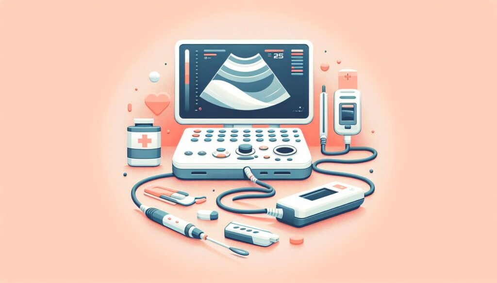
A high-frequency thyroid ultrasound is the workhorse for evaluating thyroid pathology. It provides a detailed, non-invasive look at the gland’s parenchyma, any nodules within it, and the surrounding cervical lymph nodes. There’s no radiation involved, making it the definitive first-line imaging test for most thyroid concerns.
Attendings expect a systematic and comprehensive report that answers these key clinical questions:
- Nodule Characterization: Is there a nodule? If so, what are its precise 3D measurements and its ACR TI-RADS score? This includes a detailed breakdown of its composition, echogenicity, shape, margins, and any calcifications.
- Diffuse Glandular Disease: Is the background thyroid parenchyma normal, or does it show signs of an autoimmune process like Hashimoto’s thyroiditis (heterogeneous, micronodular) or Graves’ disease (hypervascular “thyroid inferno”)?
- Cervical Lymph Node Assessment: Are there any suspicious lymph nodes in the central (level VI) or lateral (levels II-IV) compartments? We’re looking for features like a round shape, microcalcifications, cystic changes, or abnormal vascularity.
- Extrathyroidal Extension (ETE): For suspicious nodules, is there any evidence of the nodule breaking through the thyroid capsule and invading adjacent structures like the strap muscles?
Your report should be structured to address each of these points clearly, culminating in a TI-RADS category and a specific recommendation for follow-up or fine-needle aspiration (FNA) based on established size criteria.
3. Radiology Report Template for Ultrasound Thyroid
Use this template as a starting point for your macros. It’s designed to guide you through all the necessary components for a complete report that your attending and the referring clinician will appreciate.
Technique
Real-time grayscale and color Doppler ultrasound imaging of the thyroid gland and neck was performed using a high-frequency linear transducer. 3D measurements of the thyroid gland and any identified nodules were obtained. The cervical lymph node chains were surveyed.
Findings
THYROID GLAND: The thyroid gland is normal in size and echotexture. No diffuse heterogeneity or hypervascularity to suggest thyroiditis.
Right Lobe: Measures [X] x [Y] x [Z] cm.
Left Lobe: Measures [X] x [Y] x [Z] cm.
Isthmus: Measures [X] mm in AP dimension.
NODULES:
Nodule 1: [e.g., Right midpole]
Measurements: [X] x [Y] x [Z] cm.
Composition: [e.g., Solid, Mixed cystic and solid, Spongiform]
Echogenicity: [e.g., Hyperechoic, Isoechoic, Hypoechoic, Very hypoechoic]
Shape: [e.g., Wider-than-tall]
Margins: [e.g., Smooth, Ill-defined, Lobulated/Irregular, Extrathyroidal extension]
Echogenic Foci: [e.g., None, Macrocalcifications, Peripheral/rim calcifications, Punctate echogenic foci]
Color Doppler: [e.g., No significant internal vascularity, Peripheral vascularity, Internal vascularity]
ACR TI-RADS Score: [Sum of points]
ACR TI-RADS Category: [e.g., TR4 – Moderately Suspicious]
(Repeat for each additional nodule)
CERVICAL LYMPH NODES: No suspicious cervical lymph nodes identified. There are several benign-appearing lymph nodes with preserved fatty hila in the bilateral jugular chains.
Impression
1. [e.g., Single 1.6 cm moderately suspicious (TR4) nodule in the right midpole of the thyroid as described above.] According to ACR TI-RADS guidelines, FNA is recommended for TR4 nodules ≥1.5 cm.
2. [e.g., Otherwise normal thyroid gland parenchyma.]
3. [e.g., No suspicious cervical lymphadenopathy.]
4. Free Template Sources for Your On-Call Toolkit
Building your own templates is a rite of passage, but you don’t have to start from scratch. When you’re looking for a template for a study you haven’t seen in a while, two great free repositories exist that are curated by and for radiologists.
- RadReport.org: Maintained by the RSNA, this is a comprehensive library of peer-reviewed templates covering nearly every modality and subspecialty. It’s a go-to source for standardized language. (https://radreport.org/)
- Radiology Templates (AU): This is an excellent, straightforward library maintained by Australian radiologists. It’s well-organized and provides clean, practical templates that are easy to adapt. (https://www.radiologytemplates.com.au/home-page/)
Bookmark these. They’re invaluable, especially in your first couple of years of call.
5. The Next-Level Move: Free-Form Dictation to Structured Report
The template above is solid, but manually clicking through every TI-RADS feature for multiple nodules is a drag. This is where AI-powered tools can streamline your workflow without taking away your diagnostic control. Instead of filling out a form, you can simply dictate the positive findings in your own words.
For example, you might dictate: “In the left lower pole there’s a 1.8 cm solid, very hypoechoic nodule with irregular margins and punctate echogenic foci.”
Tools like GigHz Precision AI are designed to parse that free-form dictation, identify the key TI-RADS descriptors, calculate the score, and generate a perfectly structured draft segment. It applies the appropriate reporting framework, like ACR TI-RADS, and helps ensure your recommendation aligns with the guidelines. It’s about turning your natural diagnostic language into the structured data the EMR and your referring docs need, saving you clicks and mental energy.
6. When Should You Order a Thyroid Ultrasound? ACR Appropriateness Criteria
The American College of Radiology (ACR) provides evidence-based guidelines to help clinicians choose the right test for the right reason. For thyroid and parathyroid evaluation, ultrasound is almost always the place to start.
According to the ACR Appropriateness Criteria for Thyroid Disease, a thyroid ultrasound is rated “Usually Appropriate” (9/9) as the initial imaging test for a palpable thyroid nodule in a euthyroid patient, for a suspected goiter, and for thyrotoxicosis. It is also the primary modality for preoperative evaluation and post-treatment surveillance of differentiated thyroid cancer.
For suspected Parathyroid Adenoma, ultrasound is also “Usually Appropriate” (often paired with a sestamibi scan) for initial imaging in patients with primary, secondary, tertiary, or recurrent/persistent hyperparathyroidism.
While ultrasound is the first-line tool, other modalities have specific roles. A nuclear medicine thyroid uptake scan (I-123) is used to determine if a nodule is “hot” (hyperfunctioning) or “cold.” CT or MRI of the neck is better for evaluating large goiters with significant substernal extension that can’t be fully assessed with ultrasound.
7. Thyroid Ultrasound Imaging Protocol — Technique and Common Pitfalls
A high-quality thyroid ultrasound depends on meticulous technique. The goal is to use a high-frequency probe to get detailed images of the gland and surrounding structures. There is no ionizing radiation (0 mSv effective dose) and no contrast is required.
The protocol is straightforward but requires attention to detail, especially when characterizing nodules and surveying for lymph nodes.
| Phase / Component | Key Technical Parameters |
|---|---|
| Survey | Use a high-frequency linear transducer (7-15 MHz, 12+ MHz preferred). Patient is supine with neck extended. Obtain transverse and longitudinal images of both lobes and the isthmus, including measurements (Length, AP, Width). |
| Nodule Evaluation | For each significant nodule, obtain 3D measurements. Document TI-RADS features: composition, echogenicity, shape (transverse view), margins, and echogenic foci. Use color Doppler to assess vascularity. |
| Cervical Lymph Node Survey | Systematically evaluate the central (level VI) and lateral (levels II-IV) cervical lymph node compartments bilaterally. Document any abnormal nodes. |
Common protocol pitfalls:
- Misinterpreting Echogenic Foci: Distinguishing highly suspicious punctate echogenic foci (microcalcifications) from benign colloid crystals (which often produce comet-tail artifact) is critical. Microcalcifications are a key feature of papillary thyroid cancer.
- Inconsistent TI-RADS Application: The ACR TI-RADS system is standard in the US, but be aware that other systems like EU-TIRADS and K-TIRADS exist and have slightly different criteria. Stick to one system and apply it consistently.
- Overlooking Lymph Nodes: A quick or incomplete survey of the neck can miss metastatic nodes. Be systematic in checking all relevant levels, especially when a primary thyroid nodule is suspicious.
8. The 3-Months-Free Offer for Radiology Residents and Fellows
3+ months free for radiology residents and fellows
Look like a rockstar on your reports — dictate positive findings in free form, and the AI generates a structured report using ACR + SIR templates with the appropriate clinical decision support firing automatically. All we ask in return is feedback so we can keep improving the product for trainees.
Signup is simple. No credit card, no long forms. To get set up, just provide these three items:
- PGY year (e.g. PGY-2, PGY-4)
- Training type (radiology residency or specific fellowship — IR, body, MSK, neuro, peds, breast, nucs)
- Training program / hospital name
- (Optional) institutional email
Ready to give it a try? Apply for the residents free-access program and we’ll get you set up.
9. Frequently Asked Questions (FAQ)
Is it HIPAA-compliant?
Yes. The platform is designed for de-identified workflows by default. You dictate the clinical findings, not the patient’s name, MRN, or date of birth. It operates securely without requiring Protected Health Information (PHI).
Do I need my hospital’s IT department to set this up?
No. It’s a browser-based tool that works on any modern computer, including the PACS workstation or your personal laptop or call-room iPad. There’s no software to install and no integration with the EMR or PACS is needed to get started.
Does this replace PowerScribe or other dictation systems?
No, it works alongside them. You can dictate into the GigHz interface, let the AI structure your report, and then copy/paste the final, clean text into your hospital’s official dictation system. It’s an assistant, not a replacement.
Can I use this on my phone or iPad?
Yes, the web application is fully responsive and works on mobile devices, which is great for reviewing templates or prepping a report structure away from your workstation.
Can I customize the templates?
Yes. While the system comes pre-loaded with standard ACR and society-level templates, you can create, save, and modify your own templates to match your personal preferences or your institution’s specific requirements.
What happens after my residency or fellowship ends?
The free access is specifically for trainees. After you graduate, you can transition to a standard attending plan if you find the tool valuable in your practice. There’s no automatic charge or obligation.
Free GigHz Tools That Pair With This Article
Three free tools that complement the material above:
- Imaging Appropriateness Selector — Type an indication or clinical scenario in plain language and get the imaging studies the ACR rates for it, with adult and pediatric radiation levels. Built directly from 297 ACR topics, 1,336 clinical variants, and 15,823 procedure ratings.
- GigHz Imaging Protocol Library — A searchable library of 131 imaging protocols with the physics specs surfaced and the matching ACR Appropriateness Criteria alongside. Plain-English narratives readable in 60 seconds, organized by modality.
- GigHz Radiation Dose Calculator — Pick the imaging studies a patient has had and see total dose in millisieverts (mSv) with comparisons to natural background radiation, transatlantic flights, and chest X-rays. Useful for shared decision-making.
Last reviewed by Pouyan Golshani, MD — 2026-08-04.
Reviewed by Pouyan Golshani, MD, Interventional Radiologist — August 4, 2026