When to Order Imaging for Thyroid Disease: ACR Appropriateness Decoded
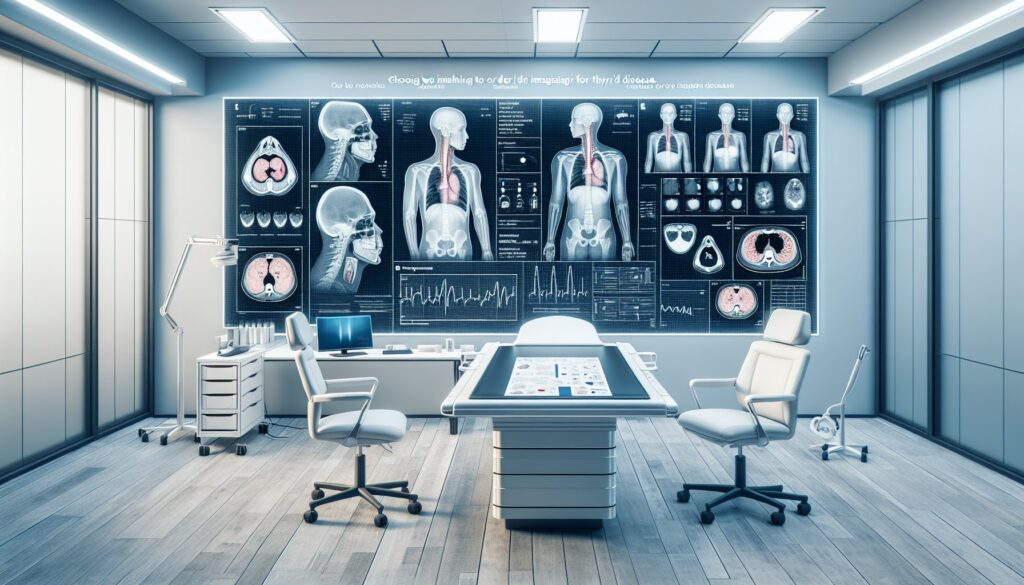
When to Order Imaging for Thyroid Disease: ACR Appropriateness Decoded
A patient presents with a palpable neck mass, and you suspect a thyroid nodule. Or perhaps their lab work returns with a TSH of 0.1 mIU/L, pointing toward thyrotoxicosis. In both scenarios, the next step often involves imaging, but the optimal study depends entirely on the clinical question. Choosing between ultrasound, a nuclear medicine uptake scan, or CT can be complex, especially when considering radiation dose and diagnostic yield. This guide decodes the American College of Radiology (ACR) Appropriateness Criteria for thyroid disease, providing a clear, evidence-based framework for ordering the right test for your patient.
What Does the American College of Radiology Guideline for Thyroid Disease Cover?
The ACR Appropriateness Criteria for Thyroid Disease provide evidence-based recommendations for imaging in specific, common clinical situations. The guidelines are designed to help referring physicians select the most appropriate imaging study for a patient’s condition while minimizing unnecessary radiation exposure. This topic focuses on the initial evaluation and follow-up of structural and functional thyroid disorders.
The covered scenarios include:
- Initial imaging for a palpable thyroid nodule or suspected goiter.
- Workup of functional disorders like thyrotoxicosis and primary hypothyroidism.
- Preoperative evaluation for known differentiated thyroid cancer.
- Post-treatment and surveillance imaging for suspected recurrence of both differentiated and medullary thyroid cancers.
These criteria do not cover the management of incidentally discovered thyroid nodules (incidentalomas) on other imaging studies, such as a CT of the chest or neck performed for other reasons. Those scenarios are addressed in separate, dedicated guidelines.
What Imaging Should I Order for Thyroid Disease? Recommendations by Clinical Scenario
The choice of imaging for thyroid disease is highly dependent on the clinical context, distinguishing between anatomical evaluation (nodules, goiter) and functional assessment (hyper- or hypothyroidism).
For the initial evaluation of a palpable thyroid nodule in a euthyroid patient, the ACR designates US thyroid as Usually appropriate. Ultrasound is the primary modality for characterizing nodules, assessing for suspicious features, and guiding fine-needle aspiration. It is non-invasive and uses no ionizing radiation. Cross-sectional imaging like CT and MRI is rated Usually not appropriate for this initial workup.
Similarly, for a suspected goiter, US thyroid is Usually appropriate to assess gland size, texture, and the presence of nodules. In cases where there is concern for substernal extension or airway compression, a CT neck without IV contrast is also considered Usually appropriate to delineate the extent of the goiter.
When evaluating thyrotoxicosis, both structural and functional imaging are key. US thyroid is Usually appropriate to look for nodules or signs of Graves’ disease. Concurrently, nuclear medicine scans like an I-123 uptake scan or an I-131 uptake scan are also Usually appropriate to differentiate between causes of hyperthyroidism, such as Graves’ disease versus a toxic adenoma. In contrast, for primary hypothyroidism, imaging is typically not indicated, and nearly all modalities, including ultrasound, are rated Usually not appropriate as the diagnosis is primarily clinical and biochemical.
In the context of malignancy, for the preoperative evaluation of differentiated thyroid cancer, both US thyroid (for detailed neck mapping of lymph nodes) and CT neck with IV contrast (to assess for local invasion and nodal metastasis) are Usually appropriate. Following treatment, surveillance for recurrence also relies heavily on ultrasound. For suspected recurrence of differentiated thyroid cancer, US thyroid, MRI neck without and with IV contrast, CT neck with IV contrast, and an I-123 whole body scan are all considered Usually appropriate depending on the specific clinical and biochemical findings (e.g., rising thyroglobulin). For suspected recurrence of medullary thyroid cancers, which do not typically take up iodine, cross-sectional imaging with MRI neck without and with IV contrast and CT of the neck and chest with IV contrast are the Usually appropriate modalities.
ACR Imaging Recommendations for Thyroid Disease: A Summary Table
| Clinical Scenario | Top Procedure | ACR Rating | Adult RRL | Pediatric RRL |
|---|---|---|---|---|
| Palpable thyroid nodule. Not goiter. Euthyroid. Initial imaging. | US thyroid | Usually appropriate | O 0 mSv | O 0 mSv [ped] |
| Suspected goiter. Initial imaging. | US thyroid | Usually appropriate | O 0 mSv | O 0 mSv [ped] |
| Thyrotoxicosis. Initial imaging. | US thyroid | Usually appropriate | O 0 mSv | O 0 mSv [ped] |
| Primary hypothyroidism. Initial imaging. | US thyroid | Usually not appropriate | O 0 mSv | O 0 mSv [ped] |
| Preoperative evaluation of differentiated thyroid cancer. | US thyroid | Usually appropriate | O 0 mSv | O 0 mSv [ped] |
| Early imaging after treatment of differentiated thyroid cancer. | US thyroid | Usually appropriate | O 0 mSv | O 0 mSv [ped] |
| Suspected recurrence of differentiated thyroid cancer. | US thyroid | Usually appropriate | O 0 mSv | O 0 mSv [ped] |
| Suspected recurrence of medullary thyroid cancers. | US thyroid | Usually appropriate | O 0 mSv | O 0 mSv [ped] |
Adult vs. Pediatric Thyroid Disease Imaging: Radiation Dose Tradeoffs
Managing radiation exposure is a critical consideration in all patients, but it is especially important in children and young adults due to their increased radiosensitivity and longer life expectancy, which allows more time for potential long-term effects of radiation to manifest. The principle of As Low As Reasonably Achievable (ALARA) is paramount in pediatric imaging.
For thyroid disease, ultrasound is the preferred first-line imaging modality for most initial evaluations in both adults and children precisely because it involves no ionizing radiation. When cross-sectional imaging like CT is necessary, pediatric protocols are tailored to minimize the dose. As reflected in the ACR data, the relative radiation level (RRL) for a pediatric CT of the neck (0.3-3 mSv) is often lower than the typical adult dose (1-10 mSv). This is achieved by adjusting technical parameters like tube current (mA) and voltage (kVp) based on the child’s size. While MRI also avoids ionizing radiation, it may require sedation or anesthesia in younger children, introducing a different set of risks to consider.
Imaging Protocol Details for Thyroid Disease
Once you’ve decided on the right study, the specific imaging protocol is essential for diagnostic quality. A well-designed protocol ensures that the radiologist receives the necessary information to answer the clinical question, covering details like patient positioning, transducer frequency for ultrasound, and contrast timing for CT or MRI. Our protocol guides cover technique, contrast parameters, and interpretation principles for the studies recommended above:
Tools to Help You Order the Right Study for Thyroid Disease
Navigating imaging guidelines and radiation safety can be streamlined with dedicated clinical decision support. GigHz offers several resources designed to help clinicians at the point of care.
The ACR Appropriateness Criteria Lookup tool provides a searchable interface to the complete ACR guidelines, allowing you to quickly find recommendations for thousands of clinical scenarios beyond thyroid disease.
For detailed procedural steps, the Imaging Protocol Library offers a comprehensive collection of standardized protocols for CT, MRI, ultrasound, and other modalities, ensuring you know the technical specifics of the test you are ordering.
To facilitate discussions with patients about radiation, the Radiation Dose Calculator helps estimate and track cumulative radiation exposure from medical imaging, providing clear, understandable context for shared decision-making.
Why is ultrasound the first-line test for a palpable thyroid nodule?
Ultrasound is the ideal initial imaging modality for a palpable thyroid nodule because it provides high-resolution anatomical detail of the thyroid gland and surrounding neck structures without using ionizing radiation. It can accurately measure the nodule’s size, determine its composition (solid, cystic, or mixed), and identify suspicious features such as microcalcifications, irregular margins, or a “taller-than-wide” shape. This information is crucial for risk stratification using systems like the ACR TI-RADS, which guides the decision on whether to perform a fine-needle aspiration (FNA) biopsy.
When is a nuclear medicine scan (like I-123) indicated for thyroid disease?
A nuclear medicine thyroid uptake and scan is indicated when assessing the functional status of the thyroid gland, particularly in cases of thyrotoxicosis (hyperthyroidism). It helps determine the cause by measuring how much iodine the gland absorbs. High uptake is characteristic of Graves’ disease or a toxic nodular goiter, while low uptake suggests thyroiditis or exogenous thyroid hormone intake. It is generally not used for the initial evaluation of a nodule in a patient with normal thyroid function tests (euthyroid).
Is imaging ever appropriate for primary hypothyroidism?
According to the ACR Appropriateness Criteria, imaging is Usually not appropriate for the initial workup of primary hypothyroidism. The diagnosis is typically made based on clinical symptoms and laboratory tests (high TSH, low free T4). The most common cause in iodine-sufficient regions is Hashimoto’s thyroiditis, an autoimmune condition that does not require imaging for diagnosis. Imaging might be considered later if a goiter or nodule develops, but it is not part of the initial diagnostic pathway.
What is the role of CT versus MRI in evaluating thyroid cancer?
Both CT with IV contrast and MRI with and without IV contrast are valuable for evaluating known or suspected thyroid cancer, particularly for assessing the extent of disease. CT is often faster and better at detecting calcifications and evaluating bone invasion. It is excellent for assessing lymph node involvement in the central and lateral neck compartments. MRI offers superior soft tissue contrast, which can be advantageous for evaluating invasion into adjacent structures like the trachea, esophagus, or recurrent laryngeal nerve. The choice between them often depends on the specific clinical question, institutional preference, and patient factors like contrast allergies or renal function.
Reviewed by Pouyan Golshani, MD, Interventional Radiologist — May 12, 2026