Should You Order MRI for Suspected Creutzfeldt-Jakob Disease? An ACR Workflow
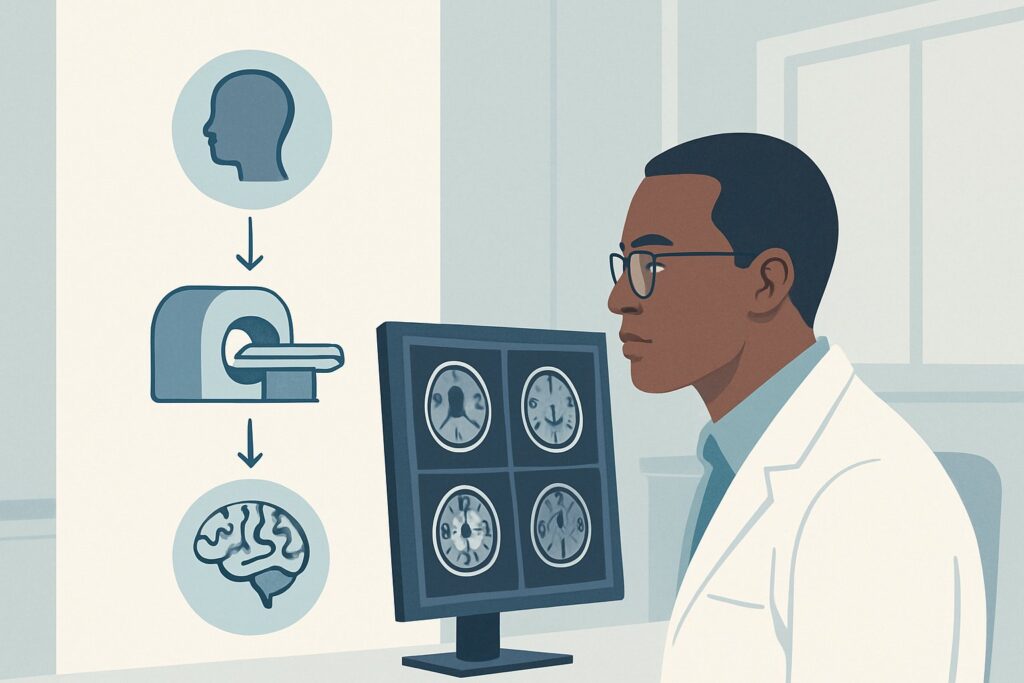
A 68-year-old patient is brought in by their family with a distressing three-month history of rapid cognitive decline, new-onset myoclonic jerks, and worsening gait instability. The constellation of symptoms raises urgent concern for a rapidly progressive dementia (RPD), with Creutzfeldt-Jakob disease (CJD) high on the differential. As the consulting physician, you must decide on the initial imaging study to both support this diagnosis and, critically, exclude its mimics. This article provides a detailed workflow for this specific clinical scenario, guiding you through the rationale for the recommended imaging, the differential diagnosis, and downstream clinical management. Based on the American College of Radiology (ACR) Appropriateness Criteria, an `MRI head without and with IV contrast` is rated Usually Appropriate as the initial imaging study.
Who Fits This Clinical Scenario?
This guidance applies to adult patients presenting with a subacute, rapidly progressive dementia. The classic clinical triad for sporadic Creutzfeldt-Jakob disease—dementia, myoclonus, and ataxia—is the core presentation. The key feature is the tempo of the decline, which occurs over weeks to months, not years. Patients in this scenario are often experiencing a profound loss of cognitive and functional abilities that is alarming to both them and their families.
This workflow is specifically for the initial imaging workup where CJD is a primary consideration. It is crucial to distinguish this scenario from others that may present differently:
- Slowly Progressive Dementia: Patients with a gradual cognitive decline over many months or years, typical of Alzheimer’s disease or frontotemporal dementia, do not fit this scenario. Their imaging workup follows a different pathway.
- Classic Parkinsonian Syndromes: Patients whose primary symptoms are tremor, rigidity, and bradykinesia should be evaluated under the Parkinsonian syndromes variant, even if cognitive changes are present.
- Predominant Chorea: If the dominant movement disorder is chorea, the workup should be directed toward suspected Huntington disease, which has its own distinct imaging recommendations.
The focus here is on the urgent need to differentiate CJD from other catastrophic but potentially treatable causes of RPD.
What Diagnoses Are You Working Up in This Scenario?
When a patient presents with rapidly progressive dementia, the differential diagnosis is broad and includes several urgent conditions. The initial imaging study is pivotal in narrowing this list and guiding subsequent tests.
Creutzfeldt-Jakob Disease (CJD): This is the primary diagnosis of concern. A fatal prion disease, CJD causes spongiform changes in the brain, leading to characteristic findings on specific MRI sequences. Imaging is a core component of the diagnostic criteria, making it an essential first step. While definitive diagnosis is via brain biopsy or autopsy, a classic clinical picture combined with characteristic MRI, electroencephalogram (EEG), and cerebrospinal fluid (CSF) findings can provide a probable diagnosis.
Autoimmune or Paraneoplastic Encephalitis: These conditions can perfectly mimic the RPD of CJD and are often treatable with immunotherapy. Conditions like anti-LGI1 or anti-NMDA receptor encephalitis can cause cognitive decline, seizures, and movement disorders. MRI in these cases may show T2/FLAIR hyperintensity, particularly in the limbic system, and may demonstrate enhancement after contrast administration—a key feature that distinguishes it from CJD.
Infectious Encephalitis: Viral encephalitides, such as Herpes Simplex Virus (HSV), can present with rapid neurologic deterioration. While the presentation is often more acute, a subacute course is possible. MRI is crucial for identifying typical patterns of involvement (e.g., temporal lobe changes in HSV) and enhancement that would point away from CJD and toward an infectious cause.
Vascular Causes: Multiple, strategically located cortical or subcortical infarcts can lead to a stepwise or rapid decline in cognitive function, sometimes mimicking RPD. MRI is highly effective at identifying acute, subacute, and chronic ischemic changes that would suggest a vascular etiology.
Metabolic or Toxic Encephalopathy: Conditions like Wernicke encephalopathy can cause ataxia and cognitive changes. While often diagnosed clinically and with lab work, MRI can show characteristic signal abnormalities in the mammillary bodies, thalami, and periaqueductal gray matter, helping to confirm the diagnosis.
Why Is MRI Head Without and With IV Contrast the Recommended Study?
The ACR panel rates `MRI head without and with IV contrast` as Usually Appropriate because it provides the highest diagnostic utility for evaluating the key differential diagnoses in rapidly progressive dementia. Its strength lies in its superior soft-tissue contrast and the power of specific advanced sequences.
The cornerstone of MRI for suspected CJD is Diffusion-Weighted Imaging (DWI). This sequence is highly sensitive for the cytotoxic edema caused by prion disease, appearing as restricted diffusion (bright signal on DWI, dark on ADC maps). These changes manifest in two classic patterns:
- Cortical Ribboning: High signal along the cerebral cortex, often in a gyriform pattern.
- Deep Gray Matter Involvement: High signal in the basal ganglia (caudate, putamen) and/or thalamus.
These DWI changes are often more conspicuous and appear earlier than corresponding abnormalities on Fluid-Attenuated Inversion Recovery (FLAIR) sequences. Therefore, a high-quality DWI sequence is non-negotiable in this workup.
The addition of IV contrast is critical not for diagnosing CJD itself (which typically does not enhance), but for evaluating the mimics. Autoimmune, infectious, and neoplastic processes frequently demonstrate enhancement, and its presence immediately redirects the diagnostic workup toward these other possibilities. Ordering the study without contrast saves time but risks missing a treatable alternative.
Why are other studies rated lower?
- CT head without IV contrast is rated May be appropriate. Its role is limited to excluding gross pathology like a large tumor, hemorrhage, or hydrocephalus. It is completely insensitive to the subtle parenchymal changes of CJD and most of its mimics. A normal CT can provide false reassurance and delay the definitive diagnosis.
- FDG-PET/CT brain is also rated May be appropriate. It can show patterns of cerebral hypometabolism that are suggestive of CJD. However, MRI is more specific, more widely available on an emergent basis, and avoids the significant radiation dose (RRL ☢☢☢ 1-10 mSv). PET is generally reserved as a secondary, problem-solving tool if the MRI is equivocal or non-diagnostic.
Ultimately, MRI without and with IV contrast offers the best balance of sensitivity for CJD and specificity for excluding its most important mimics, all without the use of ionizing radiation (RRL O 0 mSv).
What’s Next After MRI Head Without and With IV Contrast? Downstream Workflow
The results of the initial MRI will dictate the subsequent diagnostic pathway. The workflow branches based on whether the findings are classic for CJD, suggestive of an alternative diagnosis, or normal.
If the MRI is positive for CJD: When the scan reveals classic cortical ribboning or deep gray matter restricted diffusion on DWI, the probability of CJD becomes very high. The next steps are to obtain confirmatory evidence. This involves:
- Lumbar Puncture: Send cerebrospinal fluid (CSF) for 14-3-3 protein, tau protein, and, most importantly, the highly specific Real-Time Quaking-Induced Conversion (RT-QuIC) assay.
- EEG: An electroencephalogram should be performed to look for periodic sharp wave complexes (PSWCs), another classic supportive finding.
A “probable” diagnosis of sporadic CJD can be made with a characteristic clinical syndrome and a positive MRI, EEG, or RT-QuIC test.
If the MRI suggests a mimic: If the MRI shows findings like limbic enhancement, multiple scattered enhancing lesions, or changes typical of a specific infection (e.g., HSV), the workup pivots immediately. This typically involves a lumbar puncture to test for autoimmune/paraneoplastic antibody panels, infectious PCR, and cytology. A rheumatologic workup may also be indicated. This is where MRI with contrast proves its value by identifying potentially treatable conditions.
If the MRI is negative: A high-quality, technically adequate MRI that is negative for restricted diffusion makes sporadic CJD less likely but does not definitively rule it out, as findings can be subtle or absent in the very early stages. In this case, the focus should shift strongly toward the non-CJD differential. A comprehensive CSF analysis for inflammatory, infectious, and paraneoplastic markers is essential. If clinical suspicion for CJD remains high despite a negative initial scan, a repeat MRI in 2-4 weeks may be warranted to assess for evolving changes.
Pitfalls to Avoid (and When to Get Help)
In the urgent workup of rapidly progressive dementia, several common pitfalls can delay diagnosis or lead to misinterpretation.
- Accepting a “Normal” CT: A non-contrast head CT is expected to be normal in early CJD. Do not let this result lower your suspicion or delay ordering the necessary MRI.
- Omitting DWI Sequences: Ensure the ordered MRI protocol explicitly includes high-resolution Diffusion-Weighted Imaging and corresponding ADC maps. These are the most sensitive sequences and should not be skipped.
- Ignoring the Need for Contrast: Foregoing IV contrast to save time is a significant error. It is the primary way to identify treatable inflammatory mimics of CJD.
- Misinterpreting DWI Artifacts: Technical issues like motion or susceptibility artifact can sometimes mimic restricted diffusion. These findings should always be correlated with the ADC map and other sequences by an experienced neuroradiologist.
If the clinical picture is highly suggestive but the initial imaging and CSF studies are equivocal, this is the time to escalate. Consult with a neurologist and neuroradiologist to review the case, consider a repeat MRI, and discuss advanced testing like FDG-PET or a comprehensive autoimmune panel.
Related ACR Topics and Tools
This article focuses on a single, critical decision point. For a broader view of imaging for other related conditions, or to explore the tools used to make these decisions, the following resources are available.
- For breadth across all scenarios in Movement Disorders and Neurodegenerative Diseases, see our parent guide: Movement Disorders and Neurodegenerative Diseases: ACR Appropriateness Decoded.
- To look up appropriateness ratings for thousands of other clinical scenarios, use the ACR Appropriateness Criteria Lookup.
- For detailed technical parameters of the recommended study, explore the Imaging Protocol Library.
- To discuss radiation exposure from alternative studies like CT or PET/CT with patients, consult the Radiation Dose Calculator.
Frequently Asked Questions
Why is IV contrast recommended if Creutzfeldt-Jakob disease itself doesn’t typically enhance?
The intravenous contrast is not for diagnosing CJD, but for ruling out its mimics. Many treatable conditions that cause rapidly progressive dementia, such as autoimmune, paraneoplastic, or infectious encephalitis, often show characteristic patterns of enhancement. Seeing enhancement on the MRI immediately shifts the diagnostic workup toward these other possibilities.
Can I just order a non-contrast CT scan first to rule out a stroke or bleed?
While a non-contrast CT is rated as ‘May be appropriate’ by the ACR, its utility is very limited. It can exclude major events like a large hemorrhage or tumor but is insensitive to the specific brain changes of CJD and most of its inflammatory mimics. A normal CT can provide false reassurance and delay the definitive and more informative MRI study.
What specific MRI sequences are the most important for a suspected CJD workup?
Diffusion-Weighted Imaging (DWI) with its corresponding Apparent Diffusion Coefficient (ADC) map is the single most critical sequence. It is highly sensitive to the early cellular changes of CJD, often showing abnormalities before other sequences like FLAIR. The classic findings are restricted diffusion (bright on DWI, dark on ADC) in a ‘cortical ribboning’ pattern or in the deep gray matter (basal ganglia and thalamus).
What should I do if the initial MRI is negative but my clinical suspicion for CJD remains high?
A negative initial MRI does not completely exclude CJD, as imaging findings can lag behind clinical symptoms. If suspicion is high, the next steps should include a comprehensive CSF analysis with RT-QuIC testing and an EEG. Additionally, consider repeating the brain MRI in 2 to 4 weeks, as the characteristic DWI findings may evolve and become apparent over time.
Does a ‘classic’ MRI for CJD confirm the diagnosis?
No, an MRI with classic findings of cortical ribboning and/or deep gray matter restricted diffusion provides strong supportive evidence but is not, by itself, a definitive diagnosis. According to diagnostic criteria, this finding allows for a ‘probable’ diagnosis of sporadic CJD when combined with a characteristic clinical presentation. Definitive diagnosis still requires neuropathological confirmation from brain tissue.
Reviewed by Pouyan Golshani, MD, Interventional Radiologist — August 4, 2026