What Imaging Is Best for Acute Neck Pain Without Radiculopathy or Trauma?
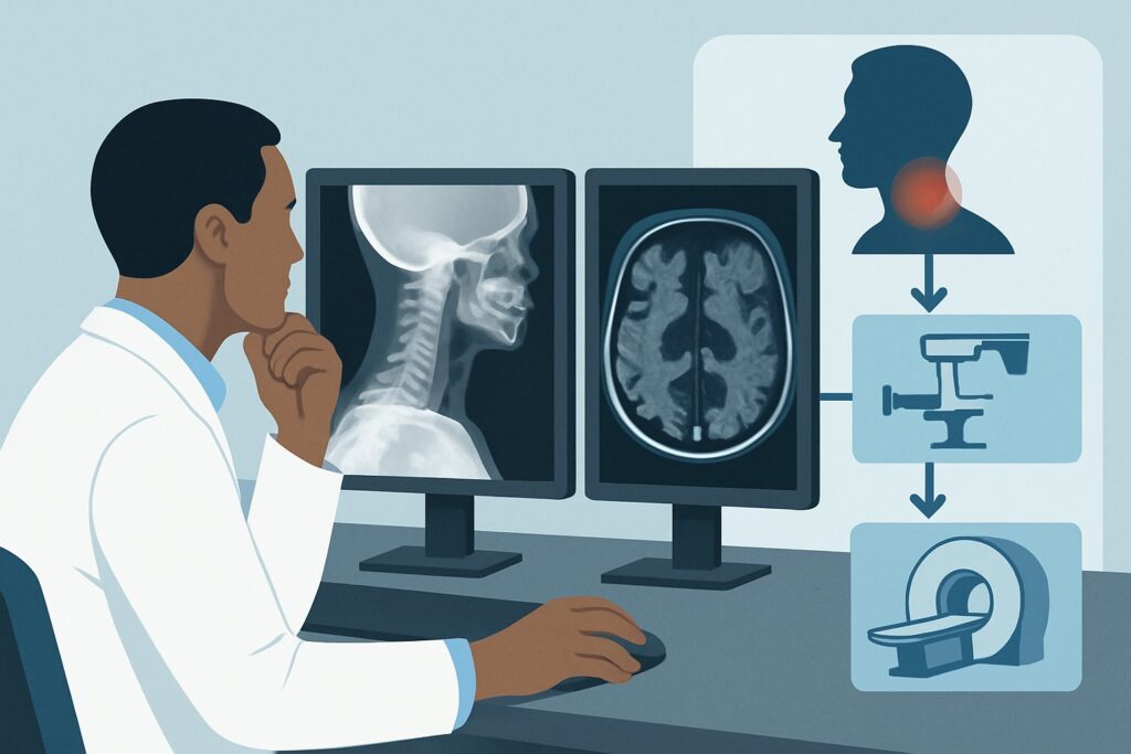
A 45-year-old patient presents to your clinic on a Tuesday morning with three days of worsening, central neck pain after sleeping awkwardly. They have full range of motion, though it is painful at the extremes. There is no history of trauma, no fever, and crucially, no pain, numbness, or weakness radiating into their arms or hands. The neurologic exam is normal. You consider ordering imaging to evaluate their axial cervical pain, but which study, if any, is the right first step? This article provides a detailed clinical workflow for this specific scenario: an adult with acute or increasing cervical pain without radiculopathy or red flags.
For this presentation, the American College of Radiology (ACR) Appropriateness Criteria rates Radiography cervical spine as May be appropriate. This nuanced rating reflects that while imaging can be useful, it is not always required, and conservative management is often the primary initial step.
Who Fits This Clinical Scenario for Acute Cervical Pain?
This guidance is tailored for a very specific and common patient presentation. Correctly identifying if your patient fits this scenario is critical to avoiding unnecessary imaging and ensuring appropriate care.
Inclusion Criteria (This article applies if your patient is):
- An adult.
- Experiencing acute (recent onset) or progressively increasing cervical pain.
- The pain is primarily axial (located in the neck itself) without radicular symptoms (no pain, paresthesia, or weakness radiating down the arm in a dermatomal pattern).
- There is no history of recent, significant trauma.
- There are no “red flags” suggesting a more serious underlying condition.
Exclusion Criteria (This workflow does NOT apply if your patient has):
- Radiculopathy: If the patient has symptoms radiating into an arm, they fit the sibling scenario for acute cervical pain with radiculopathy, where the imaging workup is different and often involves Magnetic Resonance Imaging (MRI).
- Recent Trauma: Any history of significant trauma (e.g., motor vehicle collision, fall) necessitates a different evaluation, often guided by clinical decision rules like the Canadian C-Spine Rule or NEXUS criteria to rule out fracture or ligamentous injury.
- Red Flags: The presence of systemic symptoms like fever, chills, or unexplained weight loss should raise suspicion for infection or malignancy. A known history of cancer, immunosuppression, or intravenous drug use also shifts the patient into a higher-risk category requiring a different, more urgent imaging pathway.
What Diagnoses Are You Working Up in This Scenario?
In the absence of trauma or red flags, the differential diagnosis for acute axial neck pain is primarily focused on benign, self-limiting conditions. The goal of initial imaging, when performed, is less about confirming a specific soft-tissue diagnosis and more about ruling out significant structural abnormalities and providing reassurance.
Cervical Strain or Sprain (Mechanical Neck Pain)
This is by far the most common cause. It involves injury to the muscles and ligaments of the neck, often from poor posture, awkward sleeping positions, or minor overuse. The clinical presentation is one of localized pain and stiffness that worsens with movement. Imaging is typically normal in these cases, as radiographs do not visualize soft tissues well. The diagnosis is largely clinical.
Cervical Spondylosis (Degenerative Disease)
This refers to age-related wear and tear of the cervical spine’s discs and facet joints. It is extremely common in adults and often asymptomatic. When it does cause pain, it is typically a chronic, aching discomfort. In an acute presentation, an underlying degenerative state may be exacerbated by a minor strain. Radiographs are effective at identifying spondylotic changes like disc space narrowing, osteophyte (bone spur) formation, and facet arthropathy.
Cervical Facet Arthropathy
A specific form of spondylosis, this involves degenerative arthritis of the facet joints, which guide and limit the spine’s motion. This can be a primary source of axial neck pain, sometimes with a referred pain pattern to the head or shoulders (but not true radiculopathy). Radiographs can show hypertrophic changes of the facet joints, which supports this diagnosis.
Why Are Cervical Spine X-rays Only ‘May Be Appropriate’ for This Presentation?
The ACR’s rating of May be appropriate for cervical spine radiography is a key clinical insight. It signifies that imaging is not mandatory for every patient in this scenario. For many, a trial of conservative therapy (e.g., physical therapy, NSAIDs, activity modification) for 4 to 6 weeks is the most appropriate first step. The decision to image should be based on the severity of pain, lack of improvement with initial conservative care, or the need to rule out underlying structural issues before initiating certain manual therapies.
When imaging is chosen, a standard radiographic series is the logical starting point. It is widely available, inexpensive, and provides a solid overview of bony alignment and degenerative changes. It effectively screens for less common but important causes of neck pain, such as significant spondylolisthesis (vertebral slippage) or unexpected destructive lesions. The radiation dose is relatively low (Relative Radiation Level ☢☢, 0.1-1 mSv), making it a safe initial examination.
In contrast, more advanced imaging modalities are deemed Usually not appropriate for this initial workup:
- MRI cervical spine without IV contrast: While MRI provides exquisite detail of soft tissues like intervertebral discs, the spinal cord, and nerve roots, it is not the recommended first step for uncomplicated axial neck pain. The primary reason is the high prevalence of abnormal findings (e.g., disc bulges, protrusions) in asymptomatic adults. Ordering an MRI too early can lead to the discovery of incidental findings that do not correlate with the patient’s symptoms, potentially causing patient anxiety and leading to unnecessary downstream procedures. MRI is reserved for cases where radiculopathy develops or when pain persists despite a full course of conservative therapy.
- CT cervical spine without IV contrast: CT offers superior bone detail compared to radiography and is the gold standard for evaluating suspected fractures. However, since trauma is an exclusion criterion for this scenario, its primary advantage is not relevant. Furthermore, CT involves a significantly higher radiation dose (Relative Radiation Level ☢☢☢, 1-10 mSv) than radiography without offering additional, clinically necessary information for this specific presentation.
What’s Next After Cervical Spine Radiography? Downstream Workflow
The results of the cervical spine radiograph will guide your next steps, which most often lead back to conservative management.
- If the radiograph is negative or shows only mild, age-appropriate degenerative changes: This is the most frequent outcome. It provides reassurance that there is no significant structural pathology. The appropriate next step is to proceed with or continue conservative management. This typically includes physical therapy to improve range of motion and strengthen supporting musculature, along with non-steroidal anti-inflammatory drugs (NSAIDs) or other analgesics for pain control. The patient should be re-evaluated in 4 to 6 weeks.
- If the radiograph shows moderate to severe spondylosis: The findings can help explain the patient’s symptoms and guide physical therapy. For example, the presence of severe facet arthropathy might lead a therapist to avoid certain mobilization techniques. Management remains conservative initially, but the radiographic findings provide a clearer diagnosis and prognostic information.
- If the radiograph is negative but pain persists or worsens: If the patient fails to improve after a comprehensive 4-to-6-week course of conservative therapy, or if new symptoms like radiculopathy emerge, the clinical scenario has changed. At this point, referral to a specialist (e.g., physiatry, orthopedics) and consideration of advanced imaging, such as an MRI of the cervical spine, becomes appropriate. The patient now fits a different clinical pathway, often one for chronic pain or new-onset radiculopathy.
- If the radiograph reveals an unexpected or aggressive-appearing finding: In the rare event that an X-ray shows a finding suspicious for tumor, infection, or gross instability, an immediate escalation in workup is required. This would typically involve proceeding directly to CT or MRI and obtaining an urgent specialty consultation.
Pitfalls to Avoid (and When to Get Help)
Navigating this common clinical scenario involves avoiding several potential missteps that can lead to over-testing or delayed diagnosis.
- Pitfall 1: Prematurely ordering MRI. The most common error is ordering an MRI for uncomplicated axial neck pain. This often reveals incidental degenerative findings that are not the cause of the acute pain, leading to diagnostic confusion and unnecessary interventions.
- Pitfall 2: Ignoring the “red flags.” Failing to ask about or recognize red flags (fever, night sweats, unexplained weight loss, history of cancer) can lead to a missed diagnosis of a serious underlying condition like infection or malignancy.
- Pitfall 3: Misinterpreting radicular pain. Attributing arm pain to a shoulder problem when it follows a clear dermatomal pattern is a frequent pitfall. True radiculopathy changes the scenario and warrants a different workup.
- When to Escalate: If a patient develops new, objective neurologic deficits (e.g., progressive weakness, gait instability, signs of myelopathy like hyperreflexia or clonus), immediate escalation for advanced imaging (typically MRI) and specialty consultation is mandatory.
Related ACR Topics and Tools
For a comprehensive overview of all clinical variants related to neck pain and radiculopathy, and for tools to help with study selection and patient communication, the following resources are available.
For breadth across all scenarios in Cervical Pain or Cervical Radiculopathy, see our parent guide: Cervical Pain or Cervical Radiculopathy: ACR Appropriateness Decoded.
- ACR Appropriateness Criteria Lookup — for adjacent scenarios
- Imaging Protocol Library — for technique on the recommended study
- Radiation Dose Calculator — for cumulative dose conversations
Frequently Asked Questions
Is any imaging necessary for acute neck pain without trauma or red flags?
Not always. The ACR rates radiography as ‘May be appropriate,’ indicating that for many patients, a trial of 4-6 weeks of conservative management (physical therapy, NSAIDs) is the best initial step. Imaging is typically reserved for patients with severe pain, those who fail to improve with conservative therapy, or before certain manual therapy interventions.
Why not order an MRI first to see the discs and nerves?
While MRI provides excellent soft tissue detail, it is considered ‘Usually not appropriate’ for this specific initial presentation. This is because MRI frequently shows incidental findings like disc bulges in asymptomatic individuals, which can lead to over-diagnosis and unnecessary procedures. MRI becomes appropriate if the patient develops radicular symptoms or fails to improve after a full course of conservative treatment.
If the X-ray is normal, does that mean nothing is wrong?
A normal X-ray is a reassuring finding that rules out significant bony abnormalities, malalignment, or advanced degenerative disease. However, it does not visualize soft tissues. The most common cause of acute axial neck pain is musculoligamentous strain, which will not be visible on an X-ray. A normal result supports a clinical diagnosis of mechanical neck pain and reinforces a plan for conservative management.
What if the patient has a headache with their neck pain?
If the headache is suspected to be originating from the cervical spine (cervicogenic headache) and there are no other neurologic deficits, the patient may fit a different ACR scenario. However, the initial imaging considerations are often similar, starting with conservative management before imaging. If the headache has features of a primary headache disorder (like migraine) or other red flags, a different workup is needed.
Should I order flexion-extension X-rays for this patient?
No. For this scenario of acute, non-traumatic axial neck pain, flexion-extension views are rated as ‘Usually not appropriate’ by the ACR. These views are used to assess for ligamentous instability, which is a concern after significant trauma or in patients with certain systemic conditions (like rheumatoid arthritis), but not in this routine presentation.
Reviewed by Pouyan Golshani, MD, Interventional Radiologist — May 29, 2026