Which Imaging Study Is Best for Headache with Red Flags? An ACR-Guided Workflow
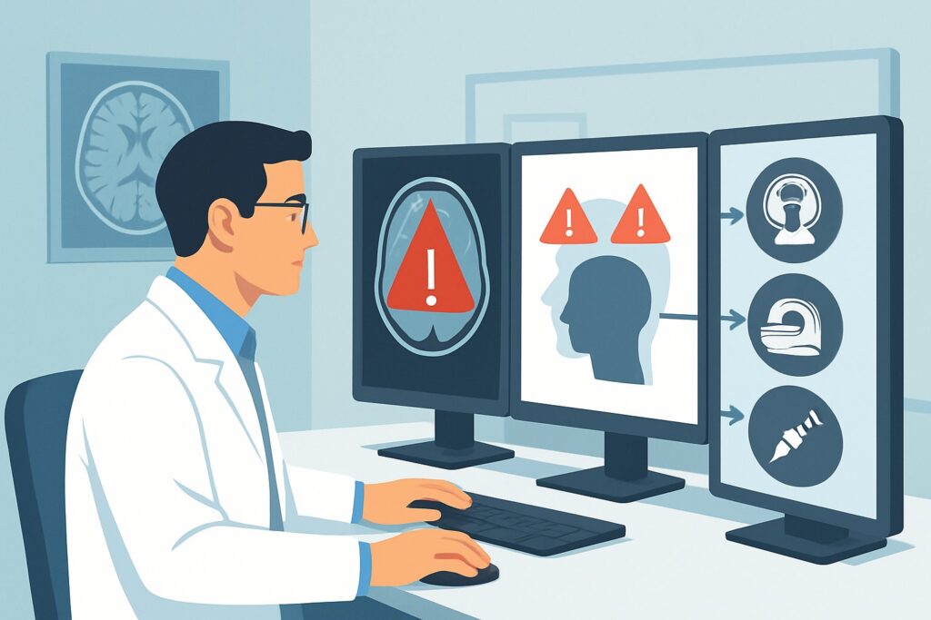
A 58-year-old woman with a history of treated breast cancer presents to your clinic with a new type of headache that has been progressively worsening over the past three weeks. She describes it as a dull, constant ache, and her neurologic examination is unremarkable. This presentation, combining older age of onset, a history of cancer, and an increasing severity pattern, raises several red flags that demand a thorough investigation beyond a simple clinical diagnosis. You need to decide on the most appropriate initial imaging study to evaluate for serious underlying pathology. According to the American College of Radiology (ACR) Appropriateness Criteria, for a patient with headache and one or more red flags, an MRI head without and with IV contrast is rated as Usually appropriate. This article details the clinical workflow for this specific scenario.
Who Fits This Clinical Scenario for a Headache with Red Flags?
This guidance applies to patients presenting with a headache accompanied by one or more of the following “red flag” features, which suggest a higher probability of secondary, structural, or otherwise serious causes. The specific red flags covered in this scenario are:
- Increasing frequency or severity of the headache
- Associated fever or a new neurologic deficit (e.g., weakness, sensory loss, cranial nerve palsy)
- A history of cancer or an immunocompromised state
- Older age of onset (typically defined as >50 years)
- Posttraumatic onset
It is crucial to distinguish this presentation from other headache scenarios that follow different diagnostic pathways. This workflow does not apply to:
- Patients with a sudden, severe “thunderclap” headache: A headache that reaches maximal severity within one hour is a distinct scenario, primarily concerned with subarachnoid hemorrhage. This presentation follows a separate ACR guideline.
- Patients with a classic primary headache and normal exam: A patient with a long-standing, stable pattern of migraine or tension-type headache and a consistently normal neurologic examination typically does not require initial imaging.
- Patients with a clearly positional headache: A headache that is significantly worse when upright and improves when lying down suggests intracranial hypotension, which has its own dedicated imaging workup.
What Diagnoses Are You Working Up in a Patient with Red Flag Headaches?
The presence of red flags broadens the differential diagnosis from primary headache disorders to serious secondary causes. The choice of imaging is driven by the need to identify or exclude these potentially life-threatening conditions.
Intracranial Mass: This is a primary concern, especially in patients with a history of cancer (metastasis), new neurologic deficits, or older age of onset. The workup must be sensitive enough to detect primary brain tumors like glioblastomas or meningiomas, as well as metastatic lesions which can be small and multifocal.
Infection or Abscess: In a patient with fever or immunocompromise, an intracranial infection is a critical consideration. This includes brain abscesses, meningitis, or encephalitis. Imaging must be able to detect parenchymal or meningeal inflammation and fluid collections.
Subdural Hematoma (SDH): Particularly relevant in older patients and those with a history of trauma (which may be minor or even forgotten), a chronic or subacute SDH can present with insidious, progressively worsening headaches. Imaging needs to reliably differentiate blood products of varying ages from adjacent brain parenchyma.
Inflammatory or Vascular Conditions: Though less common, conditions like cerebral venous sinus thrombosis (CVST) or large-vessel vasculitis (e.g., giant cell arteritis) can present with non-specific but worsening headaches. While the full workup for these extends beyond initial brain imaging, the chosen study should be able to reveal suggestive signs like venous sinus filling defects or vessel wall enhancement.
Why Is MRI Head Without and With IV Contrast the Recommended First Study?
The ACR designates MRI head without and with IV contrast as Usually appropriate because it provides the most comprehensive evaluation for the broad differential diagnosis in this red flag scenario. Its superior soft-tissue contrast and multi-sequence capabilities are essential for detecting the subtle findings associated with tumors, infections, and non-acute hemorrhages.
The non-contrast sequences (such as T1, T2, FLAIR, and DWI) are highly sensitive for detecting edema, ischemia, and blood products of different ages, which is critical for identifying a subacute subdural hematoma or an ischemic event. The addition of intravenous gadolinium-based contrast is what makes the study so powerful for this indication. Contrast enhancement is key to characterizing intracranial masses, identifying the ring-enhancing pattern of an abscess, and visualizing abnormal meningeal enhancement seen in meningitis or carcinomatosis.
While other studies are also rated for this scenario, they have significant limitations:
- CT head without IV contrast: This study is also rated Usually appropriate and is often more accessible in an emergency setting. It is excellent for detecting acute hemorrhage and large masses. However, it is significantly less sensitive than MRI for small tumors, early ischemia, inflammatory changes, and isodense subacute subdural hematomas. It also involves ionizing radiation (☢☢☢ 1-10 mSv), whereas MRI does not (O 0 mSv).
- CTA head with IV contrast: This study is rated Usually not appropriate as the initial imaging test for this scenario. A Computed Tomography Angiography (CTA) is optimized for visualizing arterial anatomy and is the primary modality for evaluating aneurysms in the setting of a thunderclap headache. For the broad differential in this red flag scenario, it provides limited information about the brain parenchyma, meninges, or venous system and would likely miss many of the target diagnoses.
Ultimately, the combination of non-contrast and contrast-enhanced sequences in a single MRI examination offers the highest diagnostic yield for the wide range of serious pathologies that must be considered in a patient with a red flag headache.
What’s the Next Step After the MRI Result? Downstream Workflow
The results of the MRI will guide the subsequent clinical pathway. The workflow branches based on whether the findings are positive, negative, or indeterminate.
If the study is positive for a specific finding:
- Mass Lesion: A finding suggestive of a primary tumor or metastasis prompts an urgent referral to neurosurgery and/or neuro-oncology. Further staging imaging (e.g., CT of the chest, abdomen, and pelvis) may be required if metastasis is suspected and no primary cancer is known.
- Abscess or Infection: This is a medical emergency requiring immediate infectious disease consultation, initiation of empiric antibiotics, and often neurosurgical evaluation for possible drainage.
- Subdural Hematoma: The disposition depends on the size, mass effect, and acuity. A small, chronic SDH may be managed conservatively, while a larger one causing neurologic symptoms requires emergent neurosurgical consultation for evacuation.
If the study is negative: A normal MRI is highly reassuring and significantly lowers the probability of a serious intracranial structural cause. The focus should shift back to diagnosing and managing a primary headache disorder (e.g., migraine, tension-type headache). If clinical suspicion for a specific condition like giant cell arteritis remains high despite a normal MRI, further targeted testing (e.g., ESR, CRP, temporal artery biopsy) is warranted.
If the study is indeterminate: Occasionally, an MRI may reveal non-specific findings, such as white matter changes or an incidental finding. In these cases, correlation with the specific clinical red flag is key. For example, if venous thrombosis was a concern and the standard MRI is equivocal, a dedicated MR Venography (MRV) may be the appropriate next step.
Pitfalls to Avoid (and When to Get Help)
Navigating the workup for a red flag headache requires careful consideration to avoid common diagnostic errors.
- Over-reliance on non-contrast CT: While a non-contrast head CT is fast and useful for ruling out acute hemorrhage, accepting a negative result as definitive in a patient with persistent red flags (like a history of cancer) can lead to a missed diagnosis of an isodense hematoma, tumor, or abscess.
- Forgetting the “V” in “VINDICATE”: Do not anchor solely on tumor or bleed. In the right clinical context (e.g., hypercoagulable state), maintain a high index of suspicion for cerebral venous sinus thrombosis, which requires dedicated venous imaging (CTV or MRV) if suspected.
- Ignoring the patient’s age: In a patient over 50 with a new headache, giant cell arteritis must be considered. A normal MRI does not rule it out, and clinical evaluation with inflammatory markers is essential.
If the clinical picture is worsening rapidly or a new, profound neurologic deficit develops, escalate care immediately to the emergency department and consult with neurology or neurosurgery, regardless of pending imaging results.
Related ACR Topics and Tools
For a comprehensive overview of imaging guidelines across all headache presentations, from primary migraine to thunderclap headache, please consult our parent topic hub article. For tools to help select the right study and understand the technical details, see the resources below.
- For breadth across all scenarios in Headache, see our parent guide: Headache: ACR Appropriateness Decoded.
- ACR Appropriateness Criteria Lookup — for adjacent scenarios
- Imaging Protocol Library — for technique on the recommended study
- Radiation Dose Calculator — for cumulative dose conversations
Frequently Asked Questions
Is a non-contrast head CT ever sufficient for a patient with a red flag headache?
A non-contrast head CT is rated ‘Usually appropriate’ and is often the first test in an emergency setting, primarily to rule out acute hemorrhage. However, it is less sensitive than MRI for many other serious causes like small tumors, abscesses, early strokes, or venous sinus thrombosis. If the CT is negative but the red flags persist, an MRI without and with contrast is typically the necessary next step to complete the workup.
Why is contrast necessary for the MRI in this scenario?
Intravenous gadolinium-based contrast is crucial because it highlights areas of blood-brain barrier breakdown. This is essential for identifying and characterizing many of the pathologies in the differential, such as the enhancement pattern of a tumor, the ring enhancement of an abscess, or the dural enhancement seen in meningitis or intracranial hypotension. A non-contrast MRI would miss or be unable to fully characterize these findings.
My patient has a history of cancer and a new headache, but the MRI was normal. What’s next?
A normal brain MRI is very reassuring against structural metastases. The next steps should focus on other potential causes. Consider leptomeningeal carcinomatosis, which can sometimes be difficult to detect on initial MRI; a lumbar puncture with cytology may be necessary if suspicion is high. Also, re-evaluate for non-neurologic causes related to their cancer or treatment, and consider management for a primary headache disorder if no secondary cause is found.
What if the patient has a contraindication to MRI, like a non-compatible pacemaker?
In cases where MRI is contraindicated, CT is the alternative. The most comprehensive CT-based study would be a CT head without and with IV contrast. This provides better characterization of potential masses or inflammatory processes than a non-contrast study alone. If vascular pathology like venous thrombosis is a specific concern, a CT Venogram (CTV) could be added.
Does a post-traumatic headache always require an MRI?
Not necessarily. The choice of imaging depends on the acuity and severity of the trauma and symptoms. In the acute setting immediately following significant head trauma, a non-contrast head CT is the standard of care to look for acute hemorrhage. This ACR scenario applies to headaches that develop or persist after the acute phase, or have a worsening pattern, where an MRI becomes more valuable for detecting subacute or chronic subdural hematomas and other subtle traumatic brain injuries.
Reviewed by Pouyan Golshani, MD, Interventional Radiologist — June 27, 2026